. 2023 Oct 27;51(19):10551-10567.
doi: 10.1093/nar/gkad740.

Rep protein accommodates together dsDNA and ssDNA which enables a loop-back mechanism to plasmid DNA replication initiation

Affiliations

Rep protein accommodates together dsDNA and ssDNA which enables a loop-back mechanism to plasmid DNA replication initiation

Katarzyna Wegrzyn et al. Nucleic Acids Res. .

Abstract

For DNA replication initiation in Bacteria, replication initiation proteins bind to double-stranded DNA (dsDNA) and interact with single-stranded DNA (ssDNA) at the replication origin. The structural-functional relationship of the nucleoprotein complex involving initiator proteins is still elusive and different models are proposed. In this work, based on crosslinking combined with mass spectrometry (MS), the analysis of mutant proteins and crystal structures, we defined amino acid residues essential for the interaction between plasmid Rep proteins, TrfA and RepE, and ssDNA. This interaction and Rep binding to dsDNA could not be provided in trans, and both are important for dsDNA melting at DNA unwinding element (DUE). We solved two crystal structures of RepE: one in a complex with ssDNA DUE, and another with both ssDNA DUE and dsDNA containing RepE-specific binding sites (iterons). The amino acid residues involved in interaction with ssDNA are located in the WH1 domain in stand β1, helices α1 and α2 and in the WH2 domain in loops preceding strands β1' and β2' and in these strands. It is on the opposite side compared to RepE dsDNA-recognition interface. Our data provide evidence for a loop-back mechanism through which the plasmid replication initiator molecule accommodates together dsDNA and ssDNA.


Figures

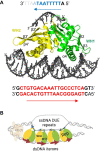
Graphical Abstract
Graphical Abstract
Figure 1.
Figure 1.
Identification of TrfA and RepE regions interacting with ssDNA. (A) Cartoon schemes of experiment with TrfA targeting ssDNA DUEoriV bottom strand (top) and of TrfA protein (bottom). The protein domains are coloured: DBD (pink), WH1 (green), WH2 (yellow). The location of the identified peptides is marked on a TrfA scheme. Sequence of detected peptides and amino acid residues numbers are put in parentheses. (B, C) MALDI-TOF spectrum of trypsin-digested TrfA crosslinked with ssDNA. Highlighted peaks indicate potential crosslinked products. (D) Cartoon schemes of RepE targeting ssDNA DUEoriS top strand (top) and RepE protein (bottom) The protein domains are coloured: WH1 (green), WH2 (yellow). The location of the identified peptides is marked on a RepE scheme. Sequence of detected peptides and amino acid residues numbers are put in parentheses. (E, F) MALDI-TOF spectrum of trypsin-digested RepE crosslinked with ssDNA. Highlighted peaks indicate potential crosslink products.
Figure 2.
Figure 2.
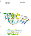
The crystal structures of RepE nucleoprotein complexes. (A) Crystal structure of RepE complexes with a single iteron dsDNA (PDB: 1REP) (29) and with ssDNA (this work). The WH1 domain in RepE is coloured in green and WH2 domain in yellow, while dsDNA is shown in blue and ssDNA in cyan. (B) Close-up view of protein fragment interacting with the thymidine-rich region of DUE ssDNA. Protein residues interacting with nucleic acid are shown in stick representation and labelled. Interacting atoms are connected with purple dotted lines. (C) Schematic representation of RepE protein–ssDNA interactions. The RepE amino acid residues interacting ssDNA are shown as ovals, coloured according to the protein domains: (green for WH1 and yellow for WH2). Radiant lines represent polar contacts and parallel lines van der Waals/stacking interactions. Black lines show contacts with amino acid side chains. Red lines show contacts with the protein backbone. Single red lines represent water-mediated contacts.
Figure 3.
Figure 3.
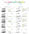
Analysis of RepE proteins variants interaction with ssDNA and dsDNA. (A) Schematic representation of RepE protein, with alpha helices (α) and beta strands (β) represented as coloured boxes and arrows, respectively. Protein domains are marked: WH1 domain in green and WH2 domain in yellow. The hydrophobic region is marked as dashed box. Peptide sequences identified by MALDI-TOF/TOF as being in contact with ssDNA (blue), amino acid substitutions predicted to affect RepE protein interaction with ssDNA (red), amino acid substitutions affecting RepE interaction with dsDNA (dark blue), amino acid substitution affecting RepE protein dimerization (orange). Analysis of the interaction of RepE protein variants (WT, F146E, Q171E, Y172E and R205E/R206E/R207E) with ssDNA (B) or dsDNA (C). The interaction was analyzed with electrophoretic mobility shift assays and surface plasmon resonance, as described in Materials and Methods. In EMSA, increasing amounts of RepE protein variants (10, 20, 40, 80, 165 nM) were incubated with 20 nM fluorescently labeled ssDNA containing DUEoriS top strand (B) and 50 nM dsDNA containing oriS sequence (C). Black arrows indicate nucleoprotein complexes. In SPR analysis, increasing amounts of protein (1, 3, 5, 10, 15, 30, 50 nM) were run over the surface of a sensor chip with immobilized ssDNA containing the sequence of DUEoriS top strand (B) or dsDNAoriS containing the sequence of iterons (C).
Figure 4.
Figure 4.
Analysis of TrfA proteins variants interaction with ssDNA and dsDNA. (A) Schematic representation of TrfA protein, with alpha helices (α) and beta strands (β) represented as coloured boxes and arrows, respectively. Protein domains are marked as follows: DBD domain in magenta, WH1 domains in green and WH2 domains in yellow. The hydrophobic region is marked as dashed boxes. Peptide sequences identified by MALDI-TOF/TOF as being in contact with ssDNA (blue), amino acid substitutions predicted to affect TrfA protein interaction with ssDNA (red), amino acid substitutions affecting TrfA protein interaction with dsDNA (dark blue), amino acid substitutions affecting TrfA dimerization (orange). (B, C) Analysis of interaction of TrfA protein variants with ssDNA (B) or dsDNA (C) was analyzed with an electrophoretic mobility shift assay and surface plasmon resonance, as described in Materials and Methods. In EMSA, increasing amounts of TrfA protein variants (WT, R156E, K303E, R327E and R347E) (10, 50, 100, 150, 250 nM) were incubated with 20 nM fluorescently labeled ssDNA containing DUEoriV bottom strand (B) or 50 nM dsDNA containing oriV sequence (C). Black arrows indicate nucleoprotein complexes. In SPR analysis, increasing amounts of protein (2, 5, 10, 15, 30, 60 nM) were run over the surface of a sensor chip with immobilized ssDNA containing the sequence of DUEoriV bottom strand (B) or dsDNAoriV containing the sequence of iterons (C).
Figure 5.
Figure 5.
Effects of the amino acid substitutions in RepE and TrfA proteins on plasmid DNA in vitro replication. Reactions were performed using crude extracts (FII) obtained from E. coli C600 cells (30) (see Materials and Methods). Reactions contained 250 ng of supercoiled DNA template, pZZ35 for RepE and pKD19L1 for TrfA, and increasing concentration of proteins variants. There are presented results from three independent repeats of each experiment.
Figure 6.
Figure 6.
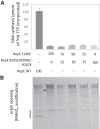
The ability to interact both dsDNA and ssDNA by RepE protein is required for efficient open complex formation and initiation of plasmid DNA synthesis. Reactions of in vitro DNA synthesis (A) and potassium permanganate footprinting followed by primer extension (B) were performed as described in Materials and Methods. Appropriate reaction mixtures contained 250 ng of supercoiled DNA template (pZZ35) and 100 ng of RepE protein variants (RepE WT, RepE F146E, RepE R205E/R206E/R207E or a mixture of these proteins) as indicated. For DNA synthesis results from three independent repeats of the experiment are presented. For KMnO4 footprinting assay the representative gel image is shown. The black line marks the region where DNA double helix destabilization and KMnO4 modification occur.
Figure 7.
Figure 7.
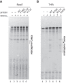
Rep-ssDNA complex formation is required for DUE melting. Potassium permanganate footprinting followed by primer extension was carried out for reaction mixtures containing RepE (A) or TrfA (B) proteins variants, as described in Materials and Methods. For reactions with RepE and TrfA protein variants, the plasmids pZZ35 and pKD19L1, respectively, were used as templates. The black line marks the region where DNA double helix destabilization and KMnO4 modification occur. In control reactions, KMnO4 and protein (A, lane 1 and B, lane 1) or just KMnO4 (A, lane 2 and B, lane 2) were omitted. The products of primer extension reaction with wild-type proteins (A, lane 3 and B, lane 3) and with mutations (A, lanes 4–6 and B, lanes 4–6), as indicated.
Figure 8.
Figure 8.
Plasmid Rep proteins form a triple nucleoprotein complex with both ssDNA and dsDNA. (A) The crystal structure of nucleoprotein complex of RepE protein with dsDNA containing the sequence of a single iteron (red) and ssDNA containing the sequence of 8-mer of DUEoriS top strand (blue). The sequence of DNA fragments detected in the crystals is bolded. The arrows indicate the direction of the specific motifs. (B) The proposed loop-back model of RepE initiator nucleoprotein complex at plasmid F oriS region. The RepE protomers bound to iterons and interacting 8-mers of DUEoriS sequences are shown. For detail description see Discussion. The WH1 domain of RepE is shown in green and the WH2 domain is in yellow. The red arrows indicate the position and direction of iteron sequences and the blue arrows indicate the position and direction of 8-mers in DUEoriS. The hypothetical position of HU protein (light orange) bound to dsDNA oriS is indicated.

Similar articles

Cited by

References

    1. Bleichert F., Botchan M.R., Berger J.M.. Mechanisms for initiating cellular DNA replication. Science. 2017; 355:eaah6317. - PubMed
    1. Hu Y., Stillman B.. Origins of DNA replication in eukaryotes. Mol. Cell. 2023; 83:352–372. - PMC - PubMed
    1. Yao N.Y., O’Donnell M.E. The DNA replication machine: structure and dynamic function. Subcell. Biochem. 2021; 96:233–258. - PubMed
    1. Rajewska M., Wegrzyn K., Konieczny I.. AT-rich region and repeated sequences - the essential elements of replication origins of bacterial replicons. FEMS Microbiol. Rev. 2012; 36:408–434. - PubMed
    1. Chatterjee S., Jha J.K., Ciaccia P., Venkova T., Chattoraj D.K.. Interactions of replication initiator RctB with single- and double-stranded DNA in origin opening of vibrio cholerae chromosome 2. Nucleic Acids Res. 2020; 48:11016–11029. - PMC - PubMed

Publication types