. 2024 Jan 4;147(1):311-324.
doi: 10.1093/brain/awad301.

TRAPPC6B biallelic variants cause a neurodevelopmental disorder with TRAPP II and trafficking disruptions

Hashem Almousa  1 Sara A Lewis  2   3 Somayeh Bakhtiari  2   3 Sandra Hinz Nordlie  2   3 Alex Pagnozzi  4 Helen Magee  2   3 Stephanie Efthymiou  5 Jennifer A Heim  2 Patricia Cornejo  6   7   8 Maha S Zaki  9   10 Najwa Anwar  11 Shazia Maqbool  11 Fatima Rahman  11 Derek E Neilson  12 Anusha Vemuri  13 Sheng Chih Jin  14 Xiao-Ru Yang  15 Abolfazl Heidari  16 Koen van Gassen  17 Aurélien Trimouille  18 Christel Thauvin-Robinet  19   20   21 James Liu  2   3 Ange-Line Bruel  20   21 Hoda Tomoum  22 Mennatallah O Shata  22 Mais O Hashem  23 Mehran Beiraghi Toosi  24   25 Ehsan Ghayoor Karimiani  26 Gözde Yeşil  27 Lokesh Lingappa  28 Debangana Baruah  28 Farnoosh Ebrahimzadeh  29 Julien Van-Gils  17 Laurence Faivre  19 Mina Zamani  30   31 Hamid Galehdari  30 Saeid Sadeghian  32 Gholamreza Shariati  31   33 Rahema Mohammad  5 Jasper van der Smagt  17 Alya Qari  34 John B Vincent  35 A Micheil Innes  15 Ali Dursun  36 R Köksal Özgül  36 Halil Tuna Akar  36 Kaya Bilguvar  37   38 Cyril Mignot  39   40 Boris Keren  39 Claudia Raveli  41 Lydie Burglen  42 Alexandra Afenjar  42 Laura Donker Kaat  43 Marjon van Slegtenhorst  43 Fowzan Alkuraya  23 Henry Houlden  5 Sergio Padilla-Lopez  2   3 Reza Maroofian  5 Michael Sacher  1   44 Michael C Kruer  2   3
Affiliations

TRAPPC6B biallelic variants cause a neurodevelopmental disorder with TRAPP II and trafficking disruptions

Hashem Almousa et al. Brain. .

Abstract

Highly conserved transport protein particle (TRAPP) complexes regulate subcellular trafficking pathways. Accurate protein trafficking has been increasingly recognized to be critically important for normal development, particularly in the nervous system. Variants in most TRAPP complex subunits have been found to lead to neurodevelopmental disorders with diverse but overlapping phenotypes. We expand on limited prior reports on TRAPPC6B with detailed clinical and neuroradiologic assessments, and studies on mechanisms of disease, and new types of variants. We describe 29 additional patients from 18 independent families with biallelic variants in TRAPPC6B. We identified seven homozygous nonsense (n = 12 patients) and eight canonical splice-site variants (n = 17 patients). In addition, we identified one patient with compound heterozygous splice-site/missense variants with a milder phenotype and one patient with homozygous missense variants. Patients displayed non-progressive microcephaly, global developmental delay/intellectual disability, epilepsy and absent expressive language. Movement disorders including stereotypies, spasticity and dystonia were also observed. Brain imaging revealed reductions in cortex, cerebellum and corpus callosum size with frequent white matter hyperintensity. Volumetric measurements indicated globally diminished volume rather than specific regional losses. We identified a reduced rate of trafficking into the Golgi apparatus and Golgi fragmentation in patient-derived fibroblasts that was rescued by wild-type TRAPPC6B. Molecular studies revealed a weakened interaction between mutant TRAPPC6B (c.454C>T, p.Q152*) and its TRAPP binding partner TRAPPC3. Patient-derived fibroblasts from the TRAPPC6B (c.454C>T, p.Q152*) variant displayed reduced levels of TRAPPC6B as well as other TRAPP II complex-specific members (TRAPPC9 and TRAPPC10). Interestingly, the levels of the TRAPPC6B homologue TRAPPC6A were found to be elevated. Moreover, co-immunoprecipitation experiments showed that TRAPPC6A co-precipitates equally with TRAPP II and TRAPP III, while TRAPPC6B co-precipitates significantly more with TRAPP II, suggesting enrichment of the protein in the TRAPP II complex. This implies that variants in TRAPPC6B may preferentially affect TRAPP II functions compared to TRAPP III functions. Finally, we assessed phenotypes in a Drosophila TRAPPC6B-deficiency model. Neuronal TRAPPC6B knockdown impaired locomotion and led to wing posture defects, supporting a role for TRAPPC6B in neuromotor function. Our findings confirm the association of damaging biallelic TRAPPC6B variants with microcephaly, intellectual disability, language impairments, and epilepsy. A subset of patients also exhibited dystonia and/or spasticity with impaired ambulation. These features overlap with disorders arising from pathogenic variants in other TRAPP subunits, particularly components of the TRAPP II complex. These findings suggest that TRAPPC6B is essential for brain development and function, and TRAPP II complex activity may be particularly relevant for mediating this function.

Keywords: ER-golgi trafficking; NEDMEBA; TRAPP-II complex; TRAPPC6B; TRAPPopathy; Trs33.


Conflict of interest statement

The authors report no competing interests.

Figures

Figure 1
Figure 1
Clinical features of patients with TRAPPC6B variants. (A) Phenogram of frequency of phenotypic features for 29 new patients and 8 previously reported patients (Supplementary Table 1). Fields with missing data were not included in calculations for that feature. Behavioural phenotypes include aggression, self-injury, anxiety and hyperactivity. ID/DD = intellectual disability/developmental disability. (BN) Photos of dysmorphic features. No unifying dysmorphic features were observed. (B and C) Patient 1 (Family 1) has gingival hypertrophy and swan neck deformity of fingers, representing dystonia. This patient also exhibited spastic-dystonic quadriplegia with contractures in the elbow flexors and plantar flexors, temporal wasting, muscle atrophy and curling of all her toes (not shown). (D) Patient 2 (Family 1) has synophrys, bitemporal narrowing, prominent cheekbones, a wide nasal root and bridge, and a papular lesion suspicious for an occult encephalocele. This patient also exhibited spastic-dystonic quadripledia, brachycephaly, trigonocephaly, temporal wasting and muscle atrophy in his proximal limb muscle (not shown). (E) Patient 5 (Family 4) has microcephaly, bitemporal narrowing, low anterior hairline, deep-set eyes, prominent ears, long nose with narrow nasal bridge and prognathia. (F) Patient 6 (Family 4) has microcephaly, bitemporal narrowing strabismus, positional plagiocephaly, deep set eyes, broad nasal root and narrow nasal bridge, and widely spaced teeth. (G) Patient 7 (Family 5) showing depressed nasal bridge, upturned nasal tip and thin upper lip. This patient also exhibited bilateral clinodactyly, tapered fingers and inverted nipples (not shown). (H and I) Patient 10 (Family 8) has microcephaly, long face, arched eyebrows, wide spaced eyes, almond shaped eye, straight nasal bridge, broad nose, broad chin and low set ears. Patient also exhibited arachnodactyly (not shown). (J and K) Patient 17 (Family 11) has narrow nasal bridge and posteriorly rotated ears. (L) Patient 18 (Family 11) has upslanting palpebral fissures and square nasal tip. (M and N) Patient 19 (Family 12) has microcephaly, bulbous nasal tip and creased earlobe.
Figure 2
Figure 2
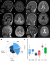
Qualitative and quantitative analysis of MRI images. (A, C, E, G, I and K) Sagittal MRI T1-weighted midline images. (B and H) Axial fluid attenuated inversion recovery (FLAIR) images. (D, F, J and L) Axial MRI T2-weighted images. (A and B) Patient 3, Family 2: 13-year-old female. (A) Small craniofacial ratio, thinning/foreshortening of the corpus callosum with greater involvement of the posterior fibres (red arrow). (B) Patchy FLAIR signal hyperintensity in the bilateral periventricular white matter (arrows) extending to the centrum semiovale, perirolandic and periatrial regions (not shown). (C and D) Patient 5, Family 4: 13-year-old female. (C) Small craniofacial ratio, diffuse thinning/foreshortening of the corpus callosum (red arrow) and brainstem and cerebellum volume loss (cyan arrow). (D) Abnormal angulation of the posterior margins of the ventricles (arrows). (E and F) Patient 6, Family 4: 9-year-old female. (E) Small craniofacial ratio, thinning/foreshortening of the corpus callosum and posterior greater than anterior parenchymal loss (arrow). (F) Diffuse parenchymal loss with preferential involvement of the posterior lobes, ex-vacuo dilatation and angulation of the ventricles (arrows). (G and H) Patient 13, Family 10: 6-year-old male. (G) Reduced frontal occipital diameter (FOD) and diffuse thinning and foreshortening of the corpus callosum (arrow). (H) Patchy FLAIR signal hyperintensity in periatrial white matter associated with abnormal square shape of the posterior margins of the lateral ventricles (arrows). (I and J) Patient 14, Family 10: 2-year-old female. (I) Reduced FOD, diffuse thinning and foreshortening of the corpus callosum (arrow). (J) Increased T2 signal in periatrial white matter associated with abnormal angulation of the posterior margins of the lateral ventricles (arrows). (K and L) Patient 16, Family 10: 5-year-old female. (K) Marginal FOD (red arrow) and inferior vermis hypoplasia (cyan arrow). (L) No signal abnormalities present. (M) Phenogram of MRI features for 16 new patients and 7 previously reported patients with MRI interpretation. Patient-specific details are provided in Supplementary Table 1. (N) Box and whisker plot of six structural measures quantified from brain MRI volumes of six patients, represented as z-scores in comparison to an age-matched control cohort of typically developing children. The threshold for significance of −1SD indicates reduced volume compared to the general population. Volume loss was identified in the cerebellum, but there was no consistent reduction in other measurements. Ventricle asymmetry measures laterality of ventricle expansion; no asymmetry was detected. Boxes represent 25th and 75th percentiles with median line; whiskers represent data range. CB = cerebellum; CC = corpus callosum; CTX = cortex; DGM = deep grey matter; GM = grey matter; Vent = ventricle asymmetry; VM = ventriculomegaly; WM = white matter; WMI = white matter hyperintensity or abnormalities.
Figure 3
Figure 3
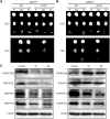
TRAPPC6B variants affect the stability of TRAPP II. TRAPPC6B (wild-type or p.Q152* variant) was cloned into either pGADT7 (A) or pGBKT7 (B); TRAPPC2L and TRAPPC3 were cloned into either pGBKT7 (A) or pGADT7 (B) and transformed into haploid yeast cells. In some cases, an empty vector (Φ) was used. The cells were mated, diploids selected and then spotted as serial dilutions on plates lacking leucine and tryptophan (DDO) or plates lacking leucine, tryptophan and histidine (TDO). (C) Fibroblasts from control or affected individuals from Family 15 (p.Q152*) were lysed and probed for the indicated TRAPP proteins or for tubulin as a loading control. WT = wild-type.
Figure 4
Figure 4
TRAPPC6B preferentially associates with the TRAPP II complex. (A) HeLa cells were either untransfected (NT) or co-transfected with TRAPPC6A-V5/TRAPPC10-FLAG, TRAPPC6A-V5/TRAPPC11-FLAG, TRAPPC6B-V5/TRAPPC10-FLAG or TRAPPC6B-V5/TRAPPC11. After 48 h, the cells were lysed and treated with anti-FLAG IgG agarose beads. The eluates from the immunoprecipitation were probed for V5, FLAG and tubulin. The blot is representative of at least three biological replicates. Inputs represent 10% of the sample subjected to immunoprecipitation (IP). (B) Quantification of the ratio of V5 immunoprecipitated with FLAG from three different experiments. The integrated density of V5, FLAG and background nearby for each band was measured using ImageJ v1.53. To obtain the corrected integrated density, the background value for each band was subtracted. The V5/FLAG ratio was then calculated using the corrected integrated densities and normalized to the highest signal detected for FLAG.
Figure 5
Figure 5
Fibroblasts from individuals with TRAPPC6B variants display membrane trafficking defects and have fragmented Golgi. (A) Fibroblasts from Families 4 (c.149+2 T>A splice-site; Patients 5 and 6) and 15 (p.Q152*; Patients 23 and 24) were transfected with sialyl transferase (ST-GFP) and incubated overnight. The next day, cells were treated with 60 µM biotin and imaged every 2 min. Golgi-associated fluorescence was quantified and plotted as a function of time. In some cases, TRAPPC6B-RFP was co-transfected to verify a rescue of the trafficking defect. The error bars represent the standard error of the mean (SEM) at each time point. (B) Representative images used for quantification of the retention using selective hooks trafficking assay at 0, 10, 20 and 30 min. n-values ranged from 48 to 61 and come from at least three biological replicates. Scale bar = 10 µm. (C) Fibroblasts were either untransfected or transfected with TRAPPC6B-RFP, fixed and stained for mannosidase II as a Golgi marker. The mannosidase II-positive structures were quantified as described in the methods section. Bars represent SEM. n-values ranged from 65 to 82 and come from at least three biological replicates. (D) Representative images used for the quantifying the number of Golgi fragments. Scale bar = 10 µm. CTRL = control.
Figure 6
Figure 6
Neuronal TRAPPC6B knockdown impairments in a Drosophila model. (A) Box and whisker plot of distance travelled in 3 s in negative geotaxis assay comparing Gal4 and UAS driver heterozygotes and shRNA neuronal-driven expression for TRAPPC6B knockdown. Statistics determined using paired t-test (n = 16 trials). Boxes represent 25th and 75th percentiles with median line; whiskers represent range of data. *P < 0.01, *P < 0.001. (B) Erect wing phenotype is significantly increased in TRAPPC6B knockdown compared to heterozygous controls. (n = 113–137 flies/genotype). Statistics determined both three-way and pairwise against individual controls using chi-squared analysis. *P < 0.01.

References

    1. Chin HF, Cai Y, Menon S, Ferro-Novick S, Reinisch KM, De La Cruz EM. Kinetic analysis of the guanine nucleotide exchange activity of TRAPP, a multimeric Ypt1p exchange factor. J Mol Biol. 2009;389:275–288. - PMC - PubMed
    1. Riedel F, Galindo A, Muschalik N, Munro S. The two TRAPP complexes of metazoans have distinct roles and act on different Rab GTPases. J Cell Biol. 2018;217:601–617. - PMC - PubMed
    1. Jenkins ML, Harris NJ, Dalwadi U, et al. . The substrate specificity of the human TRAPPII complex’s Rab-guanine nucleotide exchange factor activity. Commun Biol. 2020;3:735. - PMC - PubMed
    1. Knödler A, Feng S, Zhang J, et al. . Coordination of Rab8 and Rab11 in primary ciliogenesis. Proc Natl Acad Sci U S A. 2010;107:6346–6351. - PMC - PubMed
    1. Westlake CJ, Baye LM, Nachury MV, et al. . Primary cilia membrane assembly is initiated by Rab11 and transport protein particle II (TRAPPII) complex-dependent trafficking of Rabin8 to the centrosome. Proc Natl Acad Sci U S A. 2011;108:2759–2764. - PMC - PubMed

Publication types

Substances