Clinicopathological correlations of endometrioid and clear cell carcinomas in the uterus and ovary
- PMID: 37713813
- PMCID: PMC10508447
- DOI: 10.1097/MD.0000000000035301
Clinicopathological correlations of endometrioid and clear cell carcinomas in the uterus and ovary
Abstract
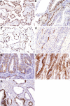
Endometrioid carcinoma (EC) and clear cell carcinoma (CC) are associated with endometrial tissue hyperplasia and endometriosis, and they occur in the endometrium and ovaries. However, detailed differences between these tumors based on immunostaining are unclear; therefore, in this study, we aimed to analyze the clinicopathological correlations between these tumors using immunostaining and to develop new treatments based on histological subtypes. Immunohistochemistry was used to investigate differentially expressed hypoxia-associated molecules (hypoxia-inducible factor-1 subunit alpha [HIF-1α], forkhead box O1, prostate-specific membrane antigen, signal transducer and activator of transcription 3 [STAT3], hepatocyte nuclear factor 1β [HNF-1β], aquaporin-3, and vimentin [VIM]) between these carcinomas because of the reported association between CC and ischemia. Immunostaining and clinicopathological data from 70 patients (21 uterine endometrioid carcinomas [UECs], 9 uterine cell carcinomas, 20 ovarian endometrioid carcinomas [OECs], and 20 ovarian cell carcinomas [OCCs]) were compared. HIF-1α and prostate-specific membrane antigen expression levels were higher in UEC and OCC than in uterine cell carcinomas and OEC. STAT3 was slightly overexpressed in UEC. Additionally, forkhead box O1 expression was either absent or significantly attenuated in all ECs. VIM and AQ3 were highly expressed in UEC, whereas HNF-1β expression was higher in OCC. UEC, OEC, and OCC were more common in the uterine fundus, left ovary, and right ovary, respectively. Ovarian endometriosis was strongly associated with EC. Our findings suggest that UEC and OCC share a carcinogenic pathway that involves HIF-1α induction under hypoxic conditions via STAT3 expression, resulting in angiogenesis. Furthermore, the anatomical position of carcinomas may contribute to their carcinogenesis. Finally, aquaporin-3 and VIM demonstrate strong potential as biomarkers for UEC, whereas HNF-1β expression is a crucial factor in CC development. These differences in tumor site and histological subtypes shown in this study will lead to the establishment of treatment based on histological and immunohistological classification.
Copyright © 2023 the Author(s). Published by Wolters Kluwer Health, Inc.
Conflict of interest statement
The authors have no funding and conflicts of interest to disclose.
Figures


Similar articles
-
Immunohistochemical characterization of prototypical endometrial clear cell carcinoma--diagnostic utility of HNF-1β and oestrogen receptor.Histopathology. 2014 Mar;64(4):585-96. doi: 10.1111/his.12286. Epub 2013 Dec 7. Histopathology. 2014. PMID: 24103020
-
TCGA molecular classification in endometriosis-associated ovarian carcinomas: Novel data on clear cell carcinoma.Gynecol Oncol. 2022 Jun;165(3):577-584. doi: 10.1016/j.ygyno.2022.03.016. Epub 2022 Mar 31. Gynecol Oncol. 2022. PMID: 35370008
-
Immunohistochemistry expression of targeted therapies biomarkers in ovarian clear cell and endometrioid carcinomas (type I) and endometriosis.Hum Pathol. 2019 Mar;85:72-81. doi: 10.1016/j.humpath.2018.10.028. Epub 2018 Nov 14. Hum Pathol. 2019. PMID: 30447298
-
Pathology of clear cell carcinoma of the ovary: A basic view based on cultured cells and modern view from comprehensive approaches.Pathol Int. 2020 Sep;70(9):591-601. doi: 10.1111/pin.12954. Epub 2020 May 31. Pathol Int. 2020. PMID: 32476214 Review.
-
[An approach to early genetic alterations in precancerous cells].Hum Cell. 2000 Sep;13(3):103-8. Hum Cell. 2000. PMID: 11197771 Review. Japanese.
Cited by
-
The mechanism of wen jing tang in the treatment of endometriosis: Insights from network pharmacology and experimental validation.Heliyon. 2024 Oct 17;10(21):e39292. doi: 10.1016/j.heliyon.2024.e39292. eCollection 2024 Nov 15. Heliyon. 2024. PMID: 39524878 Free PMC article.
-
3D visualization of uterus and ovary: tissue clearing techniques and biomedical applications.Front Bioeng Biotechnol. 2025 Jul 7;13:1610539. doi: 10.3389/fbioe.2025.1610539. eCollection 2025. Front Bioeng Biotechnol. 2025. PMID: 40692613 Free PMC article. Review.
References
-
- WHO Classification of Tumours Editorial Board. Female Genital Tumours, WHO Classification οf Tumours Series, 5th ed.; 4 [WHO website]. Lyon (France): International Agency for Research on Cancer. 2020. Available at: https://tumourclassification.iarc.who.int/chapters/34/131. [Access date Jun 8, 2023].
-
- Matias-Guiu X, Stewart CJR. Endometriosis-associated ovarian neoplasia. Pathology (Phila). 2018;50:190–204. - PubMed
-
- WHO Classification of Tumours Editorial Board. Female Genital Tumours, WHO Classification οf Tumours Series. 5th ed.; 4 [WHO website]. Lyon, France: International Agency for Research on Cancer; 2020. Available at: https://tumourclassification.iarc.who.int/chapters/34/223 [access date June 8, 2023].
-
- WHO Classification of Tumours Editorial Board. Female Genital Tumours, WHO Classification οf Tumours Series. 5th ed. 4 [WHO website]. Lyon, France: International Agency for Research on Cancer. 2020. Available at: https://tumourclassification.iarc.who.int/chapters/34/226 [access date June 8, 2023].
-
- WHO Classification of Tumours Editorial Board. Female Genital Tumours, WHO Classification οf Tumours Series, 5th ed. 4 [WHO website]. Lyon, France: International Agency for Research on Cancer. 2020. Available at: https://tumourclassification.iarc.who.int/chapters/34/29 [access date June 8, 2023].
MeSH terms
Substances
LinkOut - more resources
Full Text Sources
Medical
Research Materials
Miscellaneous

