Review
. 2023 Sep 14;186(19):4007-4037.
doi: 10.1016/j.cell.2023.07.036.

TGF-β signaling in health and disease

Affiliations
Review

TGF-β signaling in health and disease

Joan Massagué et al. Cell. .

Abstract

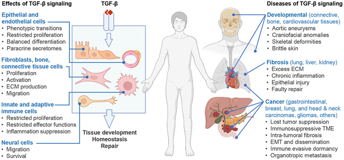
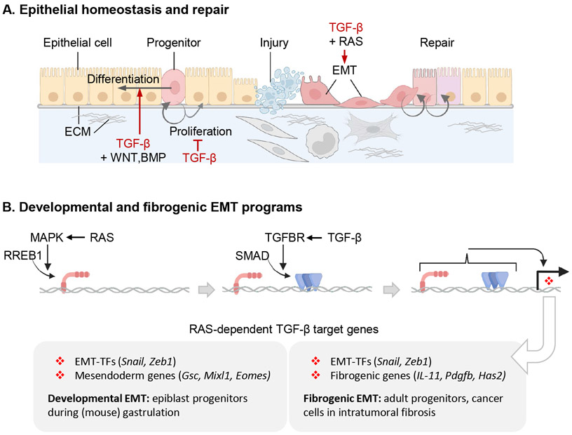
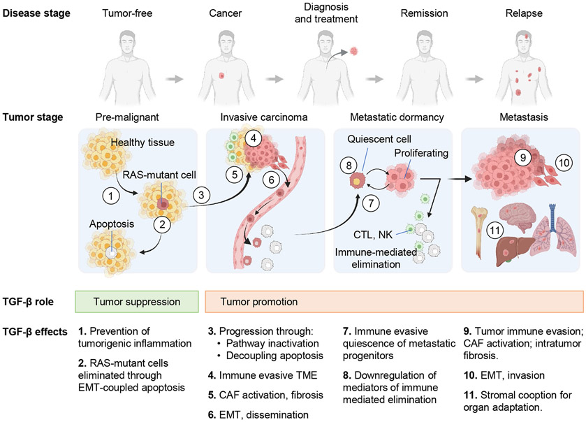
The TGF-β regulatory system plays crucial roles in the preservation of organismal integrity. TGF-β signaling controls metazoan embryo development, tissue homeostasis, and injury repair through coordinated effects on cell proliferation, phenotypic plasticity, migration, metabolic adaptation, and immune surveillance of multiple cell types in shared ecosystems. Defects of TGF-β signaling, particularly in epithelial cells, tissue fibroblasts, and immune cells, disrupt immune tolerance, promote inflammation, underlie the pathogenesis of fibrosis and cancer, and contribute to the resistance of these diseases to treatment. Here, we review how TGF-β coordinates multicellular response programs in health and disease and how this knowledge can be leveraged to develop treatments for diseases of the TGF-β system.


Conflict of interest statement

Declaration of interests J.M. holds company stock of Scholar Rock, Inc. D.S. is a founder of Pliant Therapeutics, a member of the Genentech Scientific Review Board, a member of the Amgen Immunology Scientific Advisory Board, and a member of the Scientific Review Board for Lila Biologics.

Figures

Figure 1.
Figure 1.. TGF-β in health and disease
TGF-β guards tissue homeostasis through multiple effects on different cell types. Although TGF-β signals through a common receptor and a set of transcription factors in all cells, it triggers different effects on diverse cellular functions depending on the cell type and tissue environment. Epithelial cells, fibroblasts, immune, vascular, connective, and neural cells are important TGF-β targets, and their coordinated responses determine the overall effect of TGF-β on a tissue. The whole tissue, more than any of the constituent cell types, is the target of TGF-β, and preserving tissue integrity is the ultimate output. TGF-β response programs drive embryo development and promote tissue homeostasis and injury repair in the adult. Congenital defects in TGF-β signaling cause rare yet serious developmental syndromes, and somatic alterations of this pathway underly common forms of fibrosis and cancer.
Figure 2.
Figure 2.. The TGF-β signaling pathway
A. TGF-β cytokines are generated by cleavage of the dimeric C-terminal domain of a biosynthetic precursor in the Golgi. The mature cytokine remains sequestered by non-covalent binding to the N-terminal domain of the precursor, or latency-associated peptide (LAP). LAP in this complex becomes disulfide-linked to the latent TGF-β binding protein (LTBP), which is deposited in the extracellular matrix (ECM) after secretion. Alternatively, in the indicated cell types, LAP in the TGF-β complex becomes disulfide-linked to the membrane-anchored proteins GARP or LRRC33 and retained on the cell surface. B. Activation of latent TGF-β involves binding of LAP to αv integrins on adjacent cells, leading to a conformational change that releases the captive TGF-β for binding to receptors. C. The membrane proteoglycan Betaglycan functions as a co-receptor that collects TGF-β for presentation to signaling receptors. TGF-β binds to two pairs of transmembrane serine/threonine protein kinases known as TGFBR1 (type I receptor) and TGFBR2 (type II receptor), to assemble the receptor complex. In this complex, TGFBR2 phosphorylates and activates the TGFBR1 kinase, which binds and phosphorylates (P) the transcription factors SMAD2 and SMAD3. On phosphorylation, these SMADs form trimeric complexes with SMAD4 and accumulate in the nucleus to bind and transcriptionally activate target loci. Recognition of these loci by the SMAD complex frequently requires molecular interaction with lineage-determining transcription factors (LDTF) or signal-driven transcription factors (SDTF). The signaling cycle ends with SMAD dephosphorylation and dissociation from DNA for another round of signaling, or with SMAD polyubiquitination and degradation. Each step in the pathway is controlled by different classes of regulators, the most prominent of which are listed (with examples). D. Variant versions of this pathway include: (a) TGF-β receptors links with MAPKs through TRAF adaptor proteins; (b) SMAD4 recruitment of a SKI-SKIL repressor complex to certain target genes (e.g. RORC in TH17 helper T cells) to prevent leaky transcription in the absence of TGF-β; and (c) SMAD4-independent activation of certain target genes (e.g. SOX4 in pancreatic epithelial progenitors) by SMAD2 and SMAD3.
Figure 3.
Figure 3.. TGF-β and immune regulation
Scheme of the main classes of immune cells and their regulation by TGF-β in the adult. TGF-β is a critical modulator of both adaptive and innate immunity arms, acting as a general enforcer of immune tolerance and a suppressor of inflammation. In the adaptive arm, TGF-β inhibits the maturation of naïve CD4+ T cells into TH1 and TH2 T helper cells and of naïve CD8+ T cell into cytotoxic T lymphocytes (CTL). TGF-β exerts these effects through direct inhibition of CD4+ and CD8+ maturation and through inhibition of dendritic cell subsets (DC1, DC2) that drive naïve these maturation steps. TGF-β additionally inhibits the helper functions of TH1 and TH2, and the effector functions of CTL cells, and it can do so by acting directly on these cells as well as by promoting the differentiation of CD4+ T cells into peripheral regulatory T cells (pTreg), which inhibit TH1 and TH2 cells partly through TGF-β. A specialized RORγt+ antigen-presenting cell (TC ) activates pTreg cells in the intestinal lymph nodes. TGF-β inhibits B cell proliferation but stimulates IgA class switching in B cells. In the innate immunity arm, TGF-β blunts the effector functions of natural killer (NK) cells, and the inflammatory functions of neutrophils and macrophages while favoring, in the context of tumors, the adoption of tumor-associated neutrophil (TAN) and macrophage (TAM) states which support tumor progression. In chronic infection, inflammation, and cancer, the persistent myelopoiesis includes production of myeloid-derived suppressor cells (MDSC) with TGF-β dependent immunosuppressive functions. These regulatory effects of TGF-β on the immune system occur to different extents in different tissue contexts and depending on whether the circumstance is homeostasis, acute injury or infection, or chronic inflammation, fibrosis, or cancer.
Figure 4.
Figure 4.. TGF-β regulation of fibroblasts in health and disease
A. Main effects of TGF-β on fibroblasts during injury repair and chronic fibrosis, and impact on epithelial and immune cells. TGF-β regulates fibroblast activity throughout the tissue response to injury and the return to homeostasis (left side) as well as during chronic fibrosis (right side). TGF-β potently induces the recruitment, proliferation and activation of fibroblast that produce collagens, fibronectin, and other components required for ECM assembly, as well as integrins that mediate cell adhesion to the ECM. Activated fibroblasts additionally establish paracrine communication with epithelial cells, angiogenic progenitors, and local innate and adaptive immune functions. TGF-β also induces a highly contractile myofibroblast phenotype expressing α-smooth muscle actin. These phenotypes appear to emerge at the expense of a pro-inflammatory fibroblast phenotype, while TGF-β additionally restricts inflammatory monocytes. ECM deposition and remodeling is essential for epithelial progenitors to reconstitute the barrier tissue after injury. Tissue fibrosis, characterized by chronic inflammation and accumulation of fibrillar collagens and other ECM components resulting from imbalanced production of ECM by tissue resident fibroblasts. Feed-forward loops involving TGF-β contribute to fibrosis by exaggerating normal physiologic responses and triggering further epithelial injury and inflammation. B. TGF-β potently induces expression of fibrillar collagens as well as the metabolic adaptations, enzymes, and chaperones required for the biosynthesis and ECM deposition of collagen fibrils. TGF-β induces expression of additional ECM components in fibroblasts and epithelial cells. The production and turnover of ECM is a complex process requiring inputs from epithelial cells, innate and adaptive immune cells, and other cell types. Intratumoral fibrosis contributes to the exclusion of T cells from tumors. PLOD2, procollagen-lysine,2-oxoglutarate 5-deoxygenase 2; P4HA3, prolyl-4-hydroxylase 3, catalyzes proline hydroxylation; HSP47, heat-shock protein 47; LOX, lysyl oxidase; TIMP3, tissue inhibitor of metalloproteinase 3.
Figure 5.
Figure 5.. TGF-β in epithelial cell regulation
A. TGF-β regulates the phenotypic plasticity of epithelial progenitors and their interactions with other cell types. TGF-β derived from fibroblasts, immune cells, and from the epithelial cells themselves modulates the proliferation of epithelial progenitors and regulates their differentiation, frequently with countervailing WNT, BMP and other signals. In response to injury, epithelial progenitors undergo EMT for migration to niches that provide appropriate basal lamina ECM support and signals to orchestrate injury repair and eventual resolution. TGF-β is a major inducer of EMTs, which frequently requires the cooperation of RAS-activated MAPK signals. B. RREB1 (RAS-responsive element binding protein 1) links the TGF-β-SMAD and RAS-MARK pathways and coordinates the expression of developmental and fibrogenic EMT programs. MAPK-activated RREB1 binds to target loci including in EMT-TF genes and either mesendoderm specification genes in epiblast cells or fibrogenic genes in adult epithelial progenitors and adenocarcinoma cells. DNA-bound RREB1 then enables TGF-β receptor-activated SMADs to drive expression of these genes.
Figure 6.
Figure 6.. Roles of TGF-β in cancer
During the early stages of carcinogenesis, TGF-β exerts tumor suppressive effects by inhibiting tumorigenic inflammation (1 in the graphic) or triggering EMT-coupled apoptosis in pre-malignant progenitors harboring RAS mutations (2). To escape TGF-β dependent apoptosis (3), RAS-mutant cells must acquire TGF-β pathway inactivating mutations or alterations that decouple TGF-β-dependent EMT from apoptosis. This enables carcinoma progression and turns TGF-β into a tumor promoting agonist as the disease progresses. The tumor promoting effects of TGF-β include: (4) generation of an immune evasive TME by excluding or suppressing cytotoxic T cells and NK cells and turning macrophages into TAMs and neutrophils into TANs; (5) activation of CAF fibrogenic and paracrine activities, which favor cancer cell growth, invasion, immune evasion, and angiogenesis; (6) induction of cancer cell EMTs which increase tumor invasion, entry into, and exit from the circulation for tumor dissemination; (7) induction of immune evasive dormancy in disseminated metastatic progenitors; (8) downregulation of mediators of immune clearance in dormant cancer cells; (9, 10) repeated generation of an immune evasive TME, activation of CAFs, and induction of fibrogenic EMT in dormant metastatic progenitors that resume proliferative and survive elimination by the immune system; (11) promotion of metastatic outgrowth by stimulating organ-specific cancer cell-stroma interactions. The cancer cell-intrinsic tumorigenic effects of TGF-β (effects 6, 7, 8, 10 and, partly, 11) are available to carcinoma cells that retain an active TGF-β pathway (though decoupled from apoptosis). The TME effects of TGF-β (effects 4, 5, 9 and, partly, 11) are available to carcinoma cells regardless of how the tumor suppressive effects of TGF-β are cancelled.
Figure 7.
Figure 7.. Approaches to therapeutically targeting TGF-β.
The image summarizes the main points of the TGF-β production, activation, and signaling being targeted by various agents currently under development to treat cancer, fibrosis, and other diseases. TGF-β inhibitory agents include antisense oligonucleotides targeting TGF-β expression, antibodies targeting latent TGF-β, TGF-β-activating integrins, active TGF-β or TGF-β receptors, and small-molecule compounds targeting TGF-β-activating integrins and TGF-β receptors. TGF-β receptor ectodomains fused to immune checkpoint antibodies are engineered to increase the efficacy of immunotherapeutic agents by trapping TGF-β near target cells. For the same purpose, dominant-negative TGF-β receptor constructs are overexpressed in engineered various types of anti-cancer T cells (CAR T cells, autologous CTLs).

References

    1. Roberts AB, and Sporn MB (1988). Transforming growth factor beta. Adv Cancer Res 51, 107–145. - PubMed
    1. Massague J. (1998). TGF-beta signal transduction. Annu Rev Biochem 67, 753–791. 10.1146/annurev.biochem.67.1.753. - DOI - PubMed
    1. Davis H, Raja E, Miyazono K, Tsubakihara Y, and Moustakas A (2016). Mechanisms of action of bone morphogenetic proteins in cancer. Cytokine Growth Factor Rev 27, 81–92. 10.1016/j.cytogfr.2015.11.009. - DOI - PubMed
    1. Goumans MJ, and Ten Dijke P (2018). TGF-β Signaling in Control of Cardiovascular Function. Cold Spring Harb Perspect Biol 10. 10.1101/cshperspect.a022210. - DOI - PMC - PubMed
    1. Katagiri T, and Watabe T (2016). Bone Morphogenetic Proteins. Cold Spring Harb Perspect Biol 8. 10.1101/cshperspect.a021899. - DOI - PMC - PubMed

Publication types

Substances