TDP-43 protein interactome informs about perturbed canonical pathways and may help develop personalized medicine approaches for patients with TDP-43 pathology
- PMID: 37714405
- PMCID: PMC10872580
- DOI: 10.1016/j.drudis.2023.103769
TDP-43 protein interactome informs about perturbed canonical pathways and may help develop personalized medicine approaches for patients with TDP-43 pathology
Abstract
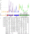
Transactive response DNA binding protein of 43 kDa (TDP-43) pathology is a common proteinopathy observed among a broad spectrum of patients with neurodegenerative disease, regardless of the mutation. This suggests that protein-protein interactions of TDP-43 with other proteins may in part be responsible for the pathology. To gain better insights, we investigated TDP-43-binding proteins in each domain and correlated these interactions with canonical pathways. These investigations revealed key cellular events that are involved and are important at each domain and suggested previously identified compounds to modulate key aspects of these canonical pathways. Our approach proposes that personalized medicine approaches, which focus on perturbed cellular mechanisms would be feasible in the near future.
Keywords: TDP-43; canonical pathways; personalized medicine; precision medicine; protein–protein interactions.
Copyright © 2023 The Author(s). Published by Elsevier Ltd.. All rights reserved.
Conflict of interest statement
Declarations of interest
No interests are declared.
Figures


Similar articles
-
Accumulation of C-terminal fragments of transactive response DNA-binding protein 43 leads to synaptic loss and cognitive deficits in human TDP-43 transgenic mice.Neurobiol Aging. 2014 Jan;35(1):79-87. doi: 10.1016/j.neurobiolaging.2013.07.006. Epub 2013 Aug 15. Neurobiol Aging. 2014. PMID: 23954172
-
The Role of TDP-43 in Neurodegenerative Disease.Mol Neurobiol. 2022 Jul;59(7):4223-4241. doi: 10.1007/s12035-022-02847-x. Epub 2022 May 2. Mol Neurobiol. 2022. PMID: 35499795 Review.
-
TDP-43: a DNA and RNA binding protein with roles in neurodegenerative diseases.Int J Biochem Cell Biol. 2010 Oct;42(10):1606-9. doi: 10.1016/j.biocel.2010.06.016. Epub 2010 Jun 25. Int J Biochem Cell Biol. 2010. PMID: 20601083 Review.
-
TAR DNA-binding protein of 43 kDa (TDP-43) and amyotrophic lateral sclerosis (ALS): a promising therapeutic target.Expert Opin Ther Targets. 2022 Jun;26(6):575-592. doi: 10.1080/14728222.2022.2083958. Epub 2022 Jun 2. Expert Opin Ther Targets. 2022. PMID: 35652285
-
TDP-43 induces mitochondrial damage and activates the mitochondrial unfolded protein response.PLoS Genet. 2019 May 17;15(5):e1007947. doi: 10.1371/journal.pgen.1007947. eCollection 2019 May. PLoS Genet. 2019. PMID: 31100073 Free PMC article.
Cited by
-
Cross-Interaction with Amyloid-β Drives Pathogenic Structural Transformation within the Amyloidogenic Core Region of TDP-43.ACS Chem Neurosci. 2025 Apr 16;16(8):1565-1581. doi: 10.1021/acschemneuro.5c00084. Epub 2025 Apr 1. ACS Chem Neurosci. 2025. PMID: 40167418
-
Spastin and alsin protein interactome analyses begin to reveal key canonical pathways and suggest novel druggable targets.Neural Regen Res. 2025 Mar 1;20(3):725-739. doi: 10.4103/NRR.NRR-D-23-02068. Epub 2024 May 13. Neural Regen Res. 2025. PMID: 38886938 Free PMC article.
-
The Regulation of TDP-43 Structure and Phase Transitions: A Review.Protein J. 2025 Apr;44(2):113-132. doi: 10.1007/s10930-025-10261-0. Epub 2025 Feb 22. Protein J. 2025. PMID: 39987392 Review.
References
Publication types
MeSH terms
Substances
Grants and funding
LinkOut - more resources
Full Text Sources
Medical

