. 2023 Sep 15;14(1):5730.
doi: 10.1038/s41467-023-41414-3.

Interlocking of co-opted developmental gene networks in Drosophila and the evolution of pre-adaptive novelty

Affiliations

Interlocking of co-opted developmental gene networks in Drosophila and the evolution of pre-adaptive novelty

Sara Molina-Gil et al. Nat Commun. .

Abstract

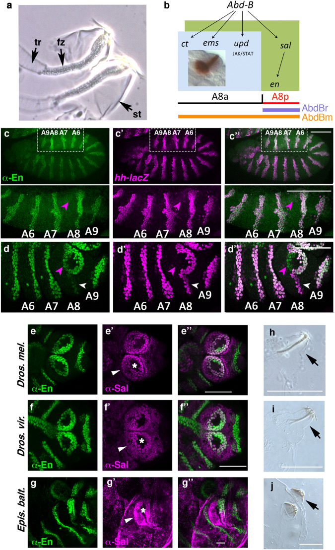
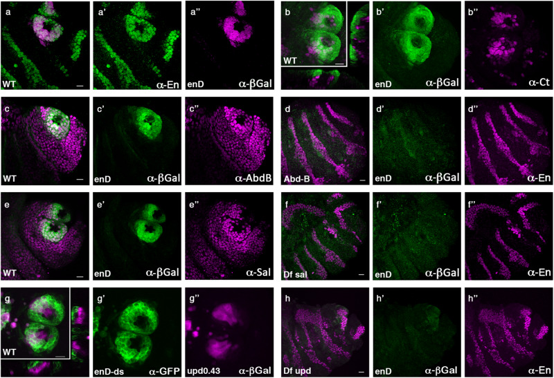
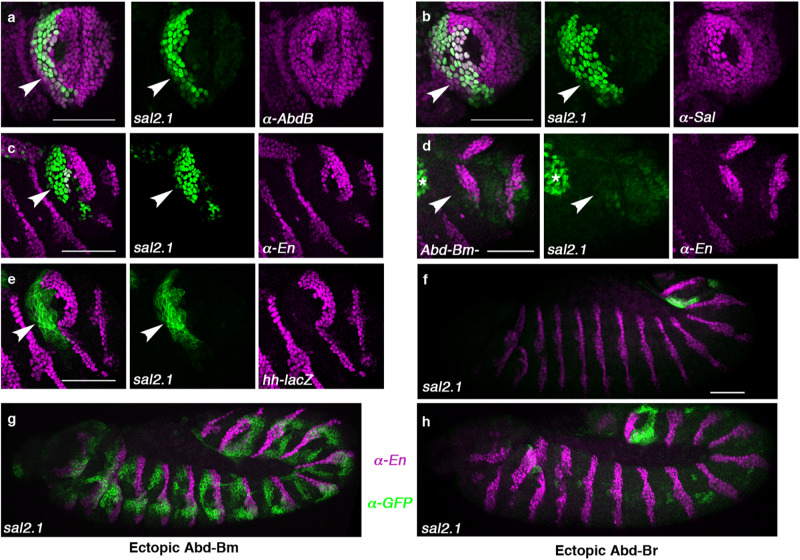
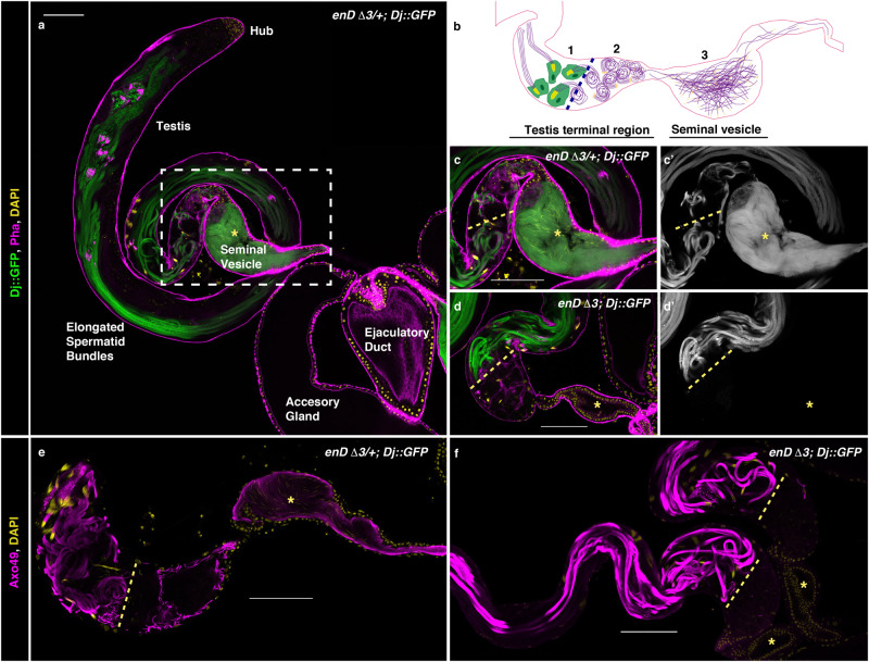
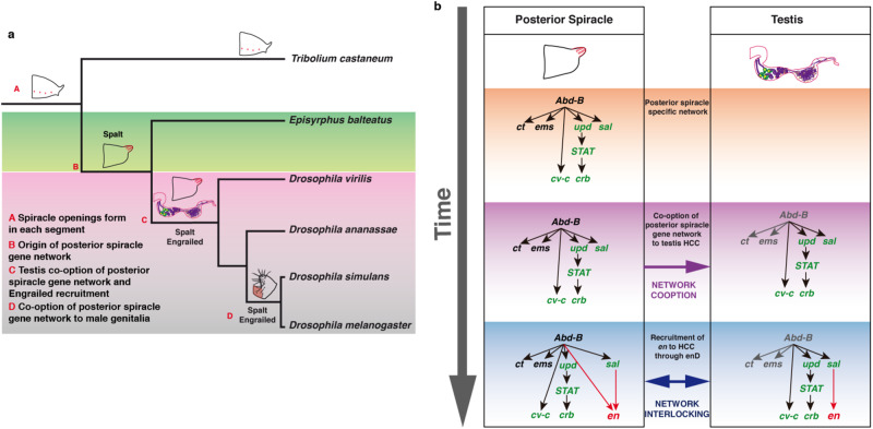
The re-use of genes in new organs forms the base of many evolutionary novelties. A well-characterised case is the recruitment of the posterior spiracle gene network to the Drosophila male genitalia. Here we find that this network has also been co-opted to the testis mesoderm where is required for sperm liberation, providing an example of sequentially repeated developmental co-options. Associated to this co-option event, an evolutionary expression novelty appeared, the activation of the posterior segment determinant Engrailed to the anterior A8 segment controlled by common testis and spiracle regulatory elements. Enhancer deletion shows that A8 anterior Engrailed activation is not required for spiracle development but only necessary in the testis. Our study presents an example of pre-adaptive developmental novelty: the activation of the Engrailed transcription factor in the anterior compartment of the A8 segment where, despite having no specific function, opens the possibility of this developmental factor acquiring one. We propose that recently co-opted networks become interlocked, so that any change to the network because of its function in one organ, will be mirrored by other organs even if it provides no selective advantage to them.


Conflict of interest statement

The authors declare no competing interests.

Figures

Fig. 1
Fig. 1. Evolution and dynamic expression of en in the anterior A8 segment.
a Close-up of the larval posterior spiracles showing the internal filters (fz) formed by the spiracular chamber cells, lodged in the protruding stigmatophores (st) and connecting to the tracheae (tr). b Scheme of the st11 early posterior spiracle organogenetic gene network activated in A8 by Abd-B. Light blue represents the spiracular chamber primordium that eventually will invaginate to form at st16 the fz (brown, inset). Olive green represents the cells of the stigmatophore primordium. The spatial expression domain of the Abd-B m (orange line) and Abd-Br (mauve line) isoforms and the location of the antero-posterior A8 compartment boundary are indicated. All spiracular chamber cells form in the Abd-Bm expressing anterior compartment, while the stigmatophore primordium comprises both anterior and posterior dorsal A8 cells. c, d hh-lacZ embryos double stained with anti-En (green) and anti-ßGal (magenta) at progressively later stages: (c-c”) st11, (d-d”) st14. Magenta arrowheads point to anterior A8 cells gaining En expression at st14, white arrowheads point at the A8 posterior cells losing En expression. The panel to the right shows both channels. To facilitate segment comparison between the extended germ band embryo (c) and the retracted germ band embryo (d), the close-up of the squared region shown in panel c, has been rotated 180°. The location of the posterior abdominal (A6–A9) compartments is labelled. eg Wild-type embryos double stained with anti-Sal (magenta) and anti-En (green) in Drosophila melanogaster (e-e”), Drosophila virilis (f-f”) and Episyrphus balteatus (g-g”). White arrowheads in (e’-g’) point at the stigmatophore Sal expression and asterisks mark the spiracle opening. Note that the cross-reactive Sal antibody staining has some background (g’) due to unspecific binding to the vitelline membrane that cannot be completely removed. hj Cuticles of recently hatched Drosophila melanogaster (h), Drosophila virilis (i) and Episyrphus balteatus larvae (j) with arrows pointing to the protruding stigmatophore. Scale bars: 50 µm in cg and 100 µm in hj.
Fig. 2
Fig. 2. Regulation of En expression in the stigmatophore of Drosophila melanogaster.
Expression of enD enhancer in wild type (ac, e, g) or posterior spiracle mutant embryos (d, f, h). a enD-lacZ embryo double stained with anti-En (a’ green) and anti-ßGal (a” magenta). b, c e enD-lacZ embryos double stained with anti-ßGal (green) and either anti-Ct (b), anti-AbdB (c), or anti-Sal (e) in magenta. g enD-GFP upd0.43-lacZ embryo double stained with anti-GFP (g’ green) and anti-ßGal (g” magenta). d, f, h enD-lacZ embryos homozygous mutant for either Abd-BM1 (d), Df(2)5 sal-salr (f) or the upd locus deficiency Df(1)os1A (h), double stained with anti-ßGal (green) and anti-En (magenta). Panels b and g also show orthogonal sections to demonstrate Ct and upd0.43 are expressed in the internal spiracular chamber cells and enD-lacZ in the external stigmatophore cells. All embryos are st14. Scale bar: 20 µm.
Fig. 3
Fig. 3. Expression and regulation of sal2.1 in the A8 segment.
ac Expression of sal2.1-lacZ in wild type st13 embryos double stained with anti-ßGal (green, arrowheads) and anti-AbdB (a), anti-Sal (b), or anti-En (c) in magenta. d Expression of sal2.1-lacZ in Abd-BM5 mutant embryos lacking the m isoform. e Expression of sal2.1-PH-GFP in hh-lacZ embryos stained with anti-GFP (green) and anti-ßGal (magenta). f Expression of sal2.1-PH-GFP (green) in wild type embryos stained with anti-En (magenta). g, h Ectopic activation with the ectoderm-specific line 69B-Gal4 of the UAS-Abd-Bm (g) or UAS-Abd-Br isoform (h) in sal2.1-PH-GFP embryos stained with anti-GFP (green) and anti-En (magenta). Arrowheads in ae point at anterior A8 cells, asterisk in panel d indicates hindgut expression. Scale bar: 50 µm.
Fig. 4
Fig. 4. Defective sperm release in enDΔ mutant testis.
Five-day-old adult testis with the sperm tails labelled with Dj::GFP (green, or grey) in control heterozygous (a, c) or homozygous enDΔ males (d). b Schematic drawing of the Drosophila melanogaster wild-type testis terminal region indicating (1) the Head Cyst Cells (HCC, green) tightly holding the sperm heads (yellow); (2) the coiled sperms and (3) the sperms after their release to the seminal vesicle. c Close-up of the squared region in a. In heterozygous males (a, c) spermiation leads to sperm accumulation in the seminal vesicle (asterisk). In homozygous enDΔ males (d) sperms cannot be detected in the seminal vesicle (asterisk) nor in the distal part of the terminal testis region (right of the discontinuous yellow line). e, f Testes with the sperm tails labelled with Axo49 (magenta) in the testis terminal region and seminal vesicle (asterisk) of control heterozygous (e) or enDΔ homozygous (f) males. All testes are counter-stained with DAPI (yellow). In (a, c, d) filamentous Actin is labelled with Rhodamine phalloidin (magenta). Scale bar: 100 µm.
Fig. 5
Fig. 5. Expression of posterior spiracle network genes in the testis terminal region.
ae Expression of enD, sal2.1 and upd0.43 enhancers in the HCC at the testis terminal region. (a) enDds-GFP (green) is expressed in the HCCs holding the 64 sperm head bundles. Sperm tails labelled with Axo49 are shown in magenta, and sperm heads are in yellow with DAPI. (a’) shows only GFP and DAPI stainings and a close-up of an HCC holding the sperm heads. b The HCC labelled in green with enDds-GFP can be distinguished by the formation of an actin basket [labelled with phalloidin (magenta or white)] holding the 64 sperm head bundles (yellow). c enDds-GFP is active in HCC cells which also express low levels of endogenous Engrailed (magenta). d sal2.1-mCherry is active in cells expressing low levels of endogenous Sal protein. e upd0.43-lacZ (magenta) is active in a subset of HCC expressing Sal (green). Wild type enD0.4-mCherry reporter expression in HCCs (f). Reporter expression is maintained in enD0.4 when the putative Abd-B DNA-binding sites are mutated (g), or when the putative STAT sites are mutated (h). In (a, b, dh) DAPI staining is yellow. Scale bar: 20 µm.
Fig. 6
Fig. 6. Expression of Sal and En in the testis of three dipteran species.
Testis terminal region of D. melanogaster (a, b), D. virilis (c, d) and whole testis of E. balteatus (e, f) stained with anti-Engrailed and anti-Spalt (a, c, e) or anti-Spalt, and the axoneme marker anti-Axo49 (b, d, f). D. virilis testes are larger but similar in shape to those of D. melanogaster and have a similar expression of En and Sal in the terminal region (a, c) where the sperms coil and individualise (b, d). The testis morphology of E. balteatus is completely different and expresses neither En nor Sal (e). Axoneme staining with anti-Axo49 and DAPI reveals the formation of shorter sperms in E. balteatus that do not coil (f). DAPI DNA staining in yellow. Scale bar: 100 µm.
Fig. 7
Fig. 7. Interlocking of co-opted gene networks.
a Simplified evolutionary tree representing the consecutive co-option events that sequentially recruited the posterior spiracle gene network to the testis and the male genital posterior lobe in Drosophila melanogaster. Temporal lines not to scale. b Schematic representation of the co-opted posterior spiracle gene network elements (green) to the testis HCC and the posterior recruitment of Engrailed function (red) to the testis that resulted in its simultaneous aphenotypic recruitment to the posterior spiracles caused by the networks’ interlocking. Genes highlighted in grey are not expressed in HCC.

Similar articles

Cited by

References

    1. True JR, Carroll SB. Gene co-option in physiological and morphological evolution. Annu. Rev. Cell Dev. Biol. 2002;18:53–80. - PubMed
    1. Murugesan, S. N. et al. Butterfly eyespots evolved via cooption of an ancestral gene-regulatory network that also patterns antennae, legs, and wings. Proc. Natl Acad. Sci. USA119, e2108661119 10.1073/pnas.2108661119 (2022). - PMC - PubMed
    1. Glassford WJ, et al. Co-option of an ancestral hox-regulated network underlies a recently evolved morphological novelty. Dev. Cell. 2015;34:520–531. - PMC - PubMed
    1. Hu N, Castelli-Gair J. Study of the posterior spiracles of Drosophila as a model to understand the genetic and cellular mechanisms controlling morphogenesis. Dev. Biol. 1999;214:197–210. - PubMed
    1. Merabet S, Hombria JC, Hu N, Pradel J, Graba Y. Hox-controlled reorganisation of intrasegmental patterning cues underlies Drosophila posterior spiracle organogenesis. Development. 2005;132:3093–3102. - PubMed

Publication types

LinkOut - more resources