. 2023 Sep 15;14(1):5732.
doi: 10.1038/s41467-023-41439-8.

Structural landscape of the respiratory syncytial virus nucleocapsids

Affiliations

Structural landscape of the respiratory syncytial virus nucleocapsids

Lorène Gonnin et al. Nat Commun. .

Abstract

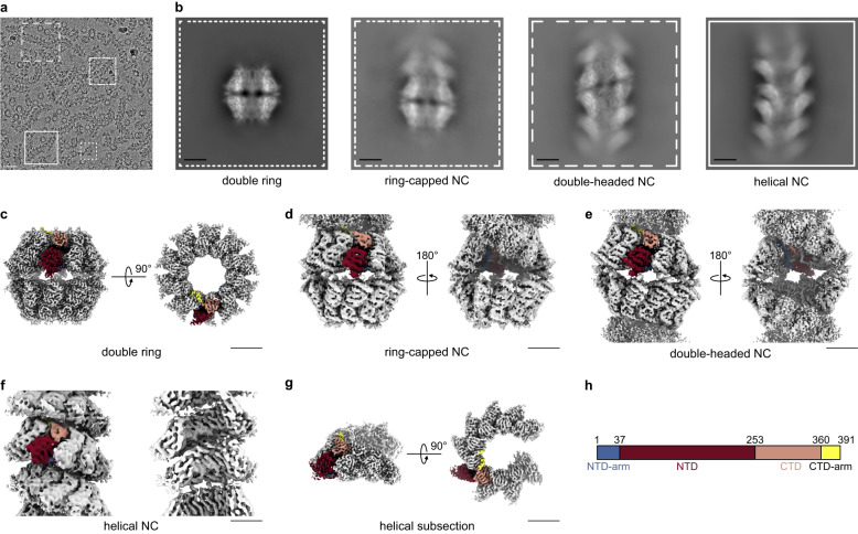
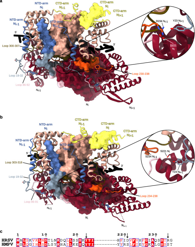
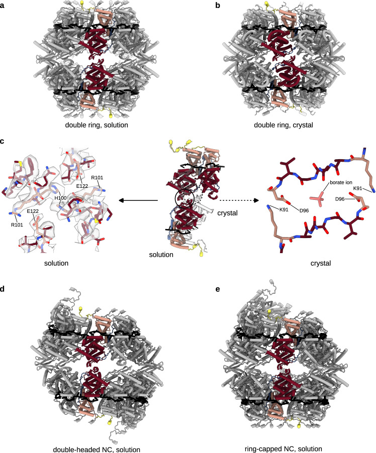
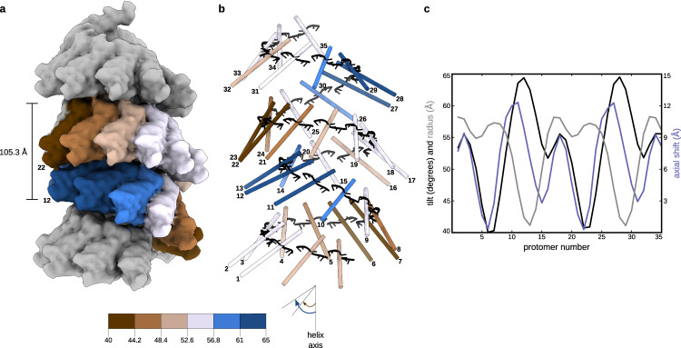
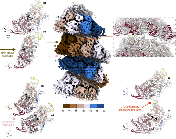
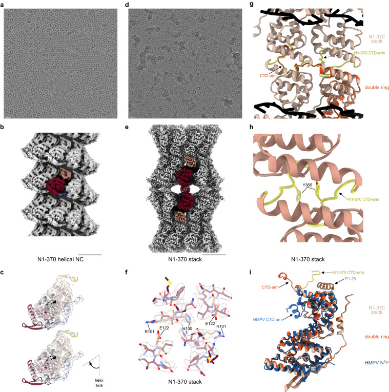
Human Respiratory Syncytial Virus (HRSV) is a prevalent cause of severe respiratory infections in children and the elderly. The helical HRSV nucleocapsid is a template for the viral RNA synthesis and a scaffold for the virion assembly. This cryo-electron microscopy analysis reveals the non-canonical arrangement of the HRSV nucleocapsid helix, composed of 16 nucleoproteins per asymmetric unit, and the resulting systematic variations in the RNA accessibility. We demonstrate that this unique helical symmetry originates from longitudinal interactions by the C-terminal arm of the HRSV nucleoprotein. We explore the polymorphism of the nucleocapsid-like assemblies, report five structures of the full-length particles and two alternative arrangements formed by a C-terminally truncated nucleoprotein mutant, and demonstrate the functional importance of the identified longitudinal interfaces. We put all these findings in the context of the HRSV RNA synthesis machinery and delineate the structural basis for its further investigation.


Conflict of interest statement

The authors declare no competing interests.

Figures

Fig. 1
Fig. 1. Cryo-EM analysis of HRSV NCs.
a A representative micrograph of HRSV NCs purified from insect cells and featuring double rings, ring-capped NCs, double-headed NCs and helical NCs. 11,386 micrographs were selected for further processing. Particles are boxed, as an illustration, scale bar 200 Å. b Representative 2D classes with the outline matching the particles highlighted in a. c Cryo-EM map of the N10 double ring (EMD:17031, side and top view). d Cryo-EM map of the ring-capped NC (EMD:17037, front and back view). e Cryo-EM map of the double-headed NC (EMD:17036, front and back view). f Cryo-EM map of the helical NC (EMD:17030, front and cut-through view). g Helical subsection (EMD:17035, side and top view). Scale bar, 50 Å in bg. h Schematic of the HRSV N sequence divided into an NTD-arm (blue-grey), NTD (rosewood), CTD (old rose) and CTD-arm (yellow). In cryo-EM maps in cg, one protomer is coloured according to this schematic, with the RNA in black.
Fig. 2
Fig. 2. Lateral interactions between N protomers in HRSV and HMPV N10 double rings.
a Atomic models of three consecutive HRSV N protomers are shown, the middle one as a surface and the edge ones as ribbons. NTD-arm, NTD, CTD and CTD-arm coloured as in Fig. 1, loop 19–32 in powder blue, loop 86–92 in mimi pink, loop 230–238 in orange and loop 300–307 in olive. The close-up of the N-hole of the middle protomer shows the tripartite Y23-D221-R234 interaction. b Same as a but for three protomers from the HMPV N-RNA ring crystal structure (PDB: 5FVC). Colouring as in Fig. 1 and in a, loop 19–32 in powder blue, loop 86–91 in mimi pink, loop 234–238 in orange and loop 303–318 loop in olive. The close-up shows the absence of a tripartite interaction in the N-hole. c Pairwise sequence alignment of HRSV and HMPV N around the residues involved in the tripartite interaction in the HRSV N oligomer. Conserved residues in red boxes; arrows pointing at residues 23, 221, and 234.
Fig. 3
Fig. 3. Longitudinal NTD-NTD interactions conserved between HRSV N10 double ring, double-headed and ring-capped NCs but different from the crystal structure of the HRSV N10 double ring.
In each panel, two opposite protomers are coloured as in Fig. 1. a Atomic model of the HRSV N10 double ring derived from the cryo-EM map shown as cartoon. b Atomic model of the HRSV N10 double ring crystal structure (PDB: 2WJ8). c Alignment of the top rings of the cryo-EM and crystal structure-based models reveals a rotation between the bottom rings. Two top-ring protomers and one opposing bottom-ring protomer are shown in the middle of the panel, with the cryo-EM-based structure coloured as in Fig. 1 and the crystal structure in white. A close-up of the cryo-EM map and the atomic model highlighting the NTD-NTD interactions is on the left, a close-up of the NTD-NTD interactions in the crystal structure is on the right. The difference between the crystallographic and the solution inter-ring interfaces may be related to the presence of a borate ion in the interaction site in PDB: 2WJ8, possibly embarked during the electrophoretic separation of decameric and undecameric HRSV N-RNA rings prior to crystallisation. d Atomic model of the double-headed NC. e Atomic model of the ring-capped NC.
Fig. 4
Fig. 4. Non-canonical helical symmetry of the HRSV NC.
a Atomic model of the NC is filtered to 10 Å resolution and displayed as surface. Protomers in one asymetric unit are coloured dependent on their axial tilt following the colour code shown at the schematic underneath, the rest of the protomers are coloured in grey. b Protomers of the model in a are shown as sticks coloured dependent on the protomer axial tilt and numbered 1 to 35. c Plot showing the axial tilt (black), the radial position (grey) and the relative axial shift of each protomer.
Fig. 5
Fig. 5. RNA accessibility and CTD-arm-mediated inter-turn interactions in the helical NC.
Cryo-EM map of the helical NC before further refinement of the asymmetric unit (EMD:17030) is shown in the middle, coloured as in Fig. 4 and as reminded in the schematic underneath the map, with protomers numbered as in Fig. 4. On the left, close-up views of two sets of opposing protomers from two successive helical turns are shown to illustrate the difference in the RNA accessibility, with the cryo-EM density in transparent grey and the atomic model represented as a ribbon and coloured as in Fig. 1. On the top right, a similarly-coloured view of the inter-turn interaction is shown to highlight the densities corresponding to the CTD-arm, with a corresponding two-protomer close-up underneath. RNA is in black.
Fig. 6
Fig. 6. Canonical helical NCs and stacked assemblies formed by the N1-370 mutant.
a A representative micrograph of the N1-370 NCs featuring mostly helical NCs and rings, scale bar 200 Å. 6312 micrographs were selected for processing. b Cryo-EM map of the canonical N1-370 helical NC (EMD:17034), with one protomer coloured as in Fig. 1, scale bar 50 Å. c Close-up view of protomers from two successive helical turns are shown to illustrate the position of the CTD-arm and the absence of inter-turn interactions, with the cryo-EM map in transparent grey and the atomic model represented as a ribbon and coloured as in Fig. 1. d A representative micrograph of the N1-370 NCs featuring helical NCs, stacks and rings, scale bar 200 Å. 6312 micrographs were selected for further processing. e Cryo-EM map of the N1-370 stack (EMD:17038), with one protomer coloured as in Fig. 1, scale bar 50 Å. f Close-up of the cryo-EM map of the N1-370 stack and the atomic model highlighting the NTD-NTD interactions (similar to the ones in the N10 double ring shown in Fig. 3c). g Alignment of the atomic models of the N10 double ring and the N1-370 stack illustrating the difference in the orientation of the CTD-arms. One protomer of the N10 double ring is shown in the bottom-right and coloured in orange, 4 protomers of the N1-370 stack are shown and coloured in beige, with the CTD-arms in yellow. RNA is in black. h Close-up of the atomic model of the N1-370 stack highlighting the CTD-CTD interactions. i Alignement of N protomers of the N10 double ring (orange), the N1-370 stack (beige) and the HMPV N0P crystal structure (N0 in blue and P1-28 in brown) (PDB: 5FVD). Positions of the CTD-arms are indicated.

Similar articles

Cited by

References

    1. Li Y, et al. Global, regional, and national disease burden estimates of acute lower respiratory infections due to respiratory syncytial virus in children younger than 5 years in 2019: a systematic analysis. Lancet. 2022;399:2047–2064. doi: 10.1016/S0140-6736(22)00478-0. - DOI - PMC - PubMed
    1. Busack B, Shorr AF. Going Viral—RSV as the neglected adult respiratory virus. Pathogens. 2022;11:1324. doi: 10.3390/pathogens11111324. - DOI - PMC - PubMed
    1. Papi A, et al. Respiratory syncytial virus prefusion f protein vaccine in older adults. N. Engl. J. Med. 2023;388:595–608. doi: 10.1056/NEJMoa2209604. - DOI - PubMed
    1. Hammitt LL, et al. Nirsevimab for prevention of RSV in healthy late-preterm and term infants. N. Engl. J. Med. 2022;386:837–846. doi: 10.1056/NEJMoa2110275. - DOI - PubMed
    1. Afonso CL, et al. Taxonomy of the order mononegavirales: update 2016. Arch. Virol. 2016;161:2351–2360. doi: 10.1007/s00705-016-2880-1. - DOI - PMC - PubMed

Publication types