

Maxillary sinus floor augmentation: a review of current evidence on anatomical factors and a decision tree
- PMID: 37714889
- PMCID: PMC10504247
- DOI: 10.1038/s41368-023-00248-x
Maxillary sinus floor augmentation: a review of current evidence on anatomical factors and a decision tree
Abstract
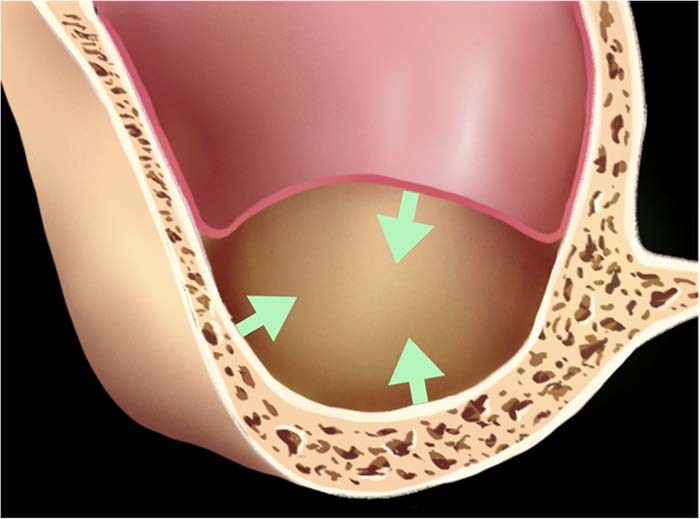
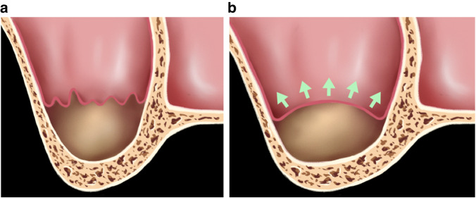
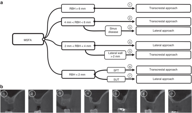
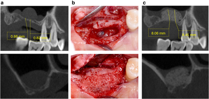
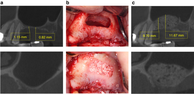
Maxillary sinus floor augmentation using lateral window and crestal technique is considered as predictable methods to increase the residual bone height; however, this surgery is commonly complicated by Schneiderian membrane perforation, which is closely related to anatomical factors. This article aimed to assess anatomical factors on successful augmentation procedures. After review of the current evidence on sinus augmentation techniques, anatomical factors related to the stretching potential of Schneiderian membrane were assessed and a decision tree for the rational choice of surgical approaches was proposed. Schneiderian membrane perforation might occur when local tension exceeds its stretching potential, which is closely related to anatomical variations of the maxillary sinus. Choice of a surgical approach and clinical outcomes are influenced by the stretching potential of Schneiderian membrane. In addition to the residual bone height, clinicians should also consider the stretching potential affected by the membrane health condition, the contours of the maxillary sinus, and the presence of antral septa when evaluating the choice of surgical approaches and clinical outcomes.
© 2023. West China School of Stomatology Sichuan University.
Conflict of interest statement
The authors declare no competing interests.
Figures
Similar articles
-
Effect of staged crestal maxillary sinus augmentation: A case series.J Periodontol. 2020 Feb;91(2):194-201. doi: 10.1002/JPER.18-0632. Epub 2019 Aug 22. J Periodontol. 2020. PMID: 31365130 Free PMC article.
-
A novel approach to manage Schneiderian membrane perforation in the maxillary sinus floor augmentation: The "Sinus Pack" technique. Anatomical factors and surgical outcomes related to perforation size and handling. Part 3/3.Am J Dent. 2024 Sept;37(SIA):21A-24A. Am J Dent. 2024. PMID: 39374507 Clinical Trial.
-
Influence of the contact area of the sub-antral space with sinus bone and the Schneiderian membrane on osteogenesis in lateral window sinus elevation surgery: a prospective experiment.BMC Oral Health. 2022 Dec 28;22(1):650. doi: 10.1186/s12903-022-02694-1. BMC Oral Health. 2022. PMID: 36578061 Free PMC article.
-
Anatomy of the Maxillary Sinus and the Role of CT Scans in Maxillary Sinus Augmentation Surgery.Clin Implant Dent Relat Res. 2025 Apr;27(2):e70019. doi: 10.1111/cid.70019. Clin Implant Dent Relat Res. 2025. PMID: 40197815 Review.
-
Perforation Risk Assessment in Maxillary Sinus Augmentation with Lateral Wall Technique.Int J Periodontics Restorative Dent. 2020 May/Jun;40(3):373-380. doi: 10.11607/prd.4179. Int J Periodontics Restorative Dent. 2020. PMID: 32233190 Review.
Cited by
-
Changes in Maxillary Sinus Structure Due to Tooth Loss and the Effects of Sex and Aging on CBCT Before Maxillary Sinus Augmentation: A Cross-Sectional Study of 120 Patients.Bioengineering (Basel). 2025 Feb 26;12(3):240. doi: 10.3390/bioengineering12030240. Bioengineering (Basel). 2025. PMID: 40150704 Free PMC article.
-
The effect of autologous platelet concentrates as solely grafting material or with bone graft materials in maxillary sinus augmentation: a meta-analysis of randomized controlled trials.Clin Oral Investig. 2025 Feb 7;29(2):120. doi: 10.1007/s00784-025-06198-4. Clin Oral Investig. 2025. PMID: 39920362
-
A platform combining automatic segmentation and automatic measurement of the maxillary sinus and adjacent structures.Clin Oral Investig. 2025 Jan 25;29(1):88. doi: 10.1007/s00784-025-06191-x. Clin Oral Investig. 2025. PMID: 39862338
-
Evaluation of thin-threaded implants primary stability in type IV bone right after maxillary sinus floor elevation: A human cadaver study.J Prosthodont. 2025 Jul;34(6):593-601. doi: 10.1111/jopr.13983. Epub 2024 Nov 14. J Prosthodont. 2025. PMID: 39539131 Free PMC article.
-
Comprehensive sinus contour classification and its characteristics from radiographic examination: a cross-sectional study.BMC Oral Health. 2024 Aug 30;24(1):1021. doi: 10.1186/s12903-024-04707-7. BMC Oral Health. 2024. PMID: 39215296 Free PMC article.
References
-
- Janner SF, et al. Characteristics and dimensions of the Schneiderian membrane: a radiographic analysis using cone beam computed tomography in patients referred for dental implant surgery in the posterior maxilla. Clin. Oral. Implants Res. 2011;22:1446–1453. - PubMed
-
- Mohan N, Wolf J, Dym H. Maxillary sinus augmentation. Dent. Clin. North Am. 2015;59:375–388. - PubMed
-
- Aghaloo TL, Moy PK. Which hard tissue augmentation techniques are the most successful in furnishing bony support for implant placement? Int. J. Oral. Maxillofac. Implants. 2007;22:49–70. - PubMed
-
- Boyne PJ, James RA. Grafting of the maxillary sinus floor with autogenous marrow and bone. J. Oral. Surg. 1980;38:613–616. - PubMed
-
- Tatum H., Jr Maxillary and sinus implant reconstructions. Dent. Clin. North Am. 1986;30:207–229. - PubMed
Publication types
MeSH terms
LinkOut - more resources
Full Text Sources
