

Subcutaneous emphysema and pneumomediastinum following orbital blowout pathological fracture in a cat with nasal lymphoma: a case report
- PMID: 37715215
- PMCID: PMC10503133
- DOI: 10.1186/s12917-023-03722-0
Subcutaneous emphysema and pneumomediastinum following orbital blowout pathological fracture in a cat with nasal lymphoma: a case report
Abstract
Background: Subcutaneous emphysema and pneumomediastinum are rare complications associated with orbital blowout pathological fracture.
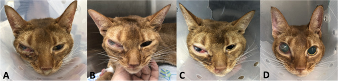
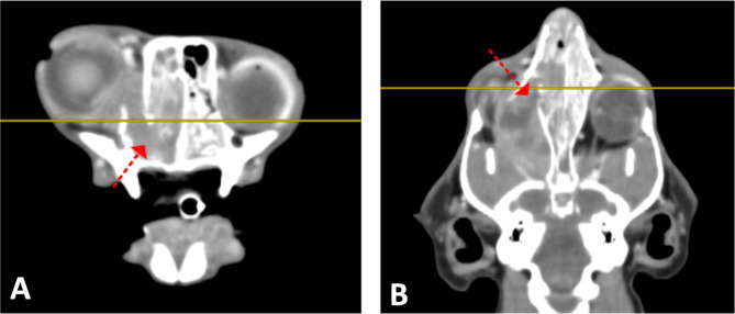
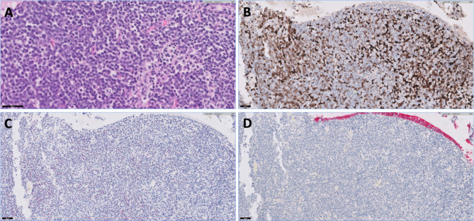
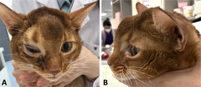
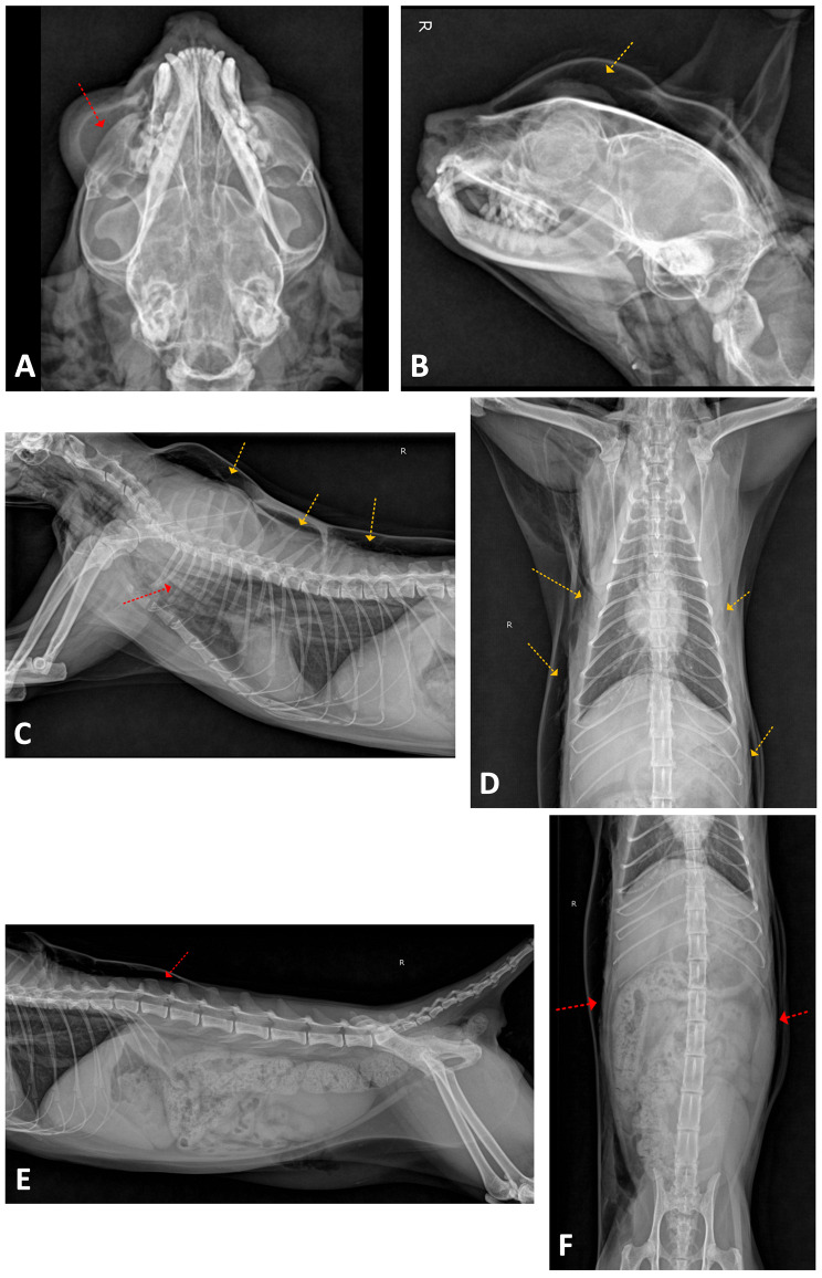
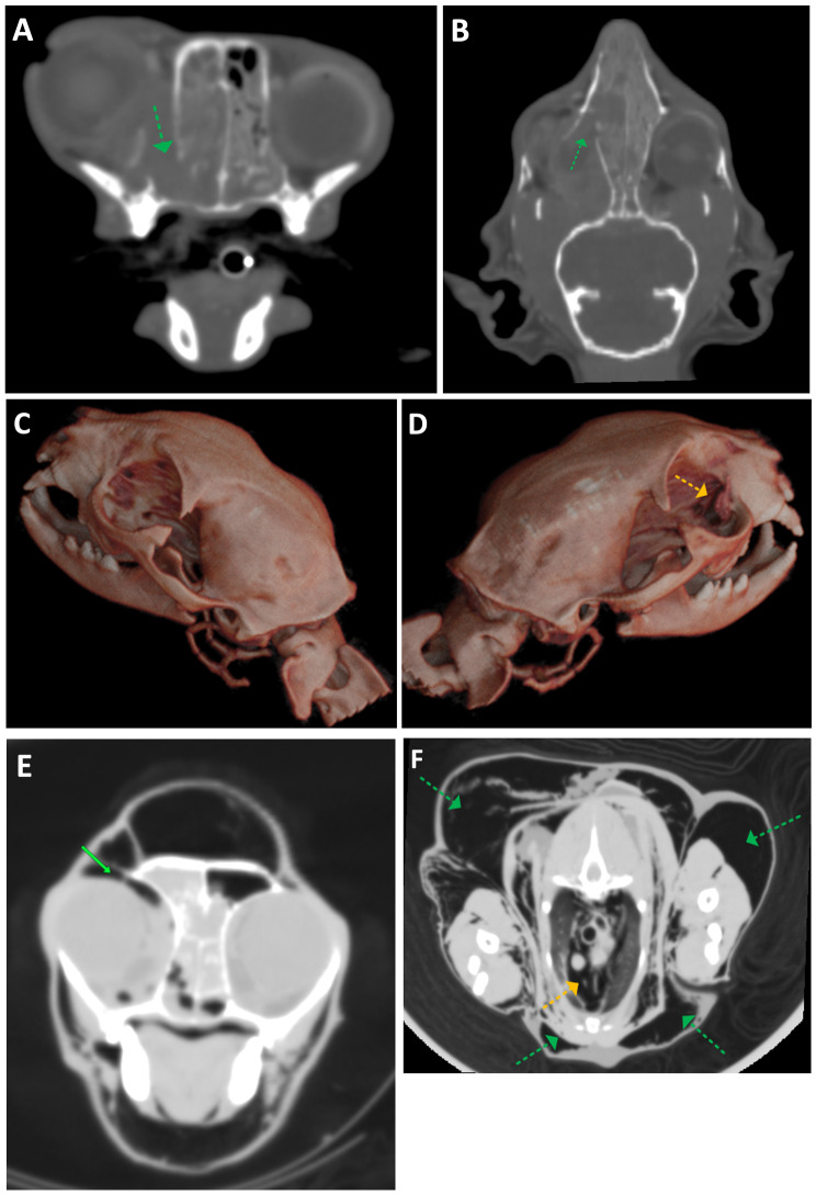
Case presentation: A 7-year old, castrated male Abbysinian cat presented with anorexia, lethargy, nausea, eyelid swelling, nasal discharge, and sneezing. Based on the clinical and diagnostic work-up, the cat was diagnosed with T cell high-grade nasal lymphoma associated with orbital pathological fracture due to the tumour invasion. After chemotherapy, the cat showed massive subcutaneous emphysema from frontal region to abdomen and pneumomediastinum due to orbital blowout pathological fracture. As the nasal mass decreased in volume; the air had moved from the maxillary sinus to the subcutaneous region and the mediastinum through fascial planes in the head and neck region.
Conclusions: This is a first case report of a massive subcutaneous emphysema and pneumomediastinum due to an orbital blowout pathological fracture following chemotherapy in feline nasal lymphoma in veterinary medicine.
Keywords: Blowout fracture; Cat; Chemotherapy; Lymphoma; Pneumomediastinum.
© 2023. BioMed Central Ltd., part of Springer Nature.
Conflict of interest statement
The authors declare no competing interests.
Figures
Similar articles
-
Anesthesia case of the month. Pneumothorax, pneumomediastinum and subcutaneous emphysema in a cat due to barotrauma after equipment failure during anesthesia.J Am Vet Med Assoc. 1998 Jan 1;212(1):30-2. J Am Vet Med Assoc. 1998. PMID: 9426773 No abstract available.
-
Pneumomediastinum and subcutaneous emphysema in a cat associated with necrotizing bronchopneumonia caused by feline herpesvirus-1.Can Vet J. 2011 Oct;52(10):1119-22. Can Vet J. 2011. PMID: 22467969 Free PMC article.
-
Subcutaneous emphysema, pneumothorax, pneumomediastinum, and pneumopericardium associated with positive-pressure ventilation in a cat.J Am Vet Med Assoc. 1995 Apr 1;206(7):997-9. J Am Vet Med Assoc. 1995. PMID: 7768723
-
Massive Self-Induced Subcutaneous Cervicofacial, Pneumomediastinum, and Pneumopericardium Emphysema Sequelae to a Nondisplaced Maxillary Wall Fracture: A Case Report and Literature Review.J Oral Maxillofac Surg. 2019 Sep;77(9):1867.e1-1867.e8. doi: 10.1016/j.joms.2019.05.010. Epub 2019 May 25. J Oral Maxillofac Surg. 2019. PMID: 31228425 Review.
-
Orbital, mediastinal, and cervicofacial subcutaneous emphysema after endodontic retreatment of a mandibular premolar: a case report.J Endod. 2014 Jun;40(6):880-3. doi: 10.1016/j.joen.2013.09.042. Epub 2013 Nov 7. J Endod. 2014. PMID: 24862722 Review.
References
-
- Felding UNA. Blowout fractures-clinic, imaging and applied anatomy of the orbit. Dan Med J. 2018;65:B5459. - PubMed
Publication types
MeSH terms
LinkOut - more resources
Full Text Sources
Research Materials
Miscellaneous
