. 2023 Jan 17:81:ftad025.
doi: 10.1093/femspd/ftad025.

Bald sea urchin disease shifts the surface microbiome on purple sea urchins in an aquarium

Affiliations

Bald sea urchin disease shifts the surface microbiome on purple sea urchins in an aquarium

Chloe G Shaw et al. Pathog Dis. .

Abstract

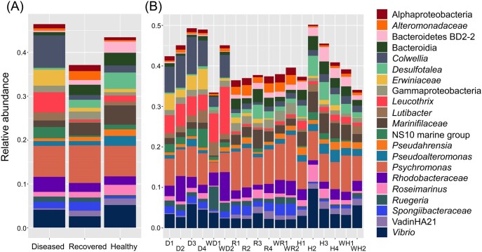
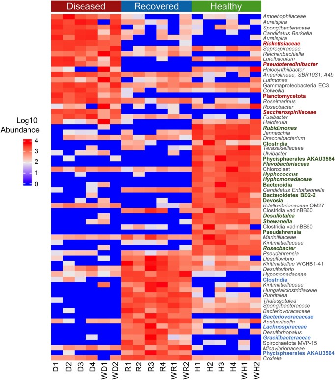
Bald sea urchin disease (BSUD) is most likely a bacterial infection that occurs in a wide range of sea urchin species and causes the loss of surface appendages. The disease has a variety of additional symptoms, which may be the result of the many bacteria that are associated with BSUD. Previous studies have investigated causative agents of BSUD, however, there are few reports on the surface microbiome associated with the infection. Here, we report changes to the surface microbiome on purple sea urchins in a closed marine aquarium that contracted and then recovered from BSUD in addition to the microbiome of healthy sea urchins in a separate aquarium. 16S rRNA gene sequencing shows that microhabitats of different aquaria are characterized by different microbial compositions, and that diseased, recovered, and healthy sea urchins have distinct microbial compositions, which indicates that there is a correlation between microbial shifts and recovery from disease.

Keywords: 16S rRNA high throughput sequencing; Strongylocentrotus purpuratus; bacterial infection; echinoderm; microbiome.


Conflict of interest statement

None declared.

Figures

Figure 1.
Figure 1.
BSUD progression and recovery in the purple sea urchin. (A) An early symptom of BSUD is drooping spines suggesting that the sea urchin may be sleeping as has been observed on rare occasions for healthy sea urchins. Alternatively, the surface infection that would impact the muscles associated with the base of the spines and the tubercles may have altered spine movement (see Video 1; Supplementary Data File 2, Video 1 Legend). (B) A sea urchin infected with BSUD that has lost many of its primary spines. (C) A sea urchin with BSUD that has lost all of its primary spines and shows the red tubercles to which primary spines are normally attached. Shorter and smaller secondary spines remain attached. (D) A magnified image of the surface of a sea urchin with BSUD that has lost all primary spines. The secondary spines (yellow arrow), pedicellariae that are too small to see in this image, and tube feet remain on the animal surface. The beginnings of newly growing spines (white arrow) are present on some tubercles. (E) A recovering sea urchin with newly regrowing primary spines that are short, pointed, and light purple. (F) A healthy sea urchin after full recovery from BSUD with the characteristic of rigid primary spines oriented perpendicular to the animal body.
Figure 2.
Figure 2.
There are many ASVs identified in the microbiomes for each group of sea urchins. Upset plot shows that the microbiomes for the diseased, recovered, and healthy groups have many ASVs that are unique. Each bar shows the ASV count and the group in which the ASVs were identified is denoted directly below each bar. Shared ASVs are indicated by the line connecting groups below the bar indicating number of ASVs counted. Of the total number of ASVs ( 40,800), 4,878 (12%) ASVs are shared among the three groups, and the diseased and healthy groups have the fewest number of shared ASVs (5.1%).
Figure 3.
Figure 3.
The surface microbiomes of sea urchins with BSUD have decreased alpha diversity compared to both recovered and healthy sea urchins. Alpha diversity was analyzed by (A) the Observed Species index, (B) the Chao1 index, and (C) the ACE index. Groups include microbiomes collected from the sea urchins and microbes in the aquarium seawater. The box plots show the average and quartile values for each group. The Chao1 index and the ACE index show significant differences between the diseased and recovered groups (ANOVA, P < .05). Error bars in the Chao1 index indicate the result as an estimate of diversity. (D) The microbial compositions of the surface microbiome are different among the three groups of sea urchins. Bray–Curtis distances estimates of beta diversity shows distinct differences for the bacterial composition and membership of ASVs in the microbiomes among the three groups of sea urchins. Ellipses show the 95% confidence intervals for the samples collected from sea urchins in each group.
Figure 4.
Figure 4.
The surface microbiomes are dominated by Proteobacteria and Bacteroidota. The phyla shown have an average relative abundance of > 0.1% for at least one sample across the groups (Supplementary Data File 2; Table S3, Supporting Information). The taxa are shown as (A) the average relative abundance in the microbiomes for each group, and (B) the relative abundance for samples collected from each sea urchin in the three groups as well as the water samples. Samples from sea urchins are indicated in (B) as diseased (D), recovered (R), and healthy (H), and the numbers (1–4) correlate with the four sea urchins within each group. Water samples are indicated with a (W).
Figure 5.
Figure 5.
The most abundant genera in the microbiomes differ among the three groups of sea urchins. The genera shown have an average relative abundance of > 1% for at least one sample (Supplementary Data File 2; Table S5, Supporting Information). The results show (A) the average relative abundance of microbial taxa from all sea urchins in each sample group and (B) the relative abundance of taxa for samples collected from each sea urchin in each of the three sample groups as well as the water samples. Taxa are listed to the right, and ASVs that could not be assigned a genus are indicated as the next matching taxonomic level. Samples from sea urchins are indicated in (B) as diseased (D), recovered (R), and healthy (H), and the numbers (1–4) correlate with the four sea urchins within each group. Water samples are indicated with a (W).
Figure 6.
Figure 6.
Many microbial taxa are significantly differentially abundant among the groups. A heatmap shows the relative abundance of the taxa in each sample within the three groups including the water samples. Identified taxa are ASVs and the lowest known taxonomic classification is listed. Taxonomic names that are colored have a notably reduced abundance or are completely absent in the other two group microbiomes. The taxa displayed have an LDA score of > 3.1 based on identification by LEfSe. All taxa with an LDA score of > 3.0 along with their LDA score and P-values are available in Supplementary Data File 2; Table S7 (Supporting Information). The threshold was increased for better visualization of the data.

Similar articles

Cited by

References

    1. Allen RL, George AN, Miranda Eet al. . Wound repair in sea urchin larvae involves pigment cells and blastocoelar cells. Dev Biol. 2022;491:56–65. 10.1016/j.ydbio.2022.08.005 - DOI - PMC - PubMed
    1. Azzolina JF, Boudouresque CHF, Nedelec H. Dynamique des populations de Paracentrotus lividus dans la baie de Port-Cros (Var): données préliminaires. Sci Rep Port-Cros Natl Park. 1985;11:61–81.
    1. Bauer J, Young C. Epidermal lesions and mortality caused by vibriosis in deep-sea Bahamian echinoids: a laboratory study. Dis Aquat Org. 2000;39:193–9. 10.3354/dao039193 - DOI - PubMed
    1. Becker PT, Egea E, Eeckhaut I. Characterization of the bacterial communities associated with the bald sea urchin disease of the echinoid Paracentrotus lividus. J Invertebr Pathol. 2008;98:136–47. 10.1016/j.jip.2007.12.002 - DOI - PubMed
    1. Becker PT, Gillan D, Eeckhaut I. Microbiological study of the body wall lesions of the echinoid Tripneustes gratilla. Dis Aquat Org. 2007;77:73–82. 10.3354/dao01821 - DOI - PubMed