. 2024 Mar;144(3):621-632.e1.
doi: 10.1016/j.jid.2023.06.212. Epub 2023 Sep 15.

Phototherapy Restores Deficient Type I IFN Production and Enhances Antitumor Responses in Mycosis Fungoides

Affiliations

Phototherapy Restores Deficient Type I IFN Production and Enhances Antitumor Responses in Mycosis Fungoides

Zizi Yu et al. J Invest Dermatol. 2024 Mar.

Abstract

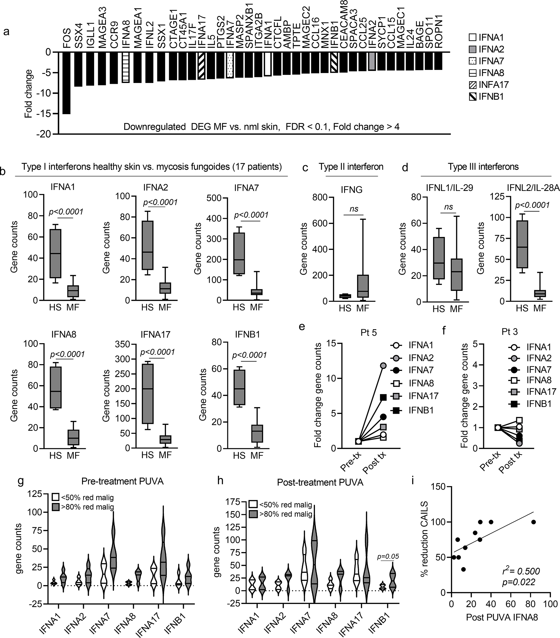
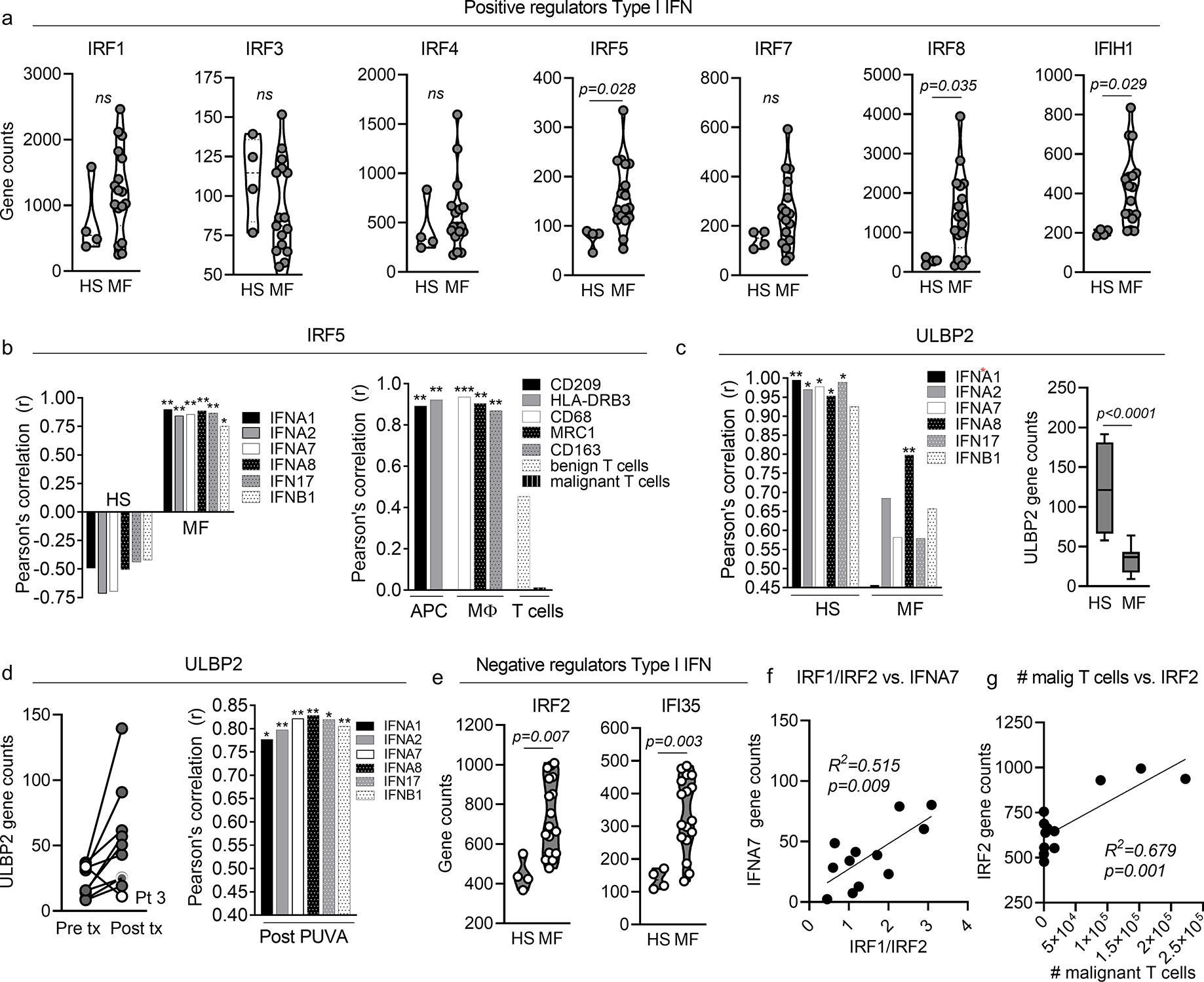
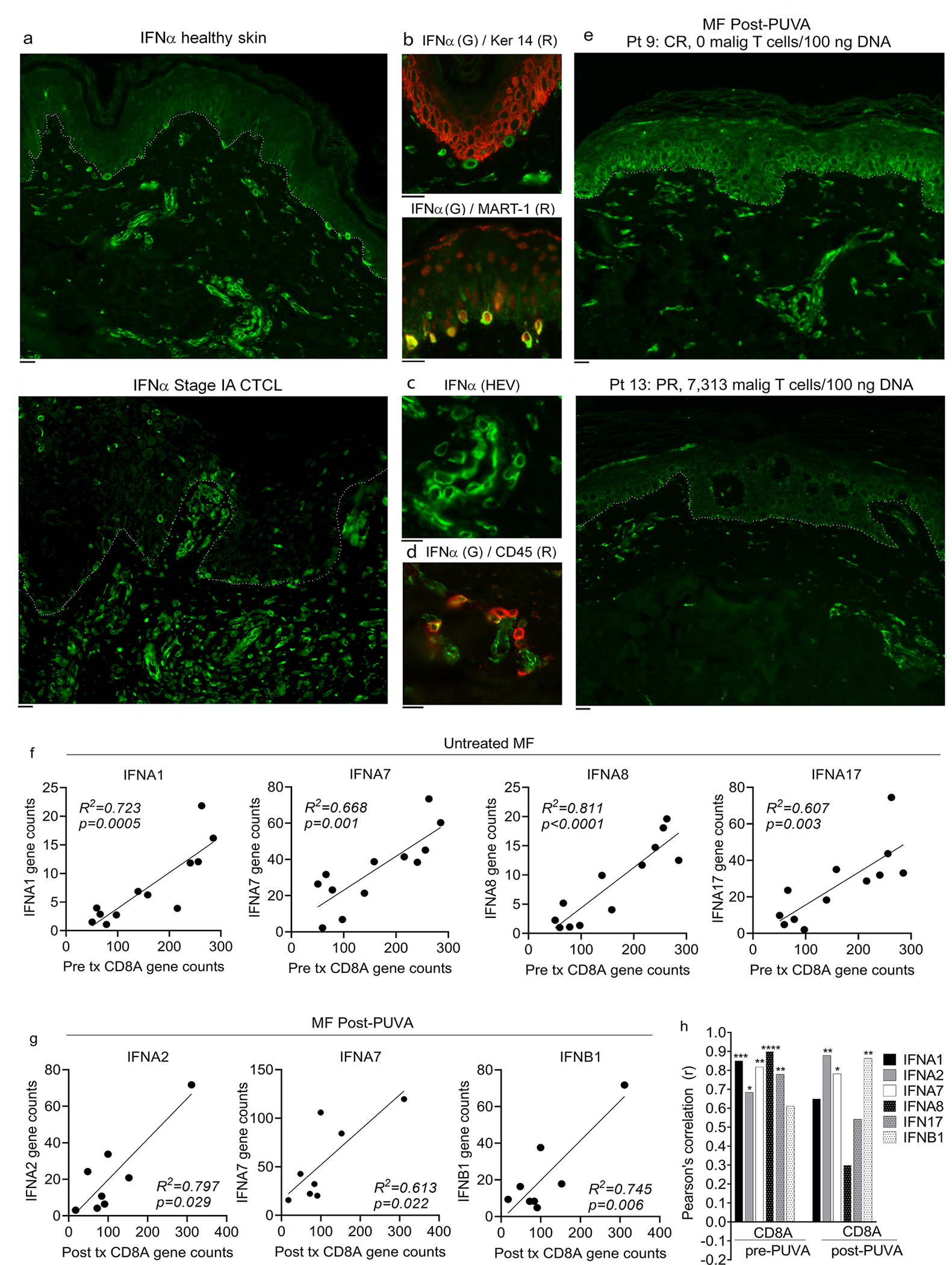
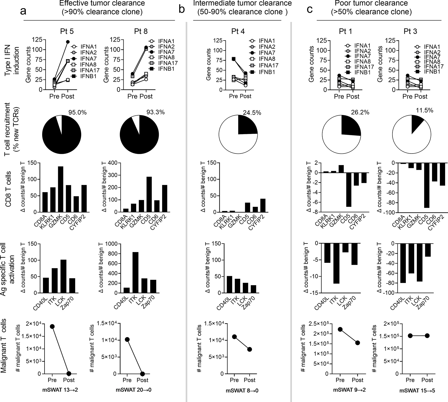
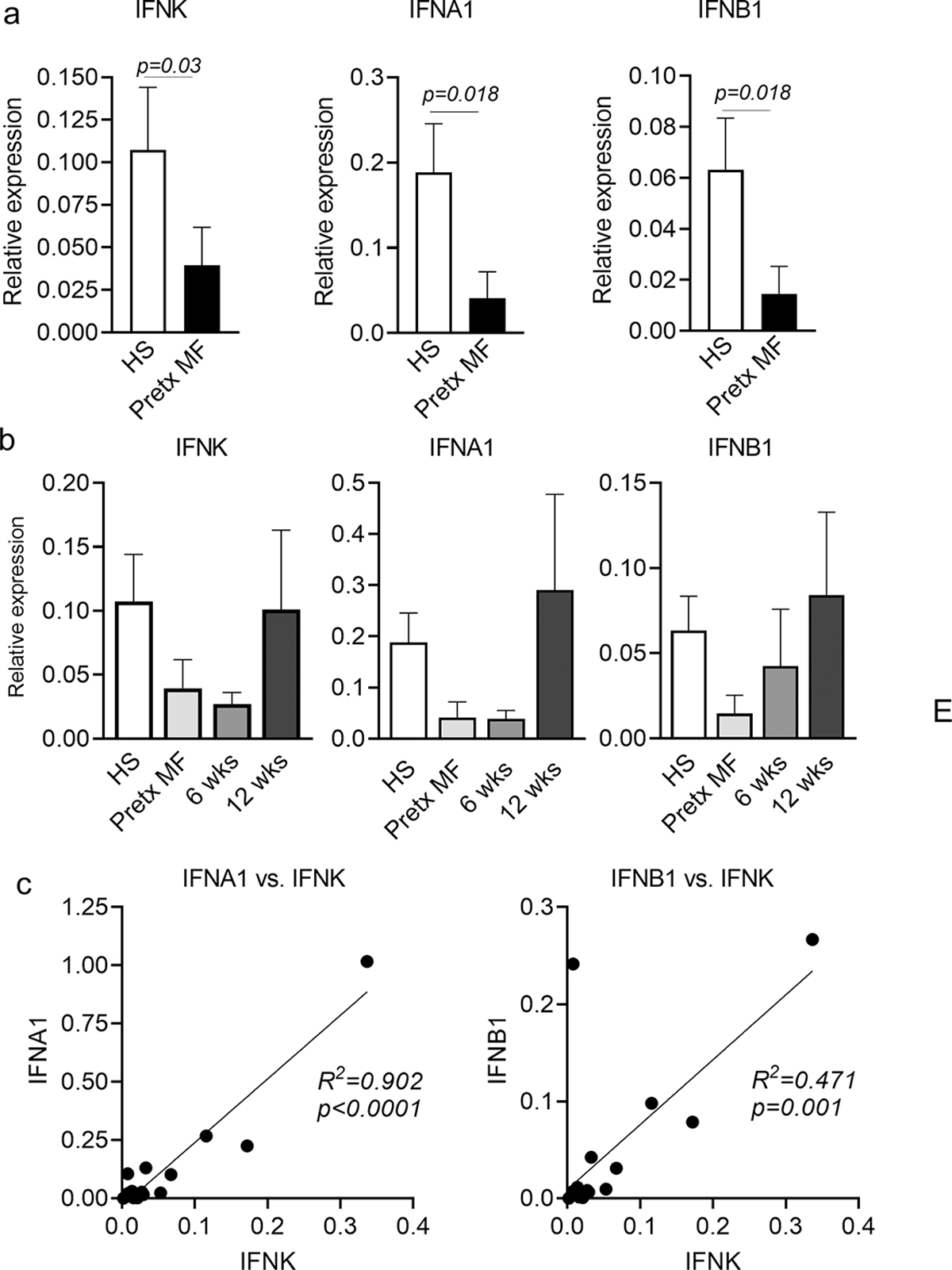
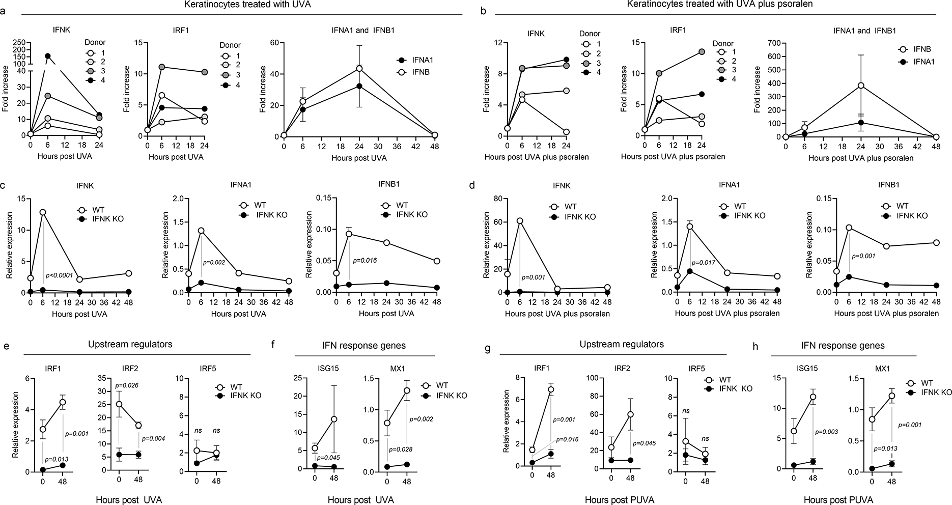
Transcriptional profiling demonstrated markedly reduced type I IFN gene expression in untreated mycosis fungoides (MF) skin lesions compared with that in healthy skin. Type I IFN expression in MF correlated with antigen-presenting cell-associated IRF5 before psoralen plus UVA therapy and epithelial ULBP2 after therapy, suggesting an enhancement of epithelial type I IFN. Immunostains confirmed reduced baseline type I IFN production in MF and increased levels after psoralen plus UVA treatment in responding patients. Effective tumor clearance was associated with increased type I IFN expression, enhanced recruitment of CD8+ T cells into skin lesions, and expression of genes associated with antigen-specific T-cell activation. IFNk, a keratinocyte-derived inducer of type I IFNs, was increased by psoralen plus UVA therapy and expression correlated with upregulation of other type I IFNs. In vitro, deletion of keratinocyte IFNk decreased baseline and UVA-induced expression of type I IFN and IFN response genes. In summary, we find a baseline deficit in type I IFN production in MF that is restored by psoralen plus UVA therapy and correlates with enhanced antitumor responses. This may explain why MF generally develops in sun-protected skin and suggests that drugs that increase epithelial type I IFNs, including topical MEK and EGFR inhibitors, may be effective therapies for MF.


Conflict of interest statement

Conflict of Interest Statement: John T. O’Malley is a current employee of Sanofi and holds stock options in the company. Phillip M. Devlin reports textbook royalties less than $5000 from Demos Medical Publishing, NYC. Nicole R. LeBoeuf is a consultant and has received honoraria from Bayer, Seattle Genetics, Sanofi, Silverback, Fortress Biotech, and Synox Therapeutics outside the submitted work. The remaining authors report no conflicts of interest.

Figures

Figure 1.
Figure 1.. A deficit in baseline type I interferon production in untreated MF can be improved by PUVA phototherapy.
(a) Gene expression levels of type I IFNs are significantly reduced in MF lesional skin as compared to skin from healthy controls. Differentially expressed genes (DEG) in lesional skin biopsies from 17 patients with IA-IIB MF compared to 4 healthy human skin donors are shown. Expression of 6/6 type I IFN genes included in the NanoString panel were significantly decreased in MF compared to healthy skin (Fig. 1a, FDR<0.1, fold change>4). (b-d) Quantitative gene count comparisons confirmed highly significant downregulation all six type I IFNs (b) and the type III IFN IFNL2/IL-28A (d) in MF; IFNG (c) and IFNL1/IL-29 (d) were not significantly different. (e-h) Studies of type I IFN production before and after PUVA phototherapy demonstrated heterogeneous patient responses. Patient 5 (e) had upregulation of all six type I IFNs and 99% clearance of the malignant T cell clone whereas Patient 3 (g) showed no IFN increases and had a 0.03% increase in malignant T cells. The mean and SEM of gene counts before (panel h, Pre treatment) and after (panel i, Post treatment) PUVA therapy in patients who cleared <50% of malignant T cells (white) and >80% of malignant T cells (gray) are shown. (i) Type I IFN levels correlate with clinical clearance of disease. The expression of IFNA8 after PUVA significantly correlated with improved CAILS score of the index lesion. Significance was determined using two-tailed T-tests (b,c,d,g,h) and linear regression analyses (i).
Figure 2.
Figure 2.. Type I IFN expression correlates with APC-associated IRF5 before PUVA phototherapy and with epithelial associated-ULBP2 after therapy, and malignant T cells are associated with IRF2, a negative regulator of epithelial IFN production.
(a) Gene expression counts of positive regulators of type I IFN production are shown for lesional skin from 17 MF patients (MF) vs. skin from four healthy donors (HS). (b) In MF, type I IFN expression was tightly associated with IRF5, which correlated with antigen presenting cell (APC) and macrophage (MΦ) specific genes. (c) In healthy skin, type I IFN production was tightly linked to expression of ULBP2, a driver of keratinocyte type I IFN production (left panel). ULBP2 gene expression was reduced in MF compared to healthy skin (c, second panel). (d) PUVA increased the expression of ULBP2 in MF skin lesions in all patients except patient 3 (white circles, left panel). Total expression all six type I IFNs correlated strongly with ULBP2 expression after PUVA (right panel). (e) IRF2 and IFI35 were up regulated in MF compared to healthy skin (HS). (f) IRF1/IRF2 correlated with expression of IFNA7 in MF skin lesions. (g) IRF2 expression levels correlated with the number of malignant T cells in skin as measured by HTS. Significance was determined using two-tailed T-tests (a,c,e), Pearson correlations with two tailed p values (b,c,d,) and linear regression analyses (f,g). * p<0.05, ** p<0.01, *** p<0.0001
Figure 3.
Figure 3.. Epithelial IFNα production is reduced in MF, can be enhanced by PUVA therapy and type I interferon levels correlate with recruitment of CD8 T cells into MF lesions.
(a) Immunostaining of healthy donor abdominal skin (top) and MF (bottom) for IFNα. Single representative donors are shown; similar results were obtained in a total of four healthy and four MF patients. The basement membrane zone is indicated by a dotted line. (b-d) Melanocytes, high endothelial venules and hematopoietic cells produce IFNα in healthy skin. Immunostaining for keratin 14 (Ker 14) and MART-1 demonstrated that melanocytes produced IFNα (b). IFNα was also produced by high endothelial venules (HEV, c) and CD45+ hematopoietic cells (d). Representative results are shown, similar results were obtained in a total of four healthy skin donors. (e) PUVA therapy increased epidermal IFNα expression in patient 9, who had eradication of malignant T cells after therapy (e, top panel) but did not improve IFNα expression in patient 13, who failed to clear disease (e, lower panel). The basement membrane zone is indicated by a dotted line. Two additional PUVA treated MF patients showed similar results. (f-h) Type I interferon levels correlate with recruitment of CD8 T cells into MF skin lesions. (a,b) Correlation of type I IFN and CD8A gene expression levels are shown in lesional skin of untreated MF patients (f) and in MF lesional skin after PUVA therapy (g). (h) The Pearson’s correlation coefficients of all type I IFN genes with CD8A in untreated MF and after PUVA are shown. A two tailed p value was utilized for all analyses. Scale bars: 10μm. * p<0.05, ** p<0.01, *** p<0.0001
Figure 4.
Figure 4.. Effective tumor clearance was associated with increased type I IFN expression, enhanced recruitment of CD8+ T cells into skin lesions and expression of genes associated with antigen specific T cell activation.
Data derived from patients with effective (a), intermediate (b) and poor (c) tumor clearance are shown. Gene expression levels of type I IFNs before (Pre) and after (Post) PUVA increased in high but not intermediate and poor responders (top row). The % of benign T cell clones newly recruited into skin after PUVA as assessed by HTS was greatest in patients with effective tumor clearance (second row). Change scores pre and post PUVA for genes associated with CD8 T cells (third row) and antigen specific activation (fourth row), normalized to the total number of benign T cells, were highest in patients with effective tumor clearance. Malignant T cell numbers in skin before (Pre) and after (Post) PUVA therapy and mSWAT clinical response scores are shown.
Figure 5.
Figure 5.. IFNκ is reduced in MF compared to healthy skin, is upregulated by PUVA phototherapy and correlates with the production of α and β type I IFNs.
(a) Quantitative RT-PCR studies demonstrated that IFNK, IFNA1 and IFNB1 were expressed at lower levels in pretreatment MF lesions (Pretx MF) compared to healthy skin (HS). (b) The expression IFNK, IFNA1 and IFNB1 in MF skin lesions tended to increase to normal levels following PUVA. (c) The expression of IFNA1 and IFNB1 were both tightly correlated with expression of IFNK; MF values both before and after PUVA are included. Significance was determined using two-tailed T-tests (a,b) and linear regression analyses (c).
Figure 6.
Figure 6.. IFNκ is responsible for induction of epithelial type I IFNs and downstream IFN response genes after UVA phototherapy.
Healthy human cultured keratinocytes were stimulated in vitro with UVA (a) or UVA plus psoralen (b), then analyzed by quantitative RT-PCR for the expression of IFNs and IRF1. IFNK and IRF1 were upregulated at 6 hours after UVA exposure (left two panels), preceding the upregulation of α and β type I interferons (IFNA1 and IFNB1, right panels), which peaked at 24 hours after light exposure. (c,d) To determine if IFNκ was responsible for the light induced increase in other type I interferons, Tert-transduced immortalized keratinocytes with CRISPR-Cas9 deletion of IFNκ were exposed to UVA and studied by qRT-PCR for their expression of IFNs. Expression of IFNK, IFNA1 and IFNB1 were significantly decreased in IFNκ knockout keratinocytes compared to wild type controls (WT) after treatment with UVA alone (c) or UVA plus psoralen (d). (e,g) Expression of IRF1 and IRF2, two upstream regulators of epithelial type I IFN production, were significantly lower both before and after treatment in IFNκ knockout keratinocytes. Expression of IRF5, an APC-associated interferon regulator, was unchanged. Results after treatment with UVA alone (e) and UVA plus psoralen (g) are shown. (f,h) Levels of the downstream interferon response genes ISG15 and MX1 were both significantly lower in IFNκ knockout keratinocytes compared to WT controls after treatment with UVA alone (f) or UVA plus psoralen (h). Significance was determined using two-tailed T-tests.

Comment in

Similar articles

Cited by

References

    1. Agar NS, Wedgeworth E, Crichton S, Mitchell TJ, Cox M, Ferreira S, et al. Survival outcomes and prognostic factors in mycosis fungoides/Sezary syndrome: validation of the revised International Society for Cutaneous Lymphomas/European Organisation for Research and Treatment of Cancer staging proposal. Journal of clinical oncology : official journal of the American Society of Clinical Oncology 2010;28(31):4730–9. - PubMed
    1. Akiyama Y, Kondou R, Iizuka A, Ohshima K, Urakami K, Nagashima T, et al. Immune response-associated gene analysis of 1,000 cancer patients using whole-exome sequencing and gene expression profiling-Project HOPE. Biomed Res 2016;37(4):233–42. - PubMed
    1. Antonczyk A, Krist B, Sajek M, Michalska A, Piaszyk-Borychowska A, Plens-Galaska M, et al. Direct inhibition of IRF-dependent transcriptional regulatory mechanisms associated with disease. Frontiers in immunology 2019. - PMC - PubMed
    1. Boukhaled GM, Harding S, Brooks DG. Opposing Roles of Type I Interferons in Cancer Immunity. Annual review of pathology 2021;16:167–98. - PMC - PubMed
    1. Buhl T, Schön MP. Peeking into immunoregulatory effects of phototherapy. Experimental dermatology 2016. - PubMed

Publication types