. 2023 Oct;299(10):105256.
doi: 10.1016/j.jbc.2023.105256. Epub 2023 Sep 15.

Structural and functional insights into the Pseudomonas aeruginosa glycosyltransferase WaaG and the implications for lipopolysaccharide biosynthesis

Affiliations

Structural and functional insights into the Pseudomonas aeruginosa glycosyltransferase WaaG and the implications for lipopolysaccharide biosynthesis

Emma R Scaletti et al. J Biol Chem. 2023 Oct.

Abstract

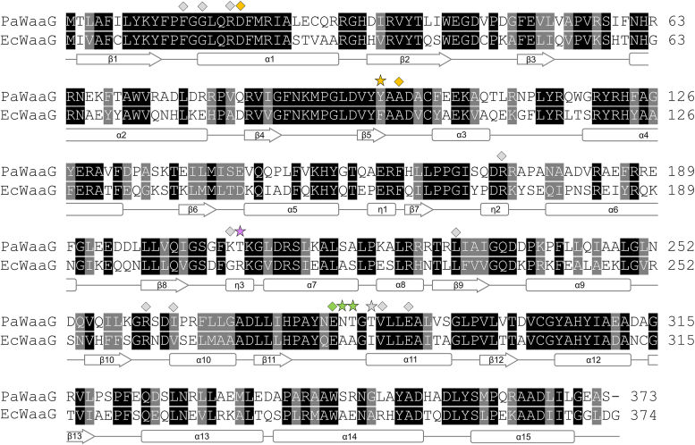
The glycosyltransferase WaaG in Pseudomonas aeruginosa (PaWaaG) is involved in the synthesis of the core region of lipopolysaccharides. It is a promising target for developing adjuvants that could help in the uptake of antibiotics. Herein, we have determined structures of PaWaaG in complex with the nucleotide-sugars UDP-glucose, UDP-galactose, and UDP-GalNAc. Structural comparison with the homolog from Escherichia coli (EcWaaG) revealed five key differences in the sugar-binding pocket. Solution-state NMR analysis showed that WT PaWaaG specifically hydrolyzes UDP-GalNAc and unlike EcWaaG, does not hydrolyze UDP-glucose. Furthermore, we found that a PaWaaG mutant (Y97F/T208R/N282A/T283A/T285I) designed to resemble the EcWaaG sugar binding site, only hydrolyzed UDP-glucose, underscoring the importance of the identified amino acids in substrate specificity. However, neither WT PaWaaG nor the PaWaaG mutant capable of hydrolyzing UDP-glucose was able to complement an E. coli ΔwaaG strain, indicating that more remains to be uncovered about the function of PaWaaG in vivo. This structural and biochemical information will guide future structure-based drug design efforts targeting PaWaaG.

Keywords: NMR; Pseudomonas aeruginosa; WaaG; X-ray crystallography; glycosyltransferase; lipopolysaccharide.


Conflict of interest statement

Conflict of interest The authors declare that they have no conflicts of interest with the contents of this article.

Figures

Figure 1
Figure 1
Lipopolysaccharide (LPS) structure ofP. aeruginosa. A, overall LPS structure represented by a snapshot from a molecular dynamics simulation of semirough LPS from P. aeruginosa serotype O10 containing core glycoform 2 (60); C atoms are colored gray, O atoms red, N atoms blue and P atoms orange. Figure produced with PyMOL (v.2.3.3, Schrödinger). B, lipopolysaccharide R1 core from Escherichia coli (7) and 1b core from P. aeruginosa (14) schematically represented using sugar residues in the Symbol Nomenclature for Glycans (SNFG) format (61) drawn by GlycanBuilder2 (62). Monosaccharide codes: d-glucose (d-Glc), d-galactose (d-Gal), d-galactosamine (d-GalN), l-Rhamnose (l-Rha), l-glycero-d-manno-heptose (ldmanHep), ketodeoxyoctonic acid (Kdo). In E. coli the glycosyltransferase WaaG transfers the first hexose residue of the outer core region to a heptose residue in the inner core region.
Figure 2
Figure 2
Crystal structure of the PaWaaG in complex with UMP.A, PaWaaG monomer is shown as a rainbow cartoon with secondary structure annotation. UMP is shown as a ball-and-stick model. B, hydrogen bond network for UMP in the PaWaaG active site. Amino acids involved in ligand coordination are depicted as sticks. Hydrogen bond interactions are shown as dashed lines. The 2Fo − Fc electron density map around UMP is contoured at 1.3 σ (blue), and the Fo − Fc electron density maps are contoured at +3.0 σ (green) and −3.0 σ (red). UMP is depicted as a stick model; C atoms are colored yellow, O atoms red, N atoms blue, and P atoms orange. Figures were produced with PyMOL (v.2.3.3, Schrödinger).
Figure 3
Figure 3
Comparison of substrate binding in PaWaaG. Hydrogen bond networks for (A) UDP-glucose, (B) UDP-galactose, and (C) UDP-GalNAc in the PaWaaG active site. In panel A, PaWaaG-UDP-glucose (amino acids in light gray color) is superimposed with PaWaaG-UMP (amino acids in cyan color). In panels (A–C) nucleotides are depicted as stick models; C atoms are colored yellow (UDP-glucose, UDP-galactose, and UDP-GalNAC) or dark teal (UMP), O atoms red, N atoms blue, and P atoms orange. Amino acids involved in ligand coordination are depicted as sticks. Hydrogen bond interactions are shown as dashed lines. The 2Fo − Fc electron density maps (blue) around the substrates are contoured at 1.0 σ (UDP-GalNAc) or 1.3 σ (UDP-glucose and UDP-galactose), and the Fo − Fc electron density maps are contoured at +3.0 σ (green) and 3.0 σ (red). D, comparison of UDP-glucose (cyan), UDP-galactose (magenta) and UDP-GalNAc (dark gray) binding in PaWaaG. The hydrogen bond interactions correspond to the PaWaaG-UDP-GalNAc structure. Figures were produced with PyMOL (v.2.3.3, Schrödinger).
Figure 4
Figure 4
Comparison of PaWaaG structures with EcWaaG.A, superposition of PaWaaG-UMP (gray) and EcWaaG-UDP-2F-glucose (dark green, PDB ID: 2iw1) monomers. Nucleotides are depicted as ball-and-stick models; C atoms are colored yellow (UMP) or green (UDP-2F-glucose), O atoms red, N atoms blue, and P atoms orange. Comparison of EcWaaG UDP-2F-glucose binding with (B) UMP, (C) UDP-glucose and (D) UDP-GalNAc binding in PaWaaG. Hydrogen bond interactions for the EcWaaG and PaWaaG complexes are shown as green and black dashed lines, respectively. Amino acids involved in ligand coordination are depicted as sticks and numbering refers to the EcWaaG complex. Where coordinating amino acids are not conserved between the structures the relevant residue from PaWaaG is labeled, with red and orange text referring to nonconserved and physiochemically conserved differences, respectively. Figures were produced with PyMOL (v.2.3.3, Schrödinger).
Figure 5
Figure 5
Structure-based sequence alignment of P. aeruginosa WaaG (UniProt:Q9HUF6) and Escherichia coli WaaG (UniProt:B7L754) performed using Clustal Omega through theEuropean Bioinformatics Institute (EBI)webserver. The resulting alignment is colored according to sequence similarity using BOXSHADE. Identical residues are shaded black, while dark gray shading indicates amino acids with conserved physicochemical properties. Residues important for the positioning of nucleotide sugars in PaWaaG are shown above the alignment. Light gray symbols indicate UMP/UDP interacting amino acids. Green symbols show residues that position the glucose, galactose, and GalNAc moieties of the nucleotide sugars. Yellow symbols indicate residues that position galactose and GalNAc only. The purple symbol indicates a residue (Thr208, PaWaaG numbering) that is proposed to be important for the specificity of PaWaaG toward UDP-GalNAc. Stars indicate five amino acid which were mutated in the PaWaaG sequence to the equivalent EcWaaG residues to produce the PaWaaG-5mut mutant. The secondary structure corresponding to the amino acid sequence of PaWaaG is displayed below the alignment.
Figure 6
Figure 6
Important residues in the PaWaaG-5mut construct. Superposition of PaWaaG-UDP-GalNAc (gray) and EcWaaG-UDP-2F-glucose (dark green, PDB ID: 2iw1) monomers. Nucleotides are depicted as stick models; C atoms are colored yellow (UDP-GalNAc) or green (UDP-2F-glucose), O atoms red, N atoms blue, and P atoms orange. Hydrogen bond interactions for the EcWaaG and PaWaaG complexes are shown as green and black dashed lines, respectively. The five mutations in the PaWaaG-5mut construct change those amino acids to the equivalent amino acids present in EcWaaG. Figures were produced with PyMOL (v.2.3.3, Schrödinger). PDB, Protein Data Bank.
Figure 7
Figure 7
Solution NMR experiments of PaWaaG with potential donor sugar substrates. Hydrolysis of (A) UDP-GalNAc by PaWaaG and (B) UDP-glucose by PaWaaG-5mut. The panels display the downfield part of the 1H NMR spectrum where deshielded protons (i.e., hydrogens in protein amide and aromatic groups, hydrogens close to electronegative atoms in sugars and nucleotides) have chemical shifts. Spectra of proteins together with sugar donors are color coded according to the number of days after substrates were added. Spectra of UDP-GalNAc or UDP-glucose in buffer only (black) and of proteins before the addition of substrate (gray) are also shown.
Figure 8
Figure 8
Complementation of the phenotype in an Escherichia coli strain lacking WaaG (ΔwaaG).A, the E. coli ΔwaaG strain grows comparably to a WT E. coli strain on LB agar (leftpanel) but is sensitive to SDS (rightpanel). B, EcWaaG, PaWaaG, and PaWaaG-5mut were expressed in E. coli ΔwaaG and plated on LB agar (top panel) or LB agar with 0.0625% (w/v) SDS (bottom panel). Plates contained 100 μg/ml ampicillin. Proteins were expressed at low levels by the inclusion of 0.02% (w/v) glucose in the agar.

References

    1. Papo N., Shai Y. A molecular mechanism for lipopolysaccharide protection of gram-negative bacteria from antimicrobial peptides. J. Biol. Chem. 2005;280:10378–10387. - PubMed
    1. Huszczynski S.M., Lam J.S., Khursigara C.M. The role of Pseudomonas aeruginosa lipopolysaccharide in bacterial pathogenesis and physiology. Pathogens. 2020;9:6. - PMC - PubMed
    1. Kim S., Patel D.S., Park S., Slusky J., Klauda J.B., Widmalm G., et al. Bilayer properties of lipid A from various gram-negative bacteria. Biophys. J. 2016;111:1750–1760. - PMC - PubMed
    1. Ståhle J., Widmalm G. Lipopolysaccharides of gram-negative bacteria: biosynthesis and structural Aspects Trends in glycoscience. Glycotechnology. 2019;31:E159–E171.
    1. Di Lorenzo F., Duda K.A., Lanzetta R., Silipo A., De Castro C., Molinaro A. A journey from structure to function of bacterial lipopolysaccharides. Chem. Rev. 2022;122:15767–15821. - PubMed

LinkOut - more resources