. 2023 Sep 16;14(9):610.
doi: 10.1038/s41419-023-06132-0.

LncRNA Neat1 targets NonO and miR-128-3p to promote antigen-specific Th17 cell responses and autoimmune inflammation

Affiliations

LncRNA Neat1 targets NonO and miR-128-3p to promote antigen-specific Th17 cell responses and autoimmune inflammation

Sisi Chen et al. Cell Death Dis. .

Abstract

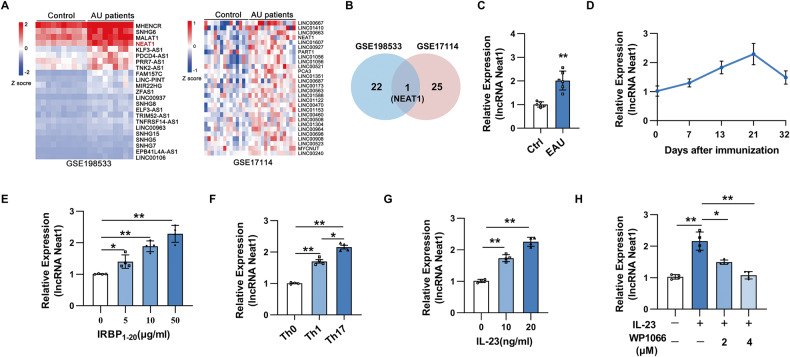
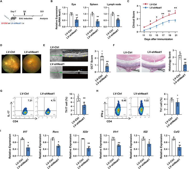
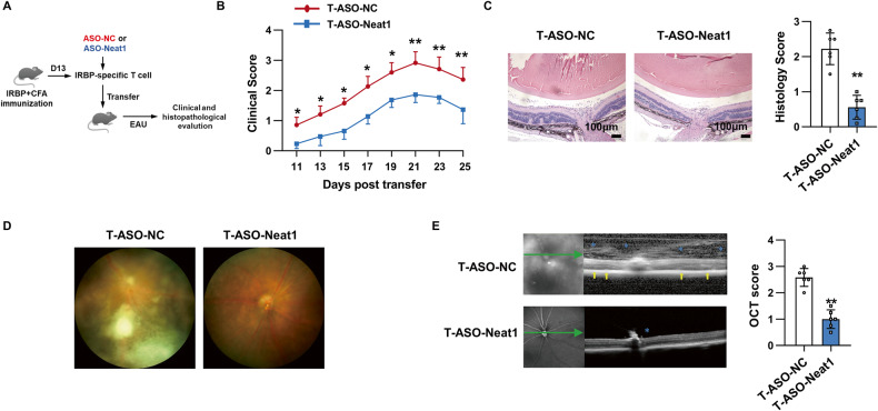
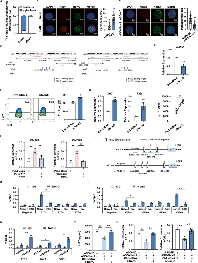
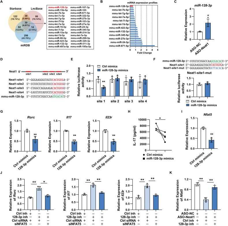
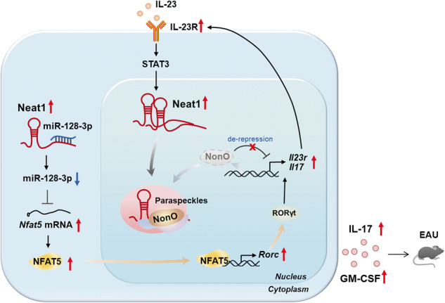
Long non-coding RNAs (lncRNAs) interaction with RNA-Binding proteins (RBPs) plays an important role in immunological processes. The generation of antigen-specific Th17 cells is closely associated with autoimmune pathogenesis. However, the function of lncRNA-RBP interactions in the regulation of pathogenic Th17 cell responses during autoimmunity remains poorly understood. Here, we found that lncRNA Neat1, highly expressed in Th17 cells, promoted antigen-specific Th17 cell responses. Both global and CD4+ T cell-specific knockdown of Neat1 protected mice against the development of experimental autoimmune uveitis (EAU). Mechanistically, Neat1 regulated RNA-Binding protein NonO, thus relieving IL-17 and IL-23R from NonO-mediated transcriptional repression and supporting antigen-specific Th17 cell responses. In addition, Neat1 also modulated miR-128-3p/NFAT5 axis to increase the expression of IL-17 and IL-23R, leading to augmented Th17 cell responses. Our findings elucidate a previously unrecognized mechanistic insight into the action of Neat1 in promoting antigen-specific Th17 responses and autoimmunity, and may facilitate the development of therapeutic targets for T cell-mediated autoimmune diseases.


Conflict of interest statement

The authors declare no competing interests.

Figures

Fig. 1
Fig. 1. Neat1 is upregulated in the PBMCs of AU patients and antigen-specific Th17 cells of EAU mice.
A Heatmap of upregulated lncRNAs in PBMCs of AU patients compared with healthy controls. Upregulated lncRNAs were sorted via analysis of dataset GSE198533 (including 10 BD patients and 10 healthy controls) and GSE17114 (containing 15 BD patients and 14 healthy controls). B Venn diagram showing the overlap between lncRNAs upregulated in two publicly available datasets of AU patients (GSE17114 and GSE198533). C Real-time qRT-PCR analysis of Neat1 expression in CD4+ T cells from naïve (Ctrl) and EAU mice (n = 6 per group). D Kinetic expression of Neat1 expression in CD4+ T cells during the course of EAU (n = 4 per group). EH CD4+ T cells were isolated from EAU mice and co-cultured with APCs and IRBP1-20 under indicated conditions for 48 h. Real-time qRT-PCR analysis of Neat1 expression (n = 4 per group). Data are presented as mean ± SD of at least three independent experiments. *p < 0.05, **p < 0.01.
Fig. 2
Fig. 2. Neat1-silenced mice developed alleviated EAU and displayed decreased Th17 cell responses.
A Schematic diagram of the experimental procedure to study the role of Neat1 in EAU. B Real-time qRT-PCR analysis of Neat1 expression in eyes, spleens, and lymph nodes of mice infected with LV-Ctrl or LV-shNeat1 (n = 6 per group). C Clinical score of EAU in mice infected with LV-Ctrl and LV-shNeat1 (n = 6 per group). D Representative images of eyes in LV-Ctrl and LV-shNeat1-infected EAU mice on day 21 after EAU induction by retinal imaging microscope. E Representative images showing fundus condition by SD-OCT. Hyper-reflective foci (blue asterisk) in the vitreous cavity and retinal folds (yellow arrows) were observed around the optical nerve. F Histopathological analysis of eyes from lentivirus-infected EAU mice (n = 6 per group). Scale bars, 100 μm. G, H Flow cytometric analysis of the percentages of Th17 (G) and Th1 (H) in T cells from lentivirus-infected EAU mice (n = 6 per group). I Real-time qRT-PCR analysis of pathogenic Th17-related genes in T cells from lentivirus-infected EAU mice (n = 4 per group). Data are presented as mean ± SD of at least three independent experiments. *p < 0.05, **p < 0.01. See also Fig. S1.
Fig. 3
Fig. 3. Neat1 downregulated in Th17 cells inhibits antigen-specific Th17 cell responses in vitro.
A, C, D CD4+ T cells isolated from immunized mice were transfected with ASO-NC or ASO-Neat1 and stimulated with IRBP1-20 in the presence of irradiated APCs under Th17-polarizing conditions. A Real-time qRT-PCR analysis of Neat1 expression (n = 4 per group). C Flow cytometric analysis of the percentages of Th17 cells (n = 4 per group). D Real-time qRT-PCR analysis of Th17-related gene expression (n = 4 per group). B ELISA analysis of IL-17 production in CD4+ T cells transfected with ASO-NC or ASO-Neat1 and restimulated with the indicated antigens and IL-23 (n = 4 per group). Data are presented as mean ± SD of at least three independent experiments. *p < 0.05, **p < 0.01. See also Fig. S2.
Fig. 4
Fig. 4. Silencing of Neat1 in Th17 cells ameliorates EAU.
A Schematic diagram of EAU induced by adoptive transfer. B Clinical scores of EAU in naïve C57BL/6 mice adoptively transferred with ASO-NC (T-ASO-NC) or ASO-Neat1 (T-ASO-Neat1)-transfected Th17 cells (n = 6 per group). C Histopathological analysis of eyes from the transferred EAU mice (n = 6 per group). Scale bars, 100 μm. D Representative images of eyes in the transferred EAU mice by retinal imaging microscope. E Representative images showing fundus condition by SD-OCT. Hyper-reflective foci (blue asterisk) in the vitreous cavity and retinal folds (yellow arrows) were observed around the optical nerve. Data are representative of the analysis of three independent experiments. Error bars represent the mean ± SD. *p < 0.05, **p < 0.01.
Fig. 5
Fig. 5. NonO is involved in Neat1 mediated antigen-specific Th17 cell responses.
A Cytoplasmic and nuclear levels of Neat1 in CD4+ T cells from EAU mice (n = 3 per group). B Representative images of paraspeckles in naïve and EAU CD4+ T cells under confocal microscopy (n = 30 cells in each group). Red, Cy3-labeled Neat1 probes; green, Alexa-488-labeled secondary antibody for NonO protein. Scale bars, 2 μm. C Representative confocal images of paraspeckles in EAU CD4+ T cells upon Neat1 knockdown (n = 30 cells in each group). D Prediction of NonO and SFPQ binding sites in the Il17 and Il23r promoter regions by MEME analysis. EH, N–O CD4+ T cells isolated from immunized mice were transfected with indicated oligonucleotides and stimulated with IRBP1-20 in the presence of irradiated APCs under Th17-polarizing conditions. E Real-time qRT-PCR analysis of NonO expression (n = 4 per group). F Flow cytometric analysis of the percentages of Th17 cells (n = 4 per group). G, O Real-time qRT-PCR analysis of Il17 and Il23r expression (n = 4 per group). H, N ELISA analysis of IL-17 secretion in the culture supernatants (n = 4 per group). I Luciferase activity analysis of reporter containing Il17 or Il23r promoter transfected into HEK293T with the indicated plasmids (n = 4 per group). JM ChIP analysis of NonO occupancy at the promoter of the Il17 or Il23r gene was performed in CD4+ T cells (n = 3 per group). Blue lines 1 to 4 indicate regions detected by ChIP-qPCR. Regions without NonO binding sites (no binding site) were used as negative control. Data present at least three independent experiments. *p < 0.05, **p < 0.01.
Fig. 6
Fig. 6. Neat1 promotes antigen-specific Th17 cell responses by regulating miR-128-3p/NFAT5 axis.
A Venn diagram analysis of putative Neat1 target miRNAs using Starbase v3.0, DIANA-LncBase, and miRDB databases. B Only 23 candidate miRNAs were expressed in CD4+ T cells isolated from EAU mice. Microarray-based bar graphs showing the expression level of candidate miRNA in CD4+ T cells isolated from EAU mice relative to those from naïve mice. C Real-time qRT-PCR analysis of miR-128-3p expression in Th17 cells transfected with ASO-NC or ASO-Neat1 (n = 4 per group). D Sequence alignment between miR-128-3p and its potential binding sites (in red letters) in the lncRNA Neat1. E, F Luciferase activity analysis of reporter carrying Neat1 binding sites or mutant site (Mut) co-transfected into HEK293T with Ctrl mimics or miR-128-3p mimics (n = 4 per group). GK CD4+ T cells isolated from immunized mice were transfected with indicated oligonucleotides and stimulated with IRBP1-20 in the presence of irradiated APCs under Th17-polarizing conditions. G, J Real-time qRT-PCR analysis of Rorc, Il17 and Il23r expression (n = 4 per group). H ELISA analysis of IL-17 secretion in the culture supernatants (n = 4 per group). I, K Real-time qRT-PCR analysis of Nfat5 expression (n = 4 per group). Data are presented as mean ± SD of at least three independent experiments. *p < 0.05, **p < 0.01. See also Fig. S3.
Fig. 7
Fig. 7. Schematic showing the molecular mechanisms by which Neat1 regulates antigen-specific Th17 cell responses in EAU.
The increased expression of Neat1 is induced by IL-23 signaling, and Neat1 in turn augments IL-23R expression by regulating NonO and miR-128-3p/NFAT5, thus forming a positive feedback loop to sustain antigen-specific Th17 cell responses that contribute to the progression of EAU.

Similar articles

Cited by

References

    1. de Smet MD, Taylor SR, Bodaghi B, Miserocchi E, Murray PI, Pleyer U, et al. Understanding uveitis: the impact of research on visual outcomes. Prog Retin Eye Res. 2011;30:452–70. doi: 10.1016/j.preteyeres.2011.06.005. - DOI - PubMed
    1. Joltikov KA, Lobo-Chan AM. Epidemiology and risk factors in non-infectious uveitis: a systematic review. Front Med (Lausanne) 2021;8:695904. doi: 10.3389/fmed.2021.695904. - DOI - PMC - PubMed
    1. Papotto PH, Marengo EB, Sardinha LR, Goldberg AC, Rizzo LV. Immunotherapeutic strategies in autoimmune uveitis. Autoimmun Rev. 2014;13:909–16. doi: 10.1016/j.autrev.2014.05.003. - DOI - PMC - PubMed
    1. Agarwal RK, Silver PB, Caspi RR. Rodent models of experimental autoimmune uveitis. Methods Mol Biol. 2012;900:443–69. doi: 10.1007/978-1-60761-720-4_22. - DOI - PMC - PubMed
    1. Amadi-Obi A, Yu CR, Liu X, Mahdi RM, Clarke GL, Nussenblatt RB, et al. TH17 cells contribute to uveitis and scleritis and are expanded by IL-2 and inhibited by IL-27/STAT1. Nat Med. 2007;13:711–8. doi: 10.1038/nm1585. - DOI - PubMed

Publication types