. 2023 Sep 16;14(1):5755.
doi: 10.1038/s41467-023-41459-4.

Ustilago maydis PR-1-like protein has evolved two distinct domains for dual virulence activities

Affiliations

Ustilago maydis PR-1-like protein has evolved two distinct domains for dual virulence activities

Yu-Han Lin et al. Nat Commun. .

Abstract

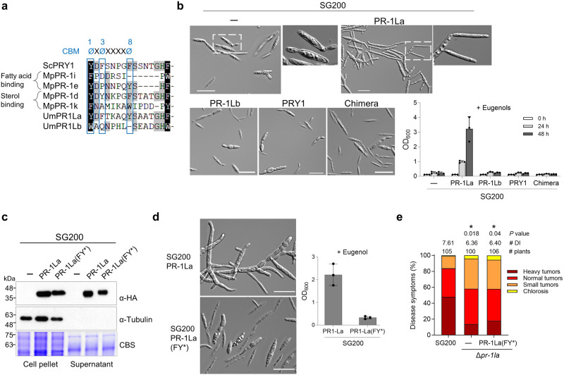
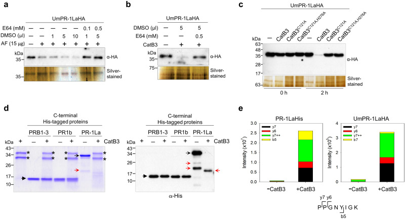
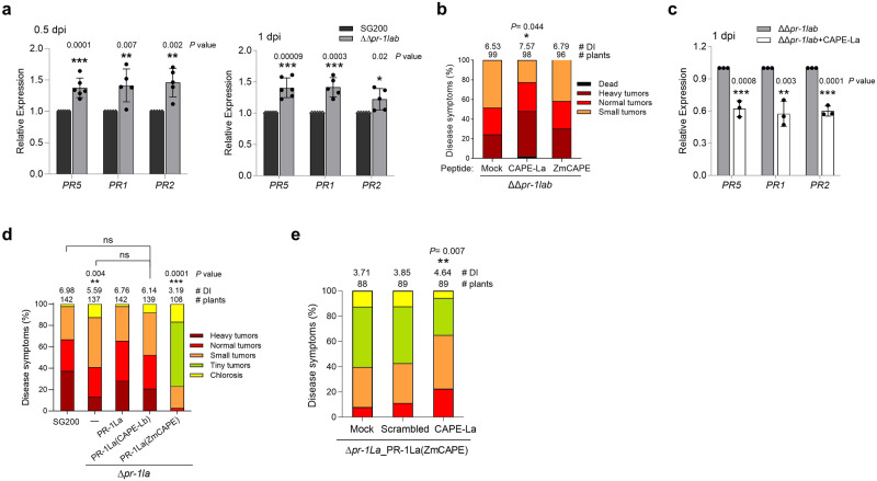
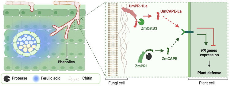
The diversification of effector function, driven by a co-evolutionary arms race, enables pathogens to establish compatible interactions with hosts. Structurally conserved plant pathogenesis-related PR-1 and PR-1-like (PR-1L) proteins are involved in plant defense and fungal virulence, respectively. It is unclear how fungal PR-1L counters plant defense. Here, we show that Ustilago maydis UmPR-1La and yeast ScPRY1, with conserved phenolic resistance functions, are Ser/Thr-rich region mediated cell-surface localization proteins. However, UmPR-1La has gained specialized activity in sensing phenolics and eliciting hyphal-like formation to guide fungal growth in plants. Additionally, U. maydis hijacks maize cathepsin B-like 3 (CatB3) to release functional CAPE-like peptides by cleaving UmPR-1La's conserved CNYD motif, subverting plant CAPE-primed immunity and promoting fungal virulence. Surprisingly, CatB3 avoids cleavage of plant PR-1s, despite the presence of the same conserved CNYD motif. Our work highlights that UmPR-1La has acquired additional dual roles to suppress plant defense and sustain the infection process of fungal pathogens.


Conflict of interest statement

The authors declare no competing interests.

Figures

Fig. 1
Fig. 1. The PR-1-like (PR-1L) family proteins in U. maydis.
a Schematic drawing displays plant PR-1 and PR-1L protein domain structure. The signal peptide (SP) is depicted as a black stripe, the Ser/Thr-rich (S/T) region as a white box, and the CNYx motif and CAPE/CAPE-like peptide (a red box) are located at the C-terminus of the CAP-domain (green box). Conserved residues shared between plant PR-1’s CAPE and PR-1L’s CAPE-like peptides are marked in green. Solanum lycopersium SlPR1b (#P04284); Saccharomyces cerevisiae ScPRY1 (#NP012456). b The sequence comparison of plant CAPEs and smut fungal CAPE-like peptides after CNYx motif. The green line indicates the MEME-derived consensus sequence P[P/V]GN[Φ/V][V/I]G. The protein accession numbers are provided. Plant CAPE peptides that have been identified and reported are indicated,. Ph Pseudozyma hubeiensis, Ss Sporisorium scitamineum, Sr Sporisorium reilianum, Uh Ustilago hordei, Ub Ustilago bromivora. c Phylogenetic analysis of plant PR-1 and PR-1-like proteins from smut fungi and yeast. Protein sequences are retrieved from the NCBI, aligned using Clustal Omega, and curated with trimAl. Analysis was carried out in MEGA 7.0 using the Maximum Likelihood method with the WAG+G+I model and 1000 bootstrap replicates. Clade I and II: smut fungal PR-1Ls; Clade III: Plant PR-1s; IV: yeast. Kb Kalmanozyma brasiliensis, Mp Melanopsichium pennsylvanicum, Man Moesziomyces antarcticus, Map Moesziomyces aphidis, Ut Ustilago trichophora, Sg Sporisorium graminicola. d qRT-PCR analysis of PR-1La expression. Total RNA was extracted from SG200-infected maize leaves harvested at the indicated days post-infection (dpi), and from cells cultured in liquid culture (A.C). Peptidylprolyl Isomerase (PPI) gene was used for normalization. The PR-1La expression level in A.C. was set to 1.0. Values are mean ± standard deviation (SD) of three biological replicates. e Virulence assay of Δpr-1l and the complementation strain. ∆pr1la_PR-1La: ∆pr1la was complemented by introducing a single allele of PR-1La under the control of its native promoter. Disease symptoms were scored at 12 dpi according to the severity depicted in the color code. The number of infected plants and the average disease index (DI) from four independent infections are shown. Significant differences between samples were determined by a two-tailed unpaired t-test (*P < 0.05; **P < 0.01; ***P < 0.001). ns no significance. Source data are provided as a Source Data file.
Fig. 2
Fig. 2. UmPR-1La is a cell-surface-associated protein.
a UmPR-1La localizes to the surface of filaments and the bud-neck of sporidial cells. Hydroxyl fatty acid-induced SG200 expressing C-terminal HA-tagged proteins under the otef promoter formed filaments on a hydrophobic parafilm surface. Immunolocalization was performed using anti-HA and anti-IgG-AF488 antibodies to locate HA-tagged proteins. SG200_PR-1La: produced the signal-peptide (SP)-containing full-length protein (279 aa; 31.5 kDa); SG200_PR-1La(FY*): produced PR-1La(FY*) proteins with the two aromatic residues at positions 3 and 8 of the caveolin binding motif replaced with alanine (see Fig. 3a); SG200_PR-1Lb: produced the SP-containing full-length protein (480 aa; 54.2 kDa); SG200_PRY1: produced the proteins (32.2 kDa) containing UmPR-1La’s SP (green strip box) fused to PRY1 (20-299 aa); SG200_NPR-1La-CAPPRY1: produced the chimera proteins (31 kDa) containing the N-terminus of UmPR1-La (1–133 aa, including SP and S/T region (green box)) fused to the PRY1’s CAP-domain (159–299 aa; orange box). Bars, 10 μm. aa: amino acid. b PRY1-mCherry localizes to the periphery of AH109 cells. Cells expressing PRY1-mCherry proteins driven by the ADH1 promoter were cultured on YPD agar and subjected to fluorescence detection. Bars: 10 μm. c UmPR-1La colocalizes with chitins. AB33 (negative control) and AB33_PR-1La strains expressing C-terminal HA-tagged PR-1La under the otef promoter, were grown in nitrate minimal medium to induce filaments. Immunolocalization of PR-1La was performed using anti-HA and anti-IgG-AF594, followed by chitin staining with WGA-AF488. Plasmolysis was performed before fluorescence detection. Plasmolysis-enlarged regions (*). d Secretion of PR-1L, PRY1, and chimeric proteins. SG200 expressing HA-tagged proteins under the otef promoter were grown in YEPSL liquid medium until reaching an OD600 of 0.6. Proteins from cell pellets and TCA-precipitated supernatants were prepared for immunoblotting analysis. Tubulin served as a cytosolic protein control, and the Coomassie blue staining (CBS)-membranes were used to verify loading. e PR-1La binds to the filamentous surface via its N-terminus. Immunolocalization of PR-1LaHis on SG200-filaments using anti-His and anti-IgG-AF488 antibodies. Bars, 10 μm. A diagram illustrates the positions of full-length and truncated PR-1La proteins. The protein purity is shown in Supplementary Fig. 2a. All Fig. 2 experiments were performed independently at least three times, yielding consistent outcomes. Source data are provided as a Source Data file.
Fig. 3
Fig. 3. UmPR-1La’s CBM is essential for conferring resistance against eugenol.
a Sequence alignment analysis of the CBM of fungal PR-1-like proteins. Moniliophthora perniciosa MpPR-1i (AEZ63368) and MpPR-1e (AEZ63364) are fatty-acid binding proteins, while MpPR-1d (AEZ63363), MpPR-1k (AEZ63370) and yeast PRY1 are sterol-binding proteins,. The positions of aromatic residues (ø) within the CBM are indicated at the top. b, d UmPR-1La protects cells against eugenol toxicity. After a 24-h treatment with 1 mM eugenol, cell morphology images of SG200 and SG200 expressing C-terminal HA-tagged proteins under the otef promoter were captured. The optical densities (OD600) of eugenol-treated cells were assessed at indicated time points (b) or 48 h (d). Values indicate mean ± sd from three independent biological assays. Bars, 20 μm. c Secretion of PR-1La and PR-1La(FY*). Proteins from cell pellets and TCA-precipitated supernatants of SG200 cells expressing HA-tagged proteins were prepared for immunoblotting analysis. Tubulin was used as an internal control for a non-secreted cytosolic protein (1:8,000 dilution, Sigma #T6199). Coomassie-blue staining of membrane served as a means of verifying loading. Similar results were observed in two independent biological experiments. e Virulence assay of SG200, ∆pr-1la, and the complementation strain ∆pr-1la_PR-1La(FY*). ∆pr-1la_PR-1La(FY*): ∆pr-1la was complemented by introducing a single allele of PR-1La(FY*) under the control of its native promoter. Disease symptoms of infected plants were evaluated at 12 dpi. The number of infected plants from three independent infections is shown above each column. The average DI is reported, and asterisks indicate statistical significance compared to SG200 as determined by a two-tailed unpaired Student’s t test (p < 0.05). Source data are provided as a Source Data file.
Fig. 4
Fig. 4. Phenolic binding of UmPR-1La triggers pseudohyphae formation.
a Structures of intermediate phenolic compounds produced in the G-type lignin pathway. The phenolic’s toxicity level is determined by the sensitivity of U. maydis to the compound. b UmPR-1La triggers pseudohyphae formation in response to other phenolics. SG200 and its derivatives expressing the indicated HA-tagged proteins under the otef promoter were grown in YEPSL medium with or without 7 mM ferulic acid (FA) for 24 h. Cell morphology images of the indicated strains were captured, and the OD600 were measured after 24 h. Values indicate mean ± sd from three independent biological assays. Bars, 20 μm. c Sr10279 localizes to the filamentous surface. SG200_Sr10279: the UmPR-1La’s SP was fused to C-terminal HA-tagged Sr10279 (accession# CBQ70610) and expressed under the otef promoter in SG200. Immunolocalization of Sr10279 on SG200_Sr10279 filaments was performed using an anti-HA and anti-IgG-AF488 antibodies. Bars, 10 μm. d Sr10279 fails to trigger pseudohyphae formation. The morphology of cells expressing the indicated proteins, grown in FA-supplemented YEPSL medium for 24 h at 28 °C, was captured. Bars, 20 μm. Similar results were observed in three independent biological experiments (c, d). e S. reilianum Sr10279 fails to restore Δpr-1la virulence. ∆pr-1la_Sr10279: a single allele of Sr10279 containing the UmPR-1La’s SP and under the control of UmPR-1La promoter was introduced into ∆pr-1la. The number of infected plants from three independent infections is indicated above each column. The DI is reported, and asterisks indicate statistical significance compared to SG200 as determined by a two-tailed unpaired t-test (p < 0.05). f Detection of PR-1La binding to ferulic acids. The fluorescence intensities of recombinant PR-1LaHis proteins were measured after incubation with or without 5 µM FA in the sodium acetate buffer (pH 5.5) for 15 min. Buffer control: sodium acetate buffer. FA control: the FA containing buffer. Tryptophan residues in PR-1La proteins were selectively excited at 290 nm, and the emission spectra were recorded in the 310–450 nm range. Values represent mean ± sd from three independent measurements in arbitrary units (arb. units). These measurements were conducted using two separate preparations of purified proteins. Source data are provided as a Source Data file.
Fig. 5
Fig. 5. UmCAPE-La peptide is released from UmPR-1La by CatB3.
a, b E64 blocks the cleavage of PR-1La. The SG200_PR-1La culture supernatant (labeled as UmPR-1LaHA) was incubated: (a) with the apoplastic fluid (AF) proteins from SA-inoculated maize, along with varying amounts of DMSO-dissolved E64 or an equivalent volume of DMSO, (b) with CatB3 in the presence of either 5 µl of E64 (final conc. of 0.5 mM) or DMSO. A representative blot from one of two independent experiments is shown. c The catalytic-site mutants of CatB3 fail to cleave UmPR-1La. The immunoblot showed the results before and after 2-hr incubations of SG200_PR-1La culture supernatant with CatB3 or its mutants. CatB3C121A: the alanine substitution at Cys121. CatB3C121A H276A: the alanine substitutions at Cys121 and His276. A representative blot from one of two independent experiments is shown. The loadings were verified using separated silver-stained PAGEs. d CatB3 does not cleave plant PR-1s. Maize PRB1-3 and tomato PR1b (C-terminal His-tagged proteins), and maize CatB3 (no tag) were purified from the apoplast of N. benthamiana. Full-length PR-1LaHis proteins was purified from E. coli. The CatB3-digested proteins were separated on SDS-PAGE and stained with Coomassie-blue or analyzed by immunoblotting. Asterisks: the CatB3 protein bands. Black arrowheads: plant PR-1 proteins. Black open arrows: PR-1La proteins. Red open arrows: truncated PR-1La proteins. A representative blot from one of three independent experiments is shown. e Detection of UmCAPE-La peptides through targeted LC-MS/MS analysis. The SG200_PR-1La culture supernatant or recombinant PR-1La protein was treated with CatB3, as described in Fig. 5c, d, before subjected to LC-MS/MS analysis. The quantity of short tryptic peptides (PPGNYIGK) was measured in both the -CatB3 and +CatB3 samples using the peak area of fragment ions at specific retention times shown in Supplementary Fig. 6b. A cartoon illustrates the fragment ions of the peptide. Fragment ion b5 extends from the N-terminus, and y6 and y7 ions extend from the C-terminus, while y7++ is a doubly charged ion. The term “intensity” refers to the amplitude of the free induction decay signal. Consistent findings were observed in two independent CatB3-cleavage experiments using recombinant PR-1LaHis and UmPR-1LaHA. Source data are provided as a Source Data file.
Fig. 6
Fig. 6. UmCAPE-La enhances plant susceptibility by suppressing plant response.
a, c Relative expression of maize PR genes at early infection. Maize seedlings inoculated with (a) SG200 and ΔΔpr-1lab without any peptides, or (c) ΔΔpr-1lab with 0.2 µM synthetic peptide CAPE-La (PPGNYIGKFKENVSPN). RNAs prepared from infected leaves harvested at indicated time point were subjected to qRT-PCR analysis. Expression levels of PR genes were normalized to the GAPDH expression. The normalized PR gene expression in (a) SG200 or (c) ΔΔpr-1lab was set to 1. Values represent mean ± sd of three independent infections. Significant differences between two samples were determined by a two-tailed unpaired Student’s t test (*P < 0.05; **P < 0.01; ***P < 0.001). PR1 (NM001147273); PR2 (HM021761); PR5 (U82201). b UmCAPE-La enhances the ΔΔpr-1lab virulence. Maize seedlings were inoculated with ΔΔpr-1lab along with 0.2 µM CAPE-La or ZmCAPE (from ZmPRB1-3). Disease symptoms were scored at 12 dpi. Total numbers of infected plants and the average DI determined from three independent infections are indicated above the respective columns. Statistical significance relative to the mock control were assessed using a two-tailed unpaired Student’s t test. d U. maydis-delivered ZmCAPEs suppress fungal virulence. Δpr-1la_PR-1La(CAPE-Lb) and Δpr-1la_PR-1La(ZmCAPE): Δpr-1la expressed the PR-1La mutant proteins carrying UmCAPE-Lb (PAGNVEGLFDAQVPAKVQPTPRLRSSCSANERHGS) or ZmCAPE peptides under the control of PR-1La promoter. The indicated strains were inoculated into maize seedlings. A description of each disease category is provided in the Method section. The average DI values determined from at least three independent infections are shown. Significant differences between SG200 and the indicated strains were determined by a two-tailed unpaired Student’s t test. Δpr-1la_PR-1La(CAPE-Lb) showed no statistically significant (ns) differences compared to the indicated strains. e UmCAPE-La mitigates the effect of ZmCAPE on fungal virulence. Maize seedlings were inoculated with Δpr-1la_PR-1La(ZmCAPE) along with 0.5 µM of CAPE-La, the scrambled peptide of CAPE-La (YFSKNIKNPPVEGNGP), or water (mock). The average DI values determined from three independent infections are shown. **P < 0.01 denotes significant difference between the CAPE-La and mock inoculation, determined by a two-tailed unpaired Student’s t test. Source data are provided as a Source Data file.
Fig. 7
Fig. 7. Hypothetical model for the PR-1La function.
Upon host entry, U. maydis employs UmPR-1La to sense phenolics, initiating signals for shielded hyphal branching as a defense against toxic phenolics. This sensing mechanism promotes fungal hyphal growth towards phenolic-enriched vascular bundles. Additionally, the exposure of UmCAPE-La on the cell surface facilitates its cleavage by CatB3 and subsequent release. UmCAPE-La presumably competes with ZmCAPE for binding to an unidentified receptor, suppressing plant immunity and enhancing U. maydis virulence. Created with BioRender.com.

Similar articles

Cited by

References

    1. Gibbs GM, Roelants K, O’Bryan MK. The CAP superfamily: cysteine-rich secretory proteins, antigen 5, and pathogenesis-related 1 proteins–roles in reproduction, cancer, and immune defense. Endocr. Rev. 2008;29:865–897. - PubMed
    1. Schneiter R, Di Pietro A. The CAP protein superfamily: function in sterol export and fungal virulence. Biomol. Concepts. 2013;4:519–525. - PubMed
    1. Wang D, Weaver ND, Kesarwani M, Dong X. Induction of protein secretory pathway is required for systemic acquired resistance. Science. 2005;308:1036–1040. - PubMed
    1. Dixon DC, Cutt JR, Klessig DF. Differential targeting of the tobacco PR-1 pathogenesis-related proteins to the extracellular space and vacuoles of crystal idioblasts. EMBO J. 1991;10:1317–1324. - PMC - PubMed
    1. Breen S, Williams SJ, Outram M, Kobe B, Solomon PS. Emerging insights into the functions of pathogenesis-related protein 1. Trends Plant Sci. 2017;22:871–879. - PubMed

Publication types

Supplementary concepts