. 2023 Sep 16;14(1):5749.
doi: 10.1038/s41467-023-41502-4.

Loss of microglial MCT4 leads to defective synaptic pruning and anxiety-like behavior in mice

Affiliations

Loss of microglial MCT4 leads to defective synaptic pruning and anxiety-like behavior in mice

Katia Monsorno et al. Nat Commun. .

Abstract

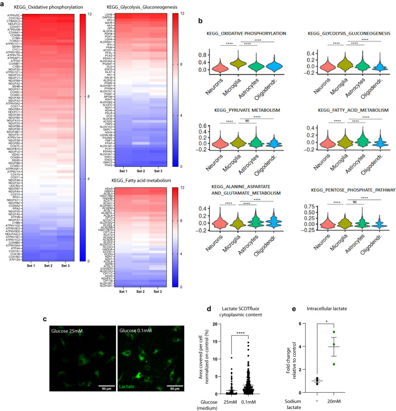
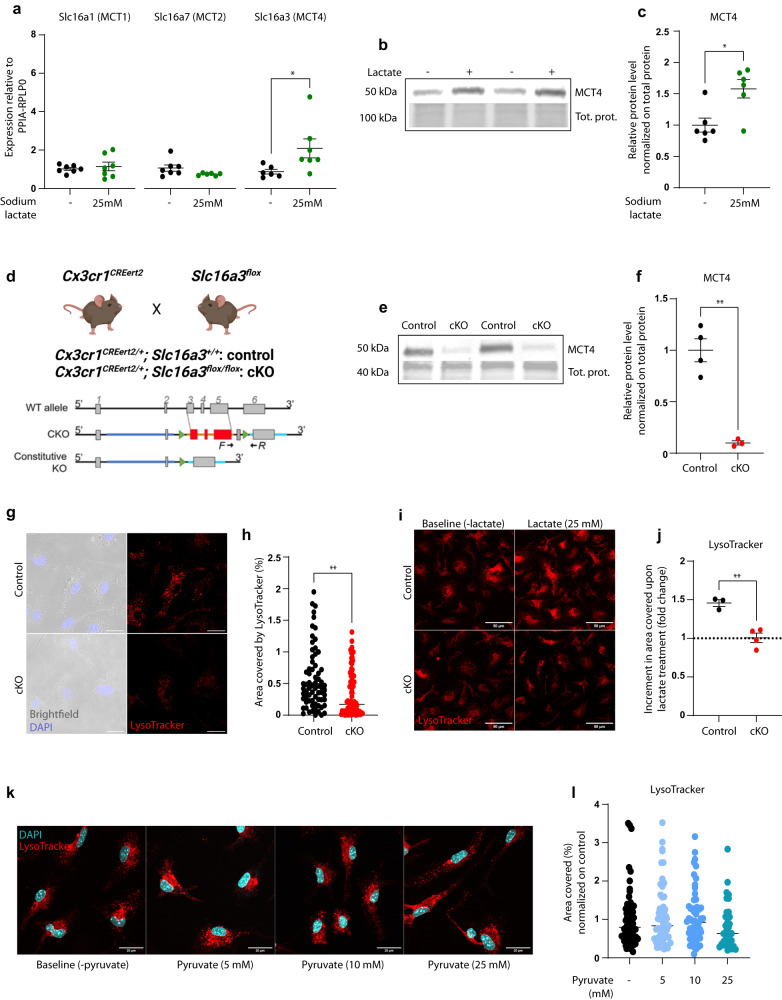
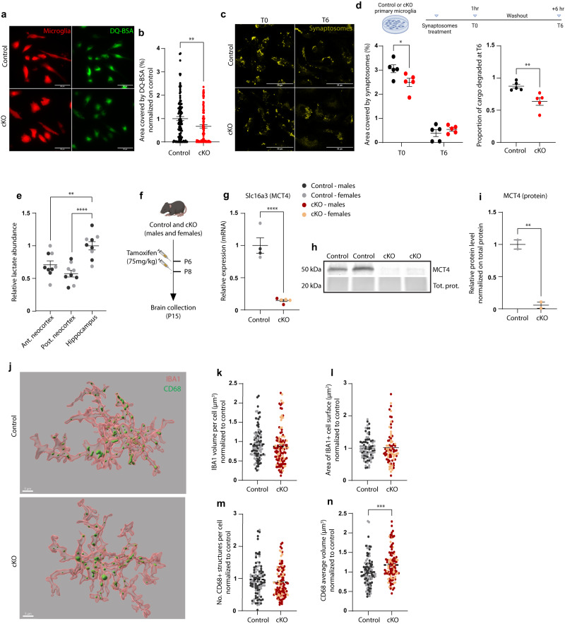
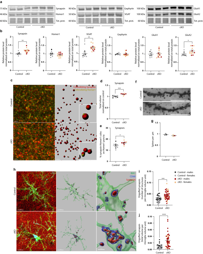
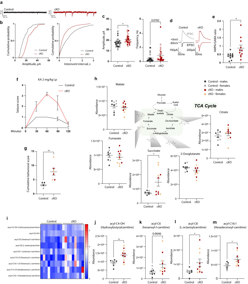
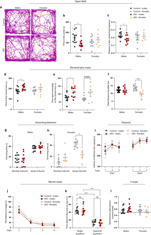
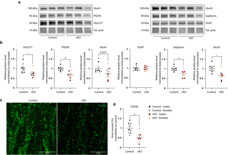
Microglia, the innate immune cells of the central nervous system, actively participate in brain development by supporting neuronal maturation and refining synaptic connections. These cells are emerging as highly metabolically flexible, able to oxidize different energetic substrates to meet their energy demand. Lactate is particularly abundant in the brain, but whether microglia use it as a metabolic fuel has been poorly explored. Here we show that microglia can import lactate, and this is coupled with increased lysosomal acidification. In vitro, loss of the monocarboxylate transporter MCT4 in microglia prevents lactate-induced lysosomal modulation and leads to defective cargo degradation. Microglial depletion of MCT4 in vivo leads to impaired synaptic pruning, associated with increased excitation in hippocampal neurons, enhanced AMPA/GABA ratio, vulnerability to seizures and anxiety-like phenotype. Overall, these findings show that selective disruption of the MCT4 transporter in microglia is sufficient to alter synapse refinement and to induce defects in mouse brain development and adult behavior.


Conflict of interest statement

Sam Benson and Marc Vendrell declare the following competing interests: the SCOTfluor-based fluorescent reagent (SCOTfluor510 lactate) is covered by a patent and it is commercialized by the company Tocris Bioscience. Patent applicant: The University of Edinburgh; Name of the inventor(s): Sam Benson and Marc Vendrell. Application number: WO/2020/187919. Status: Granted patent. All other authors declare no competing interests.

Figures

Fig. 1
Fig. 1. Microglia are metabolically flexible and can efficiently import lactate in vitro.
a Heatmaps of the microglial expression levels of genes annotated within substrate metabolism-related KEGG pathways. Data were obtained from the dataset published in Pinto et al., from three independent experiments, shown as normalized intensity values. b Violin plot showing Seurat module scores for six KEGG pathways across different cell types in the mechanically dissociated mouse hippocampus (dataset from Mattei et al.). The module scores represent the average expression of all genes in a particular cluster at a single-level, subtracted by the aggregated expression of control feature sets. Module scores in microglia are compared to other cell types; Two-sided Wilcoxon test, ****p < 0.0001; exact p values are provided in Supplementary Fig. 1b. c Representative confocal z-stack projections of SCOTfluor lactate internalized by primary microglia, in standard (25 mM) or reduced (0.1 mM) glucose conditions and d relative quantification. Data point represent individual cells; glucose 25 mM: n = 105 cells; glucose 0.1 mM: n = 168 cell from N = 3 independent experiments. Two-tailed unpaired t test, ****p < 0.0001. Scale bar: 50 µm. e Intracellular lactate content measured by LC-MS in primary microglia cells upon 6 h exposure to 20 mM Na-lactate or control in standard 25 mM glucose conditions. N = 3 independent experiments. Two-tailed unpaired t test, *p = 0.0235. d, e Data are represented as mean ± SEM. a, b, d, e Source data are provided as a Source Data file.
Fig. 2
Fig. 2. The lactate transporter MCT4 is upregulated in microglia in response to extracellular lactate and is required for lactate-dependent lysosomal acidification.
a mRNA expression of the lactate transporters Slc16a1, Slc16a7, and Slc16a3 in primary microglia in the presence or absence of 25 mM lactate (24 h treatment). Each data point represents the average of n = 3 technical replicates from N = 7 independent experiments. Two-tailed unpaired t test, *p(Slc16a3) = 0.0481. b Representative western blot of MCT4 protein expression in primary microglia in standard medium (–) or medium containing 25 mM lactate (+, 24 h treatment), and c relative quantification from N = 6 independent experiments. Two-tailed unpaired t test, *p = 0.0104. d Schematic of the breeding strategy for producing Cx3cr1CREERT2;MCT4flox mice and representation of the floxed exons in the MCT4 gene (Slc16a3). Created with BioRender.com. e Representative western blot for MCT4 depletion upon 4-hydroxytamoxifen treatment in primary microglia from control and cKO mice and f relative quantification, normalized on total protein. Data points represent primary microglia prepared from one individual, from N = 4 control and N = 3 cKO. Two-tailed unpaired t test, **p = 0.0011. g Representative confocal z-stack projections of LysoTracker labeling in control and cKO primary microglia and h relative quantification of the area covered by LysoTracker signal per cell. Data points represent individual cells, control: n = 79 cells; cKO: n = 84 cells from N = 2 independent experiments. Two-tailed unpaired t test, **p = 0.0016. Scale bar: 20 µm. i Representative confocal z-stack projections of LysoTracker staining in control and cKO primary microglia, at baseline and after 25 mM lactate exposure, and j signal quantification, displayed as fold change compared to baseline. Data point represent primary microglia prepared from single individuals (control N = 3; cKO N = 4), from three independent experiments. Two-tailed unpaired t test. **p = 0.0025. Scale bar: 50 µm. k Representative confocal z-stack projections of LysoTracker labeling in primary microglia exposed to increasing concentration of pyruvate and l relative quantification of the area covered by LysoTracker signal per cell, normalized to control. Data points represent individual cells from N = 2–3 independent experiments. Baseline (-pyruvate): n = 64 cells, Pyruvate (5 mM): n = 57 cells; Pyruvate (10 mM): n = 57 cells; Pyruvate (25 mM): n = 35 cells. a, c, f, h, j, l Data are represented as mean ± SEM. Source data are provided as a Source Data file.
Fig. 3
Fig. 3. MCT4 cKO microglia display reduced phagocytosis and lysosomal dysfunction in vitro and alteration of phagolysosomal structures in vivo.
a Representative z-stack projections of microglia (TdTomato+) treated with DQ-BSA, and b relative quantification of area covered by cleaved DQ-BSA in control and cKO cells. Data point represent individual cells, control: n = 111 cells; cKO: n = 103 cells from N = 2 independent experiments. Two-tailed unpaired t test, **p = 0.0036; Scale bar: 50 µm. c Representative confocal z-stack acquisitions of primary microglia exposed to fluorescently labeled synaptosomes (scale bar: 50 µm), and d relative quantification. Experiment timeline created with BioRender.com. Left: Microglial area covered by fluorescent signal per cell after 1 h exposure to synaptosomes (T0) and 6 h after medium washout (T6). Two-way ANOVA followed by Sidak’s post hoc multiple comparison test, *p = 0.0248. Right: proportion of cargo degraded in 6 h in control and cKO cells. Two-tailed unpaired t test, **p = 0.0074. Data points represent the average of at least 30 cells per condition, from N = 5 independent experiments. e Relative lactate abundance measured by LC-MS in different brain areas of P15 control mice. N = 5 males (black dots) and N = 4 females (gray dots). One-way ANOVA followed by Dunnett’s post hoc multiple comparison test; hippocampus vs. anterior neocortex: **p = 0.0033; hippocampus vs. posterior neocortex: ****p < 0.0001. f Schematic of experimental design for tamoxifen treatment and brain collection at P15. Created with BioRender.com. g Expression of Slc16a3 mRNA and h, i MCT4 protein in microglia acutely isolated from control and cKO mice at P15; for mRNA: Control: N = 4 (N = 2 males and N = 2 females) vs. cKO: N = 5 (N = 2 males and N = 3 females). For protein: N = 2 control females and N = 2 cKO females). Two-tailed unpaired t test, ****p < 0.0001, **p = 0.0082. j Representative 3D reconstruction of microglial cells from the CA1 stratum radiatum of control and cKO mice at P15, scale bar: 5 µm. Quantification of IBA1+ k cell volume, l surface area, m number of CD68+ structures within each microglia cell, and n average volume of CD68+ structures per cell, normalized to controls. kn Data points represent single cells from N = 4 males and N = 4 females per genotype (P15); control: n = 79–103 cells; cKO: n = 83–113 cells. Two-tailed unpaired t test, ***p = 0.0007. b, d, e, g, i, kn Data are represented as mean ± SEM. Source data are provided as a Source Data file.
Fig. 4
Fig. 4. Deletion of MCT4 in microglia leads to altered synaptic pruning in the hippocampus.
a Representative western blots of synaptic markers in hippocampal homogenate from P15 control and cKO littermates and b relative quantification, normalized to the control group. Each data point corresponds to one animal. Control: N = 3 males; N = 5 females; cKO: N = 5 males; N = 8 females. Two-tailed unpaired t test, **p(synapsin) = 0.0024; **p(VGAT) = 0.0011; *p(GluA2) = 0.0372. c Representative confocal z-stack projections and relative 3D reconstruction of synapsin- and Homer1-positive puncta in the CA1 stratum radiatum from control and cKO P15 mice. Scale bar: 2 µm and 0.3 µm in the inset. d Quantification of the total volume of synapsin-positive puncta measured in the CA1 stratum radiatum and dentate gyrus of control and cKO mice at P15, normalized to controls. Each data point corresponds to one animal. Control: N = 4 males; N = 4 females; cKO: N = 4 males; N = 2 females. Two-tailed unpaired t test, ***p = 0.0007. e Excitatory synapses quantification in the CA1 stratum radiatum of control and cKO mice at P15. Individual synapses are defined as the colocalization of the pre- and post-synaptic markers synapsin and Homer1. Each data point corresponds to one animal. Control: N = 4 males; N = 4 females; cKO: N = 4 males; N = 3 females. Two-tailed unpaired t test, *p = 0.0412. f Representative dendrites of CA1 pyramidal neurons in control and cKO mice at P15, visualized by Golgi-cox staining. Scale bar: 10 µm. g Quantification of spine density reported as absolute numbers per µm. Each data point represents one animal, as the average of n = 3–18 dendrites segments. Group size N = 3 (N = 1 male and N = 2 females per genotype). h Representative confocal z-stack projections and relative 3D surface reconstruction of IBA1-positive microglia cells, engulfing synapsin puncta within CD68-positive phagolysosomal structures, from control and cKO mice (P15, CA1 stratum radiatum). Left panel: scale bar 10 µm; Zoom-in: scale bar 3 µm. i, j Number and total volume of engulfed synapsin puncta per microglia volume unit. Two-tailed unpaired t test, ***p = 0.0001; ****p < 0.0001. Data points represent individual cells from N = 3 males and N = 4 females per genotype; n = 30 control cells, n = 42 cKO cells. b, d, e, g, i, j Data are represented as mean ± SEM. Source data are provided as a Source Data file.
Fig. 5
Fig. 5. Loss of microglial MCT4 induces functional synapse alterations and metabolic changes in the hippocampus.
ac Whole-cell recordings of CA1 pyramidal cells in brain slices from control and cKO male littermates at P15 and P30. a Representative traces from control and cKO mice. b Representative cumulative distributions of spontaneous excitatory post-synaptic currents (sEPSCs) amplitude and frequency. c Average values of sEPSCs amplitudes and frequencies (controls: n = 27 cells from N = 2 P15 and N = 3 P30 male mice; cKO: n = 24 cells from N = 2 P15 and N = 2 P30 male mice; two-tailed unpaired t test; *p = 0.0126). Data points correspond to individual cells. Filled data points indicate cells recorded at P15; empty data points are for cells analyzed at P30. d, e AMPA/GABA ratio of CA1 pyramidal neurons measured in P15 control and cKO male littermates. d Representative traces of excitatory (AMPA) and inhibitory (GABA) post-synaptic currents (EPSCs and IPSCs) recorded from control and cKO mice and e relative quantification. Data points correspond to individual cells. Control: n = 11 cells, cKO: n = 11 cells from N = 2 mice per genotype; two-tailed unpaired t test; *p = 0.0241. f, g Susceptibility to kainic acid-induced seizures at P15; N = 3 males and N = 3 females per genotype. f Seizure score according to the modified Racine scale, plotted as maximum score reported for each mouse in a 30 min period. A score of 3 corresponds to the development of clonic seizures. g Cumulative behavioral score per mouse for the total observation period (120 minutes). Two-tailed unpaired t test, **p = 0.0041. hm Abundance of metabolites measured via LC-MS in the hippocampus of P15 mice. Control: N = 5 males; N = 4 females. cKO: N = 6 males; N = 4 females. h TCA cycle metabolites. Two-tailed unpaired t test, *p(succinate) = 0.0312; i Heatmap representing the relative abundance of carnitine-related metabolites in control and cKO mice at P15. jm **p(acyl-C4-OH) = 0.0043; *p(acyl-C8) = 0.0472; *p(acyl-C16:1) = 0.0358; two-tailed unpaired t test. c, e, fm Data are represented as mean ± SEM. Source data are provided as a Source Data file.
Fig. 6
Fig. 6. Adult cKO mice display an anxiety-like behavior.
Behavioral characterization of control and cKO adult mice (7–8 months). Each data point represents one animal. Control: N = 12 males; N = 11 females; cKO: N = 11 males; N = 6 females. Statistics are calculated with two-way ANOVA followed by Sidak’s post hoc multiple comparison test. ac Open field exploration (20 minutes), with a representative tracking traces; b quantification of time spent in the center of the arena; *p(males) = 0.0105; c fraction of distance traveled in the center; *p(males) = 0.0418. df Elevated plus maze test (5 minutes), with quantification of d time spent in the closed arms, *p(males) = 0.0279; e time spent immobile in the closed arms, *p(males) = 0.0216; f total distance traveled in the maze; ****p(females) <0.0001. g, h Quantification of grooming time induced by a novel cage (novelty-induced) and upon physical stimulation (water spray on the snout, spray-induced) in g males and in h females; **p(females) = 0.0019. i Latency to fall from the accelerating rotarod. Each mouse underwent three trials per day, on two consecutive days. j, k Barnes maze test, consisting of 4 days training, four trials per day, followed by a single trial with no escape box, on day 5. j First latency to reach the escape hole. k Time spent in the quadrant where the escape hole was located (target quadrant) and at the opposite side of the maze on the test day. ****p(target-opposite) <0.0001. l Proportion of correct spontaneous alternation in the Y maze test. bl Data are represented as mean ± SEM. Source data are provided as a Source Data file.
Fig. 7
Fig. 7. Adult cKO mice display a significant decrease in the content of synaptic markers in the hippocampus.
a Representative western blots of synaptic markers in hippocampal homogenate from 7–8 months old control and cKO littermates and b relative quantification, normalized to the control group. Each data point corresponds to one animal. Control: N = 4 males; N = 5 females; cKO: N = 4 males; N = 3 females. Two-tailed unpaired t test, *p(VGLUT1) = 0.0307; **p(PSD95) = 0.0010; **p(Gephyrin) = 0.0072; *p(GluA2) = 0.0101. c Representative confocal z-stack projections of PSD95 puncta in the CA1 stratum radiatum from adult control and cKO mice (7–8 months). Scale bar: 10 µm. d Quantification of the area covered by PSD95-positive puncta, normalized to controls. Each data point corresponds to one animal. Control: N = 4 males; N = 5 females; cKO: N = 4 males; N = 3 females. Two-tailed unpaired t test, **p = 0.0038. b, d Source data are provided as a Source Data file.

References

    1. Li Q, Barres BA. Microglia and macrophages in brain homeostasis and disease. Nat. Rev. Immunol. 2018;18:225–242. doi: 10.1038/nri.2017.125. - DOI - PubMed
    1. Sierra A, Paolicelli RC, Kettenmann H. Cien Años de microglía: milestones in a century of microglial research. Trends Neurosci. 2019;42:778–792. doi: 10.1016/j.tins.2019.09.004. - DOI - PubMed
    1. Paolicelli RC, et al. Microglia states and nomenclature: a field at its crossroads. Neuron. 2022;110:3458–3483. doi: 10.1016/j.neuron.2022.10.020. - DOI - PMC - PubMed
    1. Davalos D, et al. ATP mediates rapid microglial response to local brain injury in vivo. Nat. Neurosci. 2005;8:752–758. doi: 10.1038/nn1472. - DOI - PubMed
    1. Nimmerjahn, A., Kirchhoff, F. & Helmchen, F. Resting microglial cells are highly dynamic surveillants of brain parenchyma in vivo. 308, 6 (2005). - PubMed

Publication types