. 2023 Sep 16;13(1):15360.
doi: 10.1038/s41598-023-42758-y.

Mild sleep restriction increases endothelial oxidative stress in female persons

Affiliations

Mild sleep restriction increases endothelial oxidative stress in female persons

Riddhi Shah et al. Sci Rep. .

Abstract

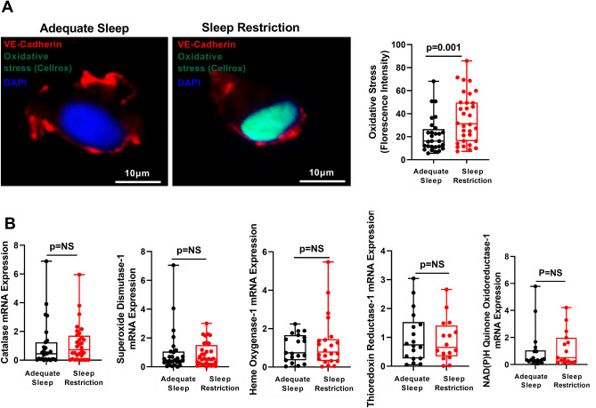
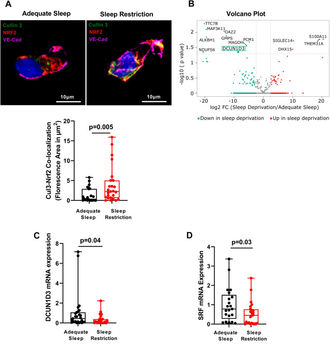
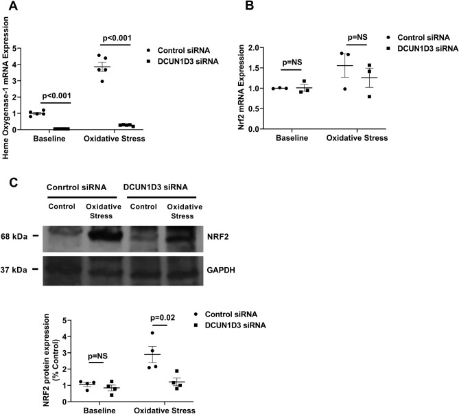
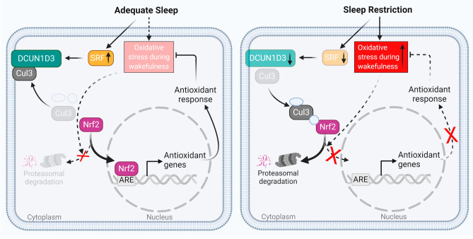
Sleep restriction is associated with increased cardiovascular risk, which is more pronounced in female than male persons. We reported recently first causal evidence that mild, prolonged sleep restriction mimicking "real-life" conditions impairs endothelial function, a key step in the development and progression of cardiovascular disease, in healthy female persons. However, the underlying mechanisms are unclear. In model organisms, sleep restriction increases oxidative stress and upregulates antioxidant response via induction of the antioxidant regulator nuclear factor (erythroid-derived 2)-like 2 (Nrf2). Here, we assessed directly endothelial cell oxidative stress and antioxidant responses in healthy female persons (n = 35) after 6 weeks of mild sleep restriction (1.5 h less than habitual sleep) using randomized crossover design. Sleep restriction markedly increased endothelial oxidative stress without upregulating antioxidant response. Using RNA-seq and a predicted protein-protein interaction database, we identified reduced expression of endothelial Defective in Cullin Neddylation-1 Domain Containing 3 (DCUN1D3), a protein that licenses Nrf2 antioxidant responses, as a mediator of impaired endothelial antioxidant response in sleep restriction. Thus, sleep restriction impairs clearance of endothelial oxidative stress that over time increases cardiovascular risk.Trial Registration: NCT02835261 .


Conflict of interest statement

The authors declare no competing interests.

Figures

Figure 1
Figure 1
Sleep restriction increases endothelial oxidative stress without upregulating antioxidant responses. (A) Representative images and Box and Whisker Plot quantitating oxidative stress (nuclear fluorescence intensity of the fluorogenic probe activated by reactive oxygen species and subsequent binding to DNA) adjusted for baseline values in endothelial cells harvested from healthy participants after adequate sleep (n = 31) and after sleep restriction (n = 32). No pre-specified covariates had moderate or strong marginal associations with oxidative stress (p-value < 0.1). (B) Box and Whisker Plot quantitating endothelial mRNA expression of Catalase, Superoxide Dismutase, Heme Oxygenase-1, Thioredoxin Reductase-1 and NAD(P)H Quinone Oxidoreductase-1 in endothelial cells harvested from participants after adequate sleep (black dots; n = 28, 28, 25, 18 and 18, respectively) and sleep restriction (red dots; n = 32, 32, 25, 16 and 15, respectively). All data throughout the figure are shown as means ± SD (linear mixed effect model). NS = not significant.
Figure 2
Figure 2
Identification of mediators of impaired antioxidant response in sleep restriction. (A) Representative images and Box and Whisker Plot quantitating endothelial Nrf2-Cul3 co-localization (indicated in yellow) after adequate sleep and sleep restriction (n = 25). (B) Volcano plot showing differential gene expression (n = 25,001 genes) in endothelial cells harvested from healthy participants (n = 5) after sleep restriction compared with adequate sleep. Horizontal dotted line separates 13 differentially expressed genes with p < 0.05 from those with p ≥ 0.05. Expression of DCUN1D3 gene (green box), the only binding target of Cul3 among genes altered by sleep restriction, was reduced − 8.46 log2 fold change (FC) after sleep restriction compared with adequate sleep (p = 0.045). (C) Box and Whisker Plot quantitating endothelial DCUN1D3 mRNA expression after adequate sleep (n = 28) and sleep restriction (n = 32). (D) Box and Whisker Plot quantitating endothelial SRF mRNA expression after adequate sleep and sleep restriction (n = 25). All data throughout the figure are shown as means ± SD (linear mixed effect model). Cul3 = Cullin-3; DCUN1D3 = defective in cullin neddylation 1 domain containing 3; Nrf2 = Nuclear factor (erythroid-derived 2)-like 2; SRF = serum response factor.
Figure 3
Figure 3
DCUN1D3 Regulates endothelial antioxidant response. (A) Scatter plot quantifying Heme Oxygenase-1mRNA expression before and after exposure to oxidative stress in HUVECs with DCUN1D3 knockdown and control (n = 5). (B) Scatter plot quantifying Nrf2 mRNA expression at baseline and after exposure to oxidative stress in HUVECs with DCUN1D3 knockdown and control (n = 3). (C) Western Blotting and Scatter Plot quantifying Nrf2 protein expression in DCUN1D3 knockdown compared to control at baseline and after exposure to oxidative stress in HUVECs (n = 4). All data throughout the figure are shown as means ± SD. NS = non-significant. Abbreviations as in Fig. 2.
Figure 4
Figure 4
Endothelial cell function during wakefulness is impaired after sleep restriction compared with adequate sleep. After adequate sleep, endothelial oxidative stress that accumulates during wakefulness is cleared by an appropriate antioxidant response. SRF mRNA expression increases after sleep pressure build-up during wakefulness, which upregulates DCUN1D3 and sequesters Cul3 toward plasma membrane. Reduced Cul3 availability in the Nrf2 ubiquitination complex releases Nrf2 and allows for its nuclear translocation and activation of antioxidant genes. After sleep restriction, endothelial antioxidant responses are not appropriately upregulated leading to increased endothelial oxidative stress. Reduced SRF mRNA expression during wakefulness after sleep restriction leads to reduction in DCUN1D3 expression and consequent increase in Cul3 availability in Nrf2 ubiquitination complex, which traps Nrf2 and precludes its nuclear translocation and activation of antioxidant genes. Impaired endothelial antioxidant response after insufficient sleep results in increased oxidative stress during wakefulness, which impairs endothelial function and may increase cardiovascular risk.

Similar articles

Cited by

References

    1. St-Onge MP, et al. Sleep duration and quality: Impact on lifestyle behaviors and cardiometabolic health: A scientific statement from the American heart association. Circulation. 2016;134:e367–e386. - PMC - PubMed
    1. Tsao CW, et al. Heart disease and stroke statistics-2022 update: A report from the American heart association. Circulation. 2022;145:e153–e639. - PubMed
    1. Makarem N, et al. Redefining cardiovascular health to include sleep: Prospective associations with cardiovascular disease in the MESA sleep study. J. Am. Heart Assoc. 2022;11:e025252. - PMC - PubMed
    1. Ferrie JE, et al. A prospective study of change in sleep duration: Associations with mortality in the Whitehall II cohort. Sleep. 2007;30:1659–1666. - PMC - PubMed
    1. Ayas NT, et al. A prospective study of sleep duration and coronary heart disease in women. Arch. Intern. Med. 2003;163:205–209. - PubMed

Publication types

Associated data