

Feasibility of administration of calcitonin gene-related peptide receptor antagonist on attenuation of pain and progression in osteoarthritis
- PMID: 37717108
- PMCID: PMC10505157
- DOI: 10.1038/s41598-023-42673-2
Feasibility of administration of calcitonin gene-related peptide receptor antagonist on attenuation of pain and progression in osteoarthritis
Abstract
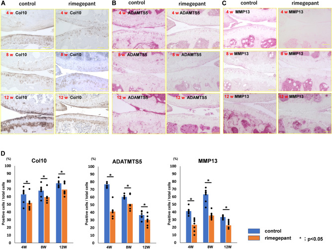
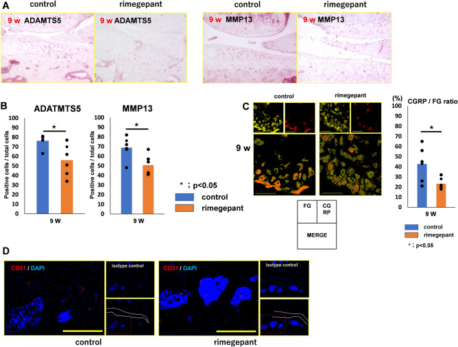
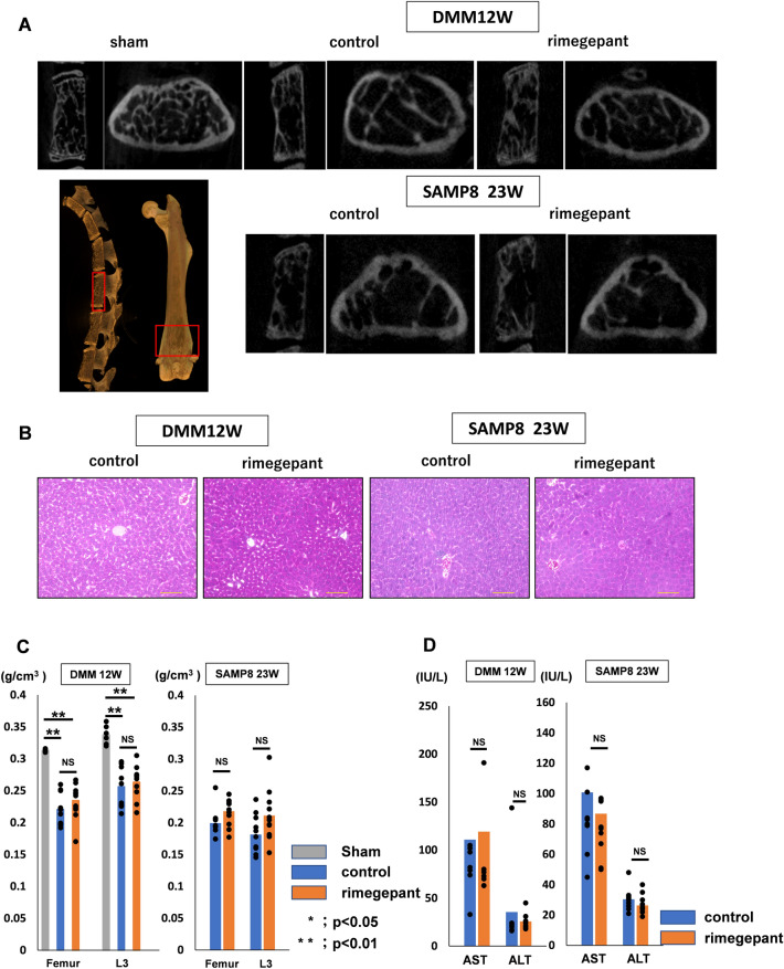
Suppressing inflammation and abnormal subchondral bone turnover is essential for reducing osteoarthritis (OA) progression and pain relief. This study focused on calcitonin gene-related peptide (CGRP), which is involved in inflammation and bone metabolism, and investigated whether a CGRP receptor antagonist (rimegepant) could suppress OA progression and relieve pain in two OA models. C57BL/6 mice (10-week-old) underwent surgical destabilization of the medial meniscus, and Rimegepant (1.0 mg/kg/100 μL) or phosphate-buffered saline (100 μL) was administered weekly intraperitoneally after OA surgery and evaluated at 4, 8, and 12 weeks. In the senescence-accelerated mice (SAM)-prone 8 (SAMP8), rimegepant was administered weekly before and after subchondral bone sclerosis and sacrificed at 9 and 23 weeks, respectively. Behavioral assessment and immunohistochemical staining (CGRP) of the dorsal root ganglion (DRG) were conducted to assess pain. In DMM mice, synovitis, cartilage degeneration, and osteosclerosis were significantly suppressed in the rimegepant group. In SAMP8, synovitis, cartilage degeneration, and osteosclerosis were significantly suppressed by rimegepant at 9 weeks; however, not at 23 weeks. Behavioral assessment shows the traveled distance and the number of standings in the rimegepant group were significantly longer and higher. In addition, CGRP expression of the DRG was significantly lower in the rimegepant group at 8 and 12 weeks of DMM and 9 weeks of SAMP8 treatment. No adverse effects were observed in either of the mouse models. Inhibition of CGRP signaling has the potential to be a therapeutic target to prevent OA progression and suppress pain through the attenuation of subchondral bone sclerosis and synovitis.
© 2023. Springer Nature Limited.
Conflict of interest statement
The authors declare no competing interests.
Figures


Similar articles
-
Attenuation of cartilage degeneration by calcitonin gene-related paptide receptor antagonist via inhibition of subchondral bone sclerosis in osteoarthritis mice.J Orthop Res. 2016 Jul;34(7):1177-84. doi: 10.1002/jor.23132. Epub 2016 Jan 12. J Orthop Res. 2016. PMID: 26686833
-
Persistent synovial inflammation plays important roles in persistent pain development in the rat knee before cartilage degradation reaches the subchondral bone.BMC Musculoskelet Disord. 2018 Aug 16;19(1):291. doi: 10.1186/s12891-018-2221-5. BMC Musculoskelet Disord. 2018. PMID: 30115046 Free PMC article.
-
Curcumin slows osteoarthritis progression and relieves osteoarthritis-associated pain symptoms in a post-traumatic osteoarthritis mouse model.Arthritis Res Ther. 2016 Jun 3;18(1):128. doi: 10.1186/s13075-016-1025-y. Arthritis Res Ther. 2016. PMID: 27260322 Free PMC article.
-
Calcitonin gene-related peptide in the joint: contributions to pain and inflammation.Br J Clin Pharmacol. 2015 Nov;80(5):965-78. doi: 10.1111/bcp.12669. Epub 2015 Jul 22. Br J Clin Pharmacol. 2015. PMID: 25923821 Free PMC article. Review.
-
Rimegepant: A Review in the Acute Treatment and Preventive Treatment of Migraine.CNS Drugs. 2023 Mar;37(3):255-265. doi: 10.1007/s40263-023-00988-8. Epub 2023 Feb 4. CNS Drugs. 2023. PMID: 36739335 Free PMC article. Review.
Cited by
-
Pathogenic Mechanisms and Therapeutic Approaches in Obesity-Related Knee Osteoarthritis.Biomedicines. 2023 Dec 20;12(1):9. doi: 10.3390/biomedicines12010009. Biomedicines. 2023. PMID: 38275369 Free PMC article. Review.
-
Osteophyte Cartilage as a Potential Source for Minced Cartilage Implantation: A Novel Approach for Articular Cartilage Repair in Osteoarthritis.Int J Mol Sci. 2024 May 20;25(10):5563. doi: 10.3390/ijms25105563. Int J Mol Sci. 2024. PMID: 38791601 Free PMC article.
-
Subchondral bone conditions influence pain in patients with osteochondral lesion of the talus.Arch Orthop Trauma Surg. 2025 Jun 9;145(1):341. doi: 10.1007/s00402-025-05956-z. Arch Orthop Trauma Surg. 2025. PMID: 40488768 No abstract available.
-
Modification of Mesenchymal Stem/Stromal Cell-Derived Small Extracellular Vesicles by Calcitonin Gene Related Peptide (CGRP) Antagonist: Potential Implications for Inflammation and Pain Reversal.Cells. 2024 Mar 10;13(6):484. doi: 10.3390/cells13060484. Cells. 2024. PMID: 38534328 Free PMC article.
-
Neural and immune roles in osteoarthritis pain: Mechanisms and intervention strategies.J Orthop Translat. 2024 Aug 7;48:123-132. doi: 10.1016/j.jot.2024.07.010. eCollection 2024 Sep. J Orthop Translat. 2024. PMID: 39220678 Free PMC article. Review.
References
-
- Mapp PI, Walsh DA. Mechanisms and targets of angiogenesis and nerve growth in osteoarthritis. Nat. Rev. Rheumatol. 2012;8:390–398. - PubMed
-
- Creamer P, Hochberg MC. Osteoarthritis. Lancet. 1997;350:503–509. - PubMed
-
- Hochberg MC. Epidemiologic considerations in the primary prevention of osteoarthritis. J. Rheumatol. 1991;18:1438–1440. - PubMed
-
- Radin EL, et al. Response of joints to impact loading. 3. Relationship between trabecular microfractures and cartilage degeneration. J. Biomech. 1973;6:51–57. - PubMed
-
- Radin EL, Rose RM. Role of subchondral bone in the initiation and progression of cartilage damage. Clin. Orthop. Relat. Res. 1986;213:34–40. - PubMed
Publication types
MeSH terms
Substances
LinkOut - more resources
Full Text Sources
Medical
Research Materials
