

Quantitative T1 mapping detects blood-brain barrier breakdown in apparently non-enhancing multiple sclerosis lesions
- PMID: 37717382
- PMCID: PMC10514220
- DOI: 10.1016/j.nicl.2023.103509
Quantitative T1 mapping detects blood-brain barrier breakdown in apparently non-enhancing multiple sclerosis lesions
Abstract
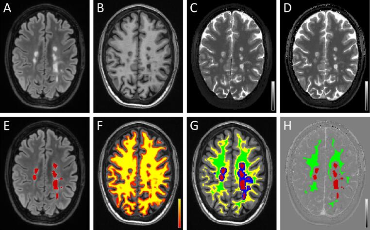
Objectives: The disruption of the blood-brain barrier (BBB) is a key and early feature in the pathogenesis of demyelinating multiple sclerosis (MS) lesions and has been neuropathologically demonstrated in both active and chronic plaques. The local overt BBB disruption in acute demyelinating lesions is captured as signal hyperintensity in post-contrast T1-weighted images because of the contrast-related shortening of the T1 relaxation time. On the contrary, the subtle BBB disruption in chronic lesions is not visible at conventional radiological evaluation but it might be of clinical relevance. Indeed, persistent, subtle BBB leakage might be linked to low-grade inflammation and plaque evolution. Here we hypothesised that 3D Quantitative Transient-state Imaging (QTI) was able to reveal and measure T1 shortening (ΔT1) reflecting small amounts of contrast media leakage in apparently non-enhancing lesions (ANELs).
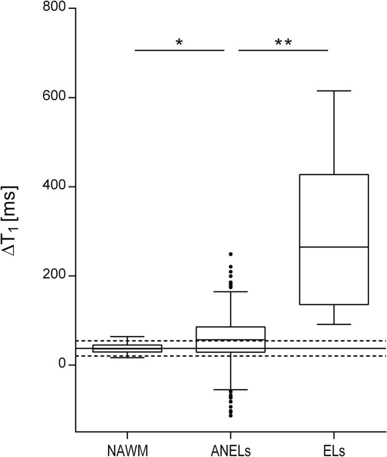
Materials and methods: Thirty-four patients with relapsing remitting MS were included in the study. All patients underwent a 3 T MRI exam of the brain including conventional sequences and QTI acquisitions (1.1 mm isotropic voxel) performed both before and after contrast media administration. For each patient, a ΔT1 map was obtained via voxel-wise subtraction of pre- and post- contrast QTI-derived T1 maps. ΔT1 values measured in ANELs were compared with those recorded in enhancing lesions and in the normal appearing white matter. A reference distribution of ΔT1 in the white matter was obtained from datasets acquired in 10 non-MS patients with unrevealing MR imaging.
Results: Mean ΔT1 in ANELs (57.45 ± 48.27 ms) was significantly lower than in enhancing lesions (297.71 ± 177.52 ms; p < 0. 0001) and higher than in the normal appearing white matter (36.57 ± 10.53 ms; p < 0.005). Fifty-two percent of ANELs exhibited ΔT1 higher than those observed in the white matter of non-MS patients.
Conclusions: QTI-derived quantitative ΔT1 mapping enabled to measure contrast-related T1 shortening in ANELs. ANELs exhibiting ΔT1 values that deviate from the reference distribution in non-MS patients may indicate persistent, subtle, BBB disruption. Access to this information may be proved useful to better characterise pathology and objectively monitor disease activity and response to therapy.
Keywords: Blood-brain barrier; Magnetic resonance fingerprinting; Magnetic resonance imaging; Multiple sclerosis; Quantitative imaging; Quantitative transient-state imaging; T1 mapping.
Copyright © 2023 The Author(s). Published by Elsevier Inc. All rights reserved.
Conflict of interest statement
Declaration of Competing Interest The authors declare that they have no known competing financial interests or personal relationships that could have appeared to influence the work reported in this paper.
Figures
References
-
- Andica C., Hagiwara A., Hori M., Kamagata K., Koshino S., Maekawa T., Suzuki M., Fujiwara H., Ikeno M., Shimizu T., Suzuki H., Sugano H., Arai H., Aoki S. Review of synthetic MRI in pediatric brains: Basic principle of MR quantification, its features, clinical applications, and limitations. J. Neuroradiol. 2019;46:268–275. doi: 10.1016/j.neurad.2019.02.005. - DOI - PubMed
Publication types
MeSH terms
Substances
LinkOut - more resources
Full Text Sources
Medical
