Review
. 2023 Sep 17;21(1):633.
doi: 10.1186/s12967-023-04515-7.

Epstein-Barr virus-acquired immunodeficiency in myalgic encephalomyelitis-Is it present in long COVID?

Affiliations
Review

Epstein-Barr virus-acquired immunodeficiency in myalgic encephalomyelitis-Is it present in long COVID?

Manuel Ruiz-Pablos et al. J Transl Med. .

Abstract

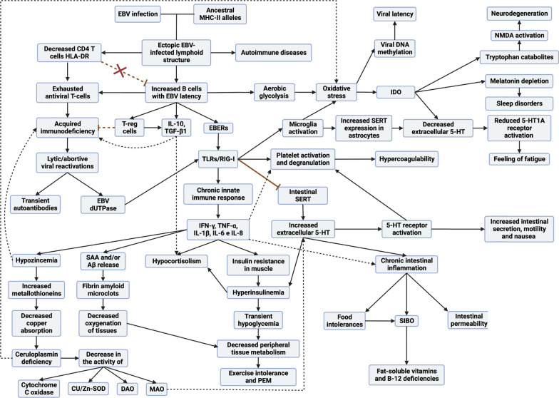
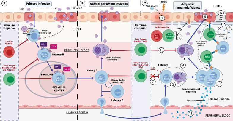
Both myalgic encephalomyelitis or chronic fatigue syndrome (ME/CFS) and long COVID (LC) are characterized by similar immunological alterations, persistence of chronic viral infection, autoimmunity, chronic inflammatory state, viral reactivation, hypocortisolism, and microclot formation. They also present with similar symptoms such as asthenia, exercise intolerance, sleep disorders, cognitive dysfunction, and neurological and gastrointestinal complaints. In addition, both pathologies present Epstein-Barr virus (EBV) reactivation, indicating the possibility of this virus being the link between both pathologies. Therefore, we propose that latency and recurrent EBV reactivation could generate an acquired immunodeficiency syndrome in three steps: first, an acquired EBV immunodeficiency develops in individuals with "weak" EBV HLA-II haplotypes, which prevents the control of latency I cells. Second, ectopic lymphoid structures with EBV latency form in different tissues (including the CNS), promoting inflammatory responses and further impairment of cell-mediated immunity. Finally, immune exhaustion occurs due to chronic exposure to viral antigens, with consolidation of the disease. In the case of LC, prior to the first step, there is the possibility of previous SARS-CoV-2 infection in individuals with "weak" HLA-II haplotypes against this virus and/or EBV.

Keywords: Chronic fatigue syndrome; EBV EBNA-1; HLA-II alleles; Immunodeficiency; Inflammation; Long COVID syndrome; Myalgic encephalomyelitis; Post-acute COVID-19 syndrome.


Conflict of interest statement

The authors declare that they have no competing interests.

Figures

Fig. 1
Fig. 1
Schematic model of the development of acquired immunodeficiency following EBV infection
Fig. 2
Fig. 2
Development of Epstein–Barr Virus (EBV)-induced acquired immunodeficiency in patients with genetic susceptibility. A Primary infection by EBV. EBV is transferred to the host via the saliva of an infected carrier, initially infecting the epithelial cells of the pharynx and subsequently naïve B lymphocytes in the tonsils, through interactions between the virus’s glycoproteins gp350 and gp42 and the host cells’ CD21 and Class II MHC (MHC-II) molecules, respectively. Lytic infection creates new viral particles that continue to infect more epithelial cells. Subsequently, the EBV-infected B cells enter a state of peripheral latency, during which they express a set of specific viral genes, including LMP1, LMP2A, EBNAs, and EBER (latency III). These latency III B cells progress through the germinal center reaction to latency II and emerge as memory B cells with latencies I/0 that establish a lifelong latent infection (B). The healthy host’s immune response is sufficient to control the EBV infection. NK cells in tonsil produce high levels of IFN-γ that withhold the transformation of B cells by EBV during earlier stages of the infection. Type III and II latency B cells are regulated by NK and T cells specific for latent proteins. However, memory B cells with type I latency are only controlled by activated EBNA-1 specific CD4 T cells. EBV-infected plasma cells can periodically enter in lytic phase, but are controlled by CD4 and CD8 T cells with specificity for EBV lytic proteins. C Development of EBV-induced acquired immunodeficiency and autoimmunity in the mucosa of genetically susceptible patients. (1) An inflammatory stimulus or other infection (SARS-CoV-2) recruit leukocytes in the mucosa, including latency I (EBNA-1) B cells and healthy B cells. (2) In the mucosa, B cells form ectopic lymphoid aggregates that enable the generation of antigen-specific immune responses. These ectopic lymphoid structures create a favorable environment for the transformation of EBV latent B cells into proliferating blasts, to become memory B cells. (3) Furthermore, activation of NK cells occurs in response to both the initial inflammatory stimulus and to restrict B cell transformation by EBV. Exposure to foreign antigens from the initial stimulus or to viral antigens from EBV leads to the activation of CD4 T cells and the release of IFN-γ, followed by upregulation of MHC-II on epithelial cells, promoting the acquisition of a non-professional antigen-presenting cell phenotype. (4) Additionally, the presence of foreign antigens could also trigger terminal differentiation and activation of latent EBV B lymphocytes, allowing the transition from the latent phase to the lytic virus phase. (5) Subsequently, newly generated viral particles infect more epithelial cells through gp42/MHC-II interaction, leading to increased inflammation and ultimately to latent EBV infection. Moreover, this chronic inflammation induces a cytokine response, leading to further recruitment of B cells and perpetuation of viral infection. (6) Latent EBV epithelial cells could enter a lytic phase, releasing new virions, undergoing lysis due to T cell response, or experiencing neoplastic transformation. (7) Immune evasion mechanisms of EBV latency (epithelial and B cells) involve a decrease in activation and cytotoxic capability of EBNA-1-specific CD4 T cells through the release of IL-10 and EBV miRNAs contained in exosomes, which could suppress the expression of target genes in the viral or host genome to maintain latent EBV infection. (8) This altered immunosurveillance leads to increased proliferation of EBV-latent B- and epithelial cells, which raises the risk of neoplastic transformation or autoimmune disease in genetically predisposed patients with ancestral MHC-II alleles susceptible to EBV. (9) The presentation of native cellular autoantigens or viral EBNA-1 through MHC-II/gp42, which can undergo post-translational modifications, such as citrullination, and form neoantigens, could trigger the activation of autoreactive CD4 T cells and the formation of autoantibodies against tissue cells. (10) Other virus latency phases or the lytic phase would be controlled by NK and CD4 and CD8 T cells, specific for the EBV lytic proteins. (11) In women, estrogens elevate the risk of developing EBV-induced acquired immunodeficiency by reducing the CD4/CD8 T lymphocyte ratio, increasing the survival of B cells, promoting the release of antibodies, and increasing the expression of the major histocompatibility complex class II
Fig. 3
Fig. 3
Sex hormones and their impact on immune responses. This figure provides an overview of the influences of estrogens and testosterone on immune responses, highlighting their effects on susceptibility and resistance to various conditions and pathogens
Fig. 4
Fig. 4
EBV infection and neuroinflammatory alterations: implications in neurotransmission and copper metabolism. A Exosomes with EBV dUTPases and EBERs from EBV-infected cells can reach the blood–brain barrier and contact endothelial cells causing activation of TLR2 receptors by dUTPases. The blood–brain barrier’s endothelial cells are activated and release proinflammatory cytokines that disrupt the blood–brain barrier’s integrity. Both EBV-infected cells crossing the blood–brain barrier and exosomes with viral genetic material can activate microglia through TLR3 receptors that detect the presence of EBERs. Activated microglia release proinflammatory cytokines IL-1β and TNF-α, which increase the expression and activity of the serotonin transporter SERT in astrocytes, causing an increase in serotonin (5-HT) reuptake and a decrease in its extracellular levels. Oligodendrocytes are particularly susceptible to inflammation. Overexposure to cytokines such as TNF-α can damage these cells, potentially leading to apoptosis and demyelination. Both increased proinflammatory cytokines and increased oxidative and nitrosative stress (ROS/RNS) from activation of microglia via TLR3 could lead to increased IDO activity in microglia, resulting in reduced tryptophan (TRP) levels, increased kynurenine catabolites and decreased 5-HT synthesis. Quinolinic acid (QUIN) stimulates glutamate (GLU) release by activating the NMDA glutamate receptor in the presynaptic neuron. On the other hand, in astrocytes it decreases the expression of glutamate transporters and increases their release, thus increasing extracellular glutamate levels. Thus, quinolinic acid can increase glutamate levels in the brain and decrease brain cells' ability to eliminate excess glutamate. Moreover, quinolinic acid has neurotoxic properties by binding to the neurons' NMDA receptor, followed by sustained calcium (Ca2+) influx leading to increased oxidative and nitrosative stress. This increase in nitrosative and oxidative stress leads to the activation of PARP-1 polymerase to prevent DNA damage. But continuous overactivation of PARP-1 leads to depletion of intracellular reserves of nicotinamide adenine dinucleotide (NAD) and ATP, with the consequent alteration in energy production and mitochondrial function. The binding of glutamate to extrasynaptic NMDA receptors can lead to a reduction in the levels of neurotrophic factors such as brain-derived neurotrophic factor (BDNF), leading to a decrease in synaptic plasticity and increased vulnerability to excitotoxicity. The decrease in 5-HT levels decreases the activation of 5-HT1A receptors allowing more glutamate to be released, as the activation of 5-HT1A receptors decreases the release of glutamate in presynaptic neurons. In addition, the activation of 5-HT1A receptors in the postsynaptic neuron inhibits the activation of NMDA receptors, so a decrease in extracellular 5-HT prevents the activation of 5-HT1A receptors allowing overexcitation of NMDA receptors by glutamate and quinolinic acid. B To the right, the same process occurs, but with alterations in dopaminergic neurons. The decrease in extracellular 5-HT levels due to increased 5-HT reuptake decreases the activation of 5-HT1A receptors in neurons allowing more dopamine (DA) to be released, as the activation of 5-HT1A receptors decreases the release of dopamine in presynaptic neurons. Additionally, the activation of 5-HT1A receptors in the postsynaptic neuron inhibits the activation of NMDA receptors, so a decrease in extracellular 5-HT prevents the activation of 5-HT1A receptors allowing overexcitation of NMDA receptors by quinolinic acid and excess glutamate released by glutaminergic neurons. In turn, the decrease in 5-HT levels decreases the activation of astrocytes’ 5-HT1A receptors, leading to less release of metallothioneins (MT) to the extracellular space. Metallothioneins are especially important to protect dopaminergic neurons from excess dopamine quinones. Consequently, a decrease in the availability of copper in these cells due to a decrease in metallothionein production and copper absorption in the intestine would lead to a decrease in dopamine breakdown via MAO, as this enzyme’s activity depends on copper. If MAO enzymes cannot efficiently break down dopamine, more dopamine may accumulate in the cytosol of neurons and undergo autoxidation. Then, in conditions of copper deficiency in dopaminergic neurons, there could be a greater propensity to form dopamine quinones and ROS due to the autoxidation of accumulated dopamine, causing mitochondrial dysfunction. In addition, the activation of microglia in response to inflammatory stimuli would increase copper uptake from the synaptic space by microglia and disrupt neuronal copper reuptake. This can decrease the amount of copper available for neurons, disrupting their ability to regulate NMDA receptor activity and leading to overactivation of NMDA receptors. Increased copper in microglia can reduce its ability to phagocytose unwanted proteins. This could lead to an accumulation of amyloid-beta (Aβ) protein released by neurons. These Aβ deposits also have a high affinity for copper, so a greater increase in Aβ in these areas would cause larger amounts of copper to deposit out of reach of neurons and even form Aβ amyloid plaques
Fig. 5
Fig. 5
EBV infection generates copper dysregulation in tissues. EBV infection increases zinc uptake, stimulating metallothionein expression, which displaces copper and affects ceruloplasmin synthesis and copper distribution in tissues

Similar articles

Cited by

References

    1. Lorusso L, Mikhaylova SV, Capelli E, Ferrari D, Ngonga GK, Ricevuti G. Immunological aspects of chronic fatigue syndrome. Autoimmun Rev. 2009;8(4):287–291. doi: 10.1016/j.autrev.2008.08.003. - DOI - PubMed
    1. Kerr JR. Epstein–Barr virus induced gene-2 upregulation identifies a particular subtype of chronic fatigue syndrome/myalgic encephalomyelitis. Front Pediatr. 2019;7(MAR):59. doi: 10.3389/fped.2019.00059. - DOI - PMC - PubMed
    1. Timbol CR, Baraniuk JN. Chronic fatigue syndrome in the emergency department. Open Access Emerg Med. 2019;11:15. - PMC - PubMed
    1. Stussman B, Williams A, Snow J, Gavin A, Scott R, Nath A, et al. Characterization of post-exertional malaise in patients with myalgic encephalomyelitis/chronic fatigue syndrome. Front Neurol. 2020;11:1025. - PMC - PubMed
    1. Poenaru S, Abdallah SJ, Corrales-Medina V, Cowan J. COVID-19 and post-infectious myalgic encephalomyelitis/chronic fatigue syndrome: a narrative review. Ther Adv Infect Dis. 2021 doi: 10.1177/20499361211009385. - DOI - PMC - PubMed

Publication types