Review
. 2023 Sep 9:31:475-496.
doi: 10.1016/j.bioactmat.2023.08.018. eCollection 2024 Jan.

Biofabrication methods for reconstructing extracellular matrix mimetics

Affiliations
Review

Biofabrication methods for reconstructing extracellular matrix mimetics

Abdellah Aazmi et al. Bioact Mater. .

Abstract

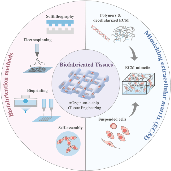
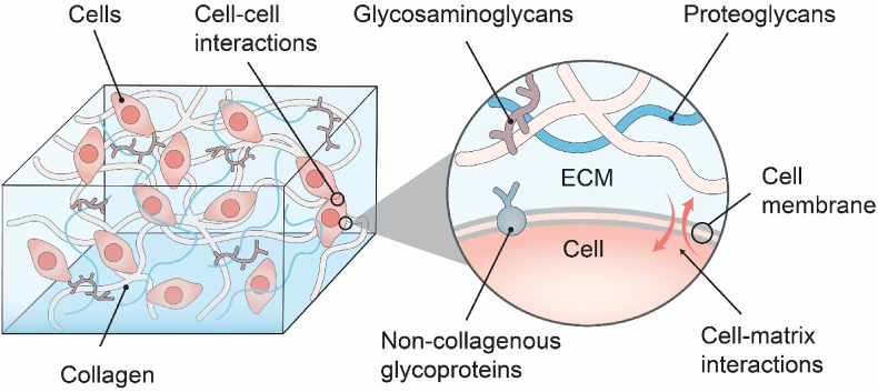
In the human body, almost all cells interact with extracellular matrices (ECMs), which have tissue and organ-specific compositions and architectures. These ECMs not only function as cellular scaffolds, providing structural support, but also play a crucial role in dynamically regulating various cellular functions. This comprehensive review delves into the examination of biofabrication strategies used to develop bioactive materials that accurately mimic one or more biophysical and biochemical properties of ECMs. We discuss the potential integration of these ECM-mimics into a range of physiological and pathological in vitro models, enhancing our understanding of cellular behavior and tissue organization. Lastly, we propose future research directions for ECM-mimics in the context of tissue engineering and organ-on-a-chip applications, offering potential advancements in therapeutic approaches and improved patient outcomes.

Keywords: Biofabrication; Bioprinting; Electrospinning; Extracellular matrix; Organ-on-a-Chip.


Conflict of interest statement

Yan Yan Shery Huang is an editorial board member for Bioactive Materials and was not involved in the editorial review or the decision to publish this article. All authors declare that there are no competing interests.

Figures

Image 1
Graphical abstract
Fig. 1
Fig. 1
Schematics of the Extracellular Matrix (ECM) illustrating the structural and chemical complexity. Different components within ECMs link together to form mechanically stable composites, and correlate to cellular metabolisms and migrations.
Fig. 2
Fig. 2
A length scale bar illustrating featured resolutions of various state-of-the-art biofabrication techniques in comparison with geometric sizes of representative cells and tissues [14,[63], [64], [65], [66], [67], [68], [69], [70], [71], [72], [73], [74]]. Herein ‘ES’ stands for electrospinning, ‘LEP’ stands for low-voltage electrospinning patterning.
Fig. 3
Fig. 3
Different biofabrication approaches towards ECM-mimicking models. (a) Soft lithography. An elastomeric stamp is first coated with an extracellular matrix (ECM) material, which is then stamped onto a substrate surface to create micropatterns of the ECM material; (b) Schematic diagram of electrospinning techniques, including far-field electrospinning, near-field electrospinning and melt electrospinning; (c) Schematic illustrations of different hydrogel 3D-printing setups, including extrusion, inkjet, laser-assisted and stereolithography.
Fig. 4
Fig. 4
Demonstration of microfluidic organ-on-chip models with functionality in Tissue Engineering. (a) Schematic of an alveolar lung-on-a-chip model and micrographs showing the expansion of the sacs under a strain of 8% [87]; (b) Schematic of organ-on-a-chip and the confocal image of liver cancer microspheres cultured in 7 days [91]; (c) Seeding of podocytes and human glomerular endothelial cells in microfluidic chip with confocal images after 28 days [94]; (d) Intestine-on-chip model, establishing co-culture of HeLa cells and bacteria [95]; (e) biodegradable vasculature-on-chip model [101]; (f) On-chip perivascular niche with patient-derived glioma model [103]. (a) Reproduced from Ref. [87] with permission ofNational Academy of Sciences, ©2021; (b) Reproduced from Ref. [91] with permission of Springer Nature, ©202022; (c) Reproduced from Ref. [94] with permission of Springer Nature, ©2019; (d) Reproduced from Ref. [95] with permission of Royal Society of Chemistry, ©2010; (e) Reproduced from Ref. [101] with permission of Springer Nature, ©2018; (f) Reproduced from Ref. [103] with permission of Royal Society of Chemistry, ©2021.
Fig. 5
Fig. 5
Demonstration of electrospun Nano/Micro-fibril structures with functionality in Tissue Engineering. (a) Fiber topography influences cell adhesion and morphology [140]; (b) Single cell behaviours differs in featured wavy and loop fibre patterns, as demonstrated in fluorescent channel [141]; (c) Internal fibrous microporosity creates an external coating of Chondrocytes on scaffold [145]; (d) Fibrous membrane creates two distinct but interacting tissue layers [146]; (e) Melt electrospun structure supports cell growth within large pores [147]; (f) Suspended fibers provide topographic guidance for fusion and expansion of cell aggregates over 10 days [148]; (a) Reproduced from Ref. [140] with permission of American Chemical Society, ©2014; (b) Reproduced from Ref. [141] with permission of IOP Publishing, ©2022; (c) Reproduced from Ref. [145] with permission of Elsevier, ©2008; (d) Reproduced from Ref. [146] with permission of Elsevier ©2018; (e) Reproduced from Ref. [147] with permission of IOP Publishing, ©2015; (f) Reproduced from Ref. [148] with permission of American Chemical Society, © 2022.
Fig. 6
Fig. 6
Demonstrations of 3D-bioprinted in vitro tissue/organ models with functionality in Tissue Engineering. (a) Schematics of vascular channel created using gelatin as sacrificial ink; fuorescence image of the printed vascular channel, with HUVECs (in red) and beads flow (in green) [202]; (b) photographs of the agarose-template created microchannels perfused with a fluorescent microbead suspension; longitudinal confocal images of a huvec-lined microchannel [210]; (c) Fabrication of a carotid artery structure using ‘SLAM’ strategy, demonstrating material durability and perfusion [211]; (d) Top view of FRESH-printed collagen heart valve using ‘FRESH’ strategy, and a sequence of valve opening under pulsatile flow over ∼1s [206]; (e) A printed heart within a support bath; 3D confocal image of the printed heart (CMs in pink, ECs in orange; sarcomeric actinin in green); [215]; (f) Entangled vascular networks with mathematical space-filling curves to entangled vessel topologies within hydrogels; Photograph of a printed hydrogel containing the distal lung subunit [216]. (a) Reproduced from Ref. [207] with permission of Wiley-VCH, ©2019; (b) Reproduced from Ref. [210] with permission of Royal Society of Chemistry, ©2014; (c) Reproduced from Ref. [211] with permission of Wiley-VCH, ©2019; (d) Reproduced from Ref. [206] with permission of American Association for the Advancement of Science, ©2019; (e) Reproduced from Ref. [215] with permission of Wiley-VCH, ©2019; (f) Reproduced from Ref. [216] with permission of American Association for the Advancement of Science, ©2019.
Fig. 7
Fig. 7
Applications of the biofabrication of ECM-like substrates for organ and cancer models: (a) High magnification micrographs show the healthy morphology of single neurons grown on the different substrates including poly-ornithine, PEDOT:PSS 1% ethylene glycol (EG) and PEDOT:PSS 3% EG Ref. [252]; (b) Fluorescence images showing fluorescein-HA (green) on the HA-binding peptide (HS-Pep-1) printed areas [253]; (c) Schematic of (c1) the one-step bioprinting method of the liver-on-a-chip model and (c2) a side view of the live-on-a-chip model [254]; (d) Illustration of 3D-bioprinted hybrid implant made of CNT-incorporated alginate and photo-cross-linked cell-laden hydrogel at 5 μg CNT/mg hydrogel [255]; (e) Schematics of vascular channel created using gelatin as sacrificial ink; fluorescence image of the printed vascular channel, with HUVECs (in red) and beads flow (in green) [199]; (f) Micro-CT scanning demonstrating a merged 3-D image of reconstructed open lumen construct in a thick collagen scaffold [207]; (g) Schematics of the embedded 3D printing strategy to produce the electro-mimetic bone matrices and the biomimetic cochleae (g1 and g2) [256]; (h) Scanning electron microscopy imaging of bioinks (comprised of hyaluronic acid, sodium alginate and gelatin), as well as Hematoxylin–eosin staining of Human glial cells within such bioinks [257]. (a) Reproduced from Ref. [252] with permission of Frontiers Media S.A., ©2015; (b) Reproduced from Ref. [253] with permission of Royal Society of Chemistry, ©2019; (c) Reproduced from Ref. [254] with permission of Royal Society of Chemistry, ©2016; (e) Reproduced from Ref. [199] with permission of Springer Nature, ©2012; (f) Reproduced from Ref. [207] with permission of Elsevier, ©2012 (g) Reproduced from Ref. [256] with permission of Springer Nature, ©2021; (h) Reproduced from Ref. [257] with permission of Springer Singapore, ©2020.

Similar articles

Cited by

References

    1. Discher D.E., Janmey P., Wang Y.L. Tissue cells feel and respond to the stiffness of their substrate. Science. 2005;310(5751):1139–1143. - PubMed
    1. Buxboim A., Ivanovska I.L., Discher D.E. Matrix elasticity, cytoskeletal forces and physics of the nucleus: how deeply do cells 'feel' outside and in? J. Cell Sci. 2010;123(Pt 3):297–308. - PMC - PubMed
    1. Nelson C.M. Geometric control of tissue morphogenesis. Biochim. Biophys. Acta. 2009;1793(5):903–910. - PMC - PubMed
    1. Thery M. Micropatterning as a tool to decipher cell morphogenesis and functions. J. Cell Sci. 2010;123(Pt 24):4201–4213. - PubMed
    1. Lutolf M.P., Hubbell J.A. Synthetic biomaterials as instructive extracellular microenvironments for morphogenesis in tissue engineering. Nat. Biotechnol. 2005;23(1):47–55. - PubMed

LinkOut - more resources