Review
. 2023 Aug 31:14:1260987.
doi: 10.3389/fphys.2023.1260987. eCollection 2023.

The hypoxia-inducible factor 1 pathway plays a critical role in the development of breast muscle myopathies in broiler chickens: a comprehensive review

Affiliations
Review

The hypoxia-inducible factor 1 pathway plays a critical role in the development of breast muscle myopathies in broiler chickens: a comprehensive review

Nabeel Alnahhas et al. Front Physiol. .

Abstract

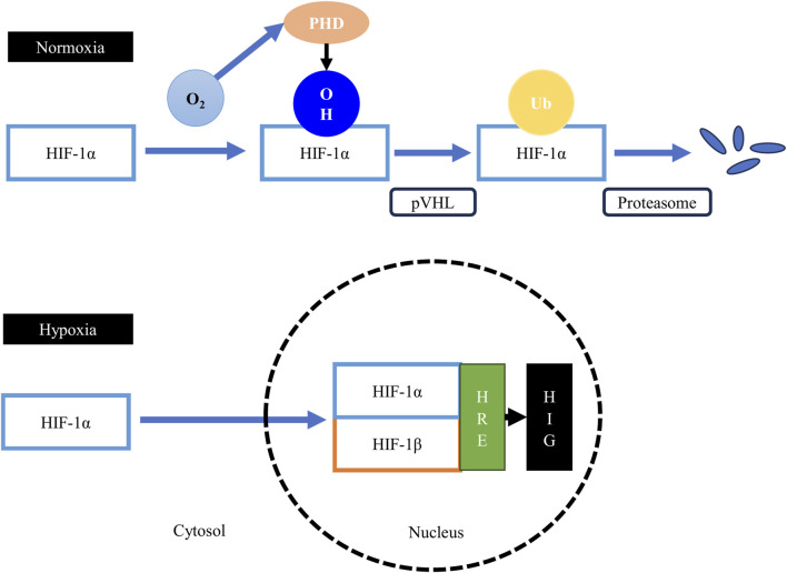
In light of the increased worldwide demand for poultry meat, genetic selection efforts have intensified to produce broiler strains that grow at a higher rate, have greater breast meat yield (BMY), and convert feed to meat more efficiently. The increased selection pressure for these traits, BMY in particular, has produced multiple breast meat quality defects collectively known as breast muscle myopathies (BMM). Hypoxia has been proposed as one of the major mechanisms triggering the onset and occurrence of these myopathies. In this review, the relevant literature on the causes and consequences of hypoxia in broiler breast muscles is reviewed and discussed, with a special focus on the hypoxia-inducible factor 1 (HIF-1) pathway. Muscle fiber hypertrophy induced by selective breeding for greater BMY reduces the space available in the perimysium and endomysium for blood vessels and capillaries. The hypoxic state that results from the lack of circulation in muscle tissue activates the HIF-1 pathway. This pathway alters energy metabolism by promoting anaerobic glycolysis, suppressing the tricarboxylic acid cycle and damaging mitochondrial function. These changes lead to oxidative stress that further exacerbate the progression of BMM. In addition, activating the HIF-1 pathway promotes fatty acid synthesis, lipogenesis, and lipid accumulation in myopathic muscle tissue, and interacts with profibrotic growth factors leading to increased deposition of matrix proteins in muscle tissue. By promoting lipidosis and fibrosis, the HIF-1 pathway contributes to the development of the distinctive phenotypes of BMM, including white striations in white striping-affected muscles and the increased hardness of wooden breast-affected muscles.

Keywords: HIF-1; broiler chickens; hypoxia; spaghetti meat; white striping; wooden breast.


Conflict of interest statement

Author EP was employed by the company Olymel S.E.C./L.P. The remaining authors declare that the research was conducted in the absence of any commercial or financial relationships that could be construed as a potential conflict of interest.

Figures

FIGURE 1
FIGURE 1
Consequences of muscle fiber hypertrophy. Genetic selection for increased breast meat yield (BMY) operates by increasing muscle fiber diameter. Myofiber hypertrophy leads to a decrease in the interstitial spaces (perimysium and endomysium) in which blood vessels and capillaries are found, resulting in a reduced density of the vascular network. Myofiber hypertrophy is also associated with decreased energy or glycogen content in muscle tissue. The reduced capillary density and decreased glycogen reserves predispose the Pectoralis major muscle to the development of breast muscle myopathies.
FIGURE 2
FIGURE 2
Examples of breast muscles exhibiting severe white striping (A), wooden breast (B) and spaghetti meat (C). Severe white striping is characterized by the occurrence of white striations with a diameter greater than 1 mm, running in parallel to the direction of muscle fibers, covering the entire ventral surface of the muscle. Severe wooden breast is associated with increased muscle hardness from the cranial to the caudal end of the Pectoralis major muscle, coupled with petechia. Severe spaghetti meat is associated with disintegration and separation of muscle bundles appearing like spaghetti.
FIGURE 3
FIGURE 3
Biochemical changes in myopathic muscles. Breast muscle myopathies are associated with a higher ultimate pH than unaffected muscles. The increase in ultimate pH can be explained by the lower glycolytic reserves in these muscles at death, coupled with decreased mitochondrial activity, leading to lower ATP levels and early cessation of post-mortem acidification. The lower lactate level in myopathic muscles is the result of the upregulation of the monocarboxylate transporter 4 (MTC4), which increases lactate export from cells.
FIGURE 4
FIGURE 4
The hypoxia-inducible factor 1 (HIF-1) pathway. Under normoxic conditions, the proline residues on the HIF-1α subunit are hydroxylated by the oxygen-dependent prolyl-4-hydroxylases (PHD), leading to the binding of the von Hippel–Lindau tumor suppressor protein (pVHL), the substrate recognition component of an E3 ubiquitin ligase, to the HIF-1α protein. The pVHL protein tags the sarcoplasmic HIF-1α protein for ubiquitination and subsequent rapid proteasomal degradation. Therefore, formation of the HIF-1 heterodimer is obstructed, suppressing the transcription of hypoxia-inducible genes (HIG). Under hypoxic conditions, the activity of the prolyl-4-hydroxylases is suppressed, which allows the stabilization of the HIF-1α subunit and its subsequent translocation to the nucleus to form the HIF-1 heterodimer with the HIF-1β subunit. The heterodimer then binds to the hypoxia-responsive elements (HRE), upregulating the expression of HIG.
FIGURE 5
FIGURE 5
Hypoxia alters muscle energy metabolism through the upregulation of the HIF-1 pathway. Under hypoxic conditions, HIF-1α upregulates pyruvate dehydrogenase kinase 1 (PDK1), leading to the inactivation of the enzyme pyruvate dehydrogenase (PDH) through phosphorylation. Consequently, the conversion of pyruvate to acetyl-CoA is decreased. Concurrently, HIF-1α upregulates lactate dehydrogenase (LDH), increasing the conversion of pyruvate to lactate, and upregulates the monocarboxylate transporter 4 (MTC4), promoting lactate export from cells. The decrease in the conversion of pyruvate to acetyl-CoA slows the tricarboxylic acid cycle (TCA), leading to the accumulation of its intermediates. These intermediates downregulate the activity of prolyl-4-hydroxylases (PHD), further upregulating the HIF-1 pathway by allowing the stabilization of HIF-1α protein in the sarcoplasm. Black arrows: upregulation, blunt red arrows: downregulation.
FIGURE 6
FIGURE 6
Hypoxia alters muscle lipid metabolism. Upregulation of HIF-1α downregulates myogenic factors while upregulating lipogenic factors. The increased expression of acetyl-CoA carboxylase a (ACCα) and fatty acid synthase (FAS) promotes the synthesis of long-chain fatty acids in myopathic muscles. The upregulation of HIF-1α is also associated with increased expression of peroxisome proliferator-activated receptor gamma (PPARγ), leading to increased adipogenesis and accumulation of lipids in myopathic muscles. Hypoxia also upregulates the expression of fatty acid binding proteins (FABPs), further stimulating lipogenesis and lipid storage in muscle tissues. These hypoxia-induced changes are responsible for the observed lipidosis in myopathic muscles. Black arrows: upregulation, blunt red arrows: downregulation.
FIGURE 7
FIGURE 7
Hypoxia promotes fibrosis in myopathic muscles. The upregulation of HIF-1α promotes the synthesis and deposition of extracellular matrix (ECM) proteins (collagen and fibronectin) in myopathic muscles through its interaction with the transforming growth factor β1/SMAD signaling pathway and its upregulation of connective tissue growth factor (CTGF).
FIGURE 8
FIGURE 8
Hypoxia impairs mitochondrial morphology and functions. The perturbation of the tricarboxylic acid cycle (TCA) induced by the upregulation of HIF-1α slows the electron transport chain (ETC.), increasing the probability of electron leakage at complex I (CI) and III (CIII). The leaked electrons bind to molecular oxygen to form reactive oxygen species (ROS). High concentrations of ROS and prolonged exposure of cellular components to ROS lead to oxidative stress and subsequently to lipid peroxidation and protein oxidation. The products from oxidative damage (MDA, carbonyls, thiols, 15-HETE, and 15-KETE) accumulate in myopathic muscles. Furthermore, the upregulation of HIF-1α upregulates genes responsible for mitochondrial fission (DRP1) and fusion (VEGF-A, MFN1, and MFN2). The stimulation of mitochondrial dynamics is a response mechanism aiming to alleviate cellular stress and eliminate defective mitochondria induced by hypoxia. Black arrows: upregulation, blunt red arrows: downregulation.
FIGURE 9
FIGURE 9
Hypoxia and ischemia stimulate apoptosis in myopathic muscles. The ischemic and hypoxic state of myopathic muscle is responsible for necrosis and myofiber degeneration, inducing an inflammatory response in affected muscles. Moreover, hypoxia stresses the sarcoplasmic reticulum (SR) and damages the nuclei of affected muscle tissue. The inflammation, SR stress, and nuclei damage upregulate the caspase signaling pathway, ultimately dismantling cellular proteins, and leading to apoptosis as a response mechanism to limit further damage to muscle tissue.
FIGURE 10
FIGURE 10
Hypoxia plays a critical role in the development of breast muscle myopathies through the activation of the hypoxia-inducible factor 1 (HIF-1) pathway. The hypertrophic growth of breast muscles is associated with decreased capillary density, leading to decreased circulation (ischemia) and oxygenation (hypoxia) in muscle tissue, coupled with lower nutrient supply. The histopathological changes associated with breast muscle myopathies, including necrosis, myofiber degeneration, and inflammation, are consequences of the ischemic and hypoxic states. Hypoxia can stress the sarcoplasmic reticulum and damage the nuclei, leading to the activation of the caspase signaling pathway, triggering apoptosis. Moreover, HIF-1α can further stimulate antemortem anaerobic glycolysis in the Pectoralis major muscle, depleting glycogen reserves by the time birds reach market age, contributing to the early cessation of post-mortem acidification in this muscle which explain the higher ultimate pH in these muscles. Furthermore, HIF-1α slows the TCA cycle and impedes mitochondrial function, leading to increased ROS production and accumulation, triggering a state of oxidative stress in the muscle and damaging its cellular components. HIF-1α also alters lipid metabolism in the Pectoralis major muscle, leading to increased synthesis of long-chain fatty acids, lipogenesis, and lipid storage, ultimately causing lipidosis. Finally, by interacting with the TGF-β1/SMAD pathway and with the connective tissue growth factor, HIF-1α increases the deposition of extracellular matrix (ECM) proteins in muscle tissue, leading to fibrosis. These structural and metabolic changes trigger the onset and development of breast muscle myopathies and give them their distinctive phenotypes. Black arrows: upregulation, blunt red arrows: downregulation.

Similar articles

Cited by

References

    1. Abasht B., Mutryn M. F., Michalek R. D., Lee W. R. (2016). Oxidative stress and metabolic perturbations in wooden breast disorder in chickens. Plos One 11 (4), e0153750. 10.1371/journal.pone.0153750 - DOI - PMC - PubMed
    1. Alnahhas N., Berri C., Boulay M., Baéza E., Jégo Y., Baumard Y., et al. (2014). Selecting broiler chickens for ultimate pH of breast muscle: analysis of divergent selection experiment and phenotypic consequences on meat quality, growth, and body composition traits. J. Anim. Sci. 92 (9), 3816–3824. 10.2527/jas.2014-7597 - DOI - PubMed
    1. Alnahhas N., Berri C., Chabault M., Chartrin P., Boulay M., Bourin M. C., et al. (2016). Genetic parameters of white striping in relation to body weight, carcass composition, and meat quality traits in two broiler lines divergently selected for the ultimate pH of the pectoralis major muscle. BMC Genet. 17, 61. 10.1186/s12863-016-0369-2 - DOI - PMC - PubMed
    1. Baldi G., Soglia F., Laghi L., Tappi S., Rocculi P., Tavaniello S., et al. (2019). Comparison of quality traits among breast meat affected by current muscle abnormalities. Food Res. Int. 115, 369–376. 10.1016/j.foodres.2018.11.020 - DOI - PubMed
    1. Baldi G., Soglia F., Mazzoni M., Sirri F., Canonico L., Babini E., et al. (2018). Implications of white striping and spaghetti meat abnormalities on meat quality and histological features in broilers. Animal 12 (1), 164–173. 10.1017/s1751731117001069 - DOI - PubMed