

Trick or Treat: An Intrahepatic Intraductal Papillary Neoplasm of the Bile Duct
- PMID: 37719498
- PMCID: PMC10500955
- DOI: 10.7759/cureus.43494
Trick or Treat: An Intrahepatic Intraductal Papillary Neoplasm of the Bile Duct
Abstract
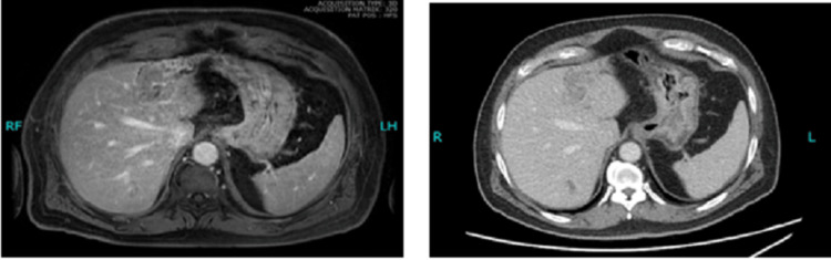
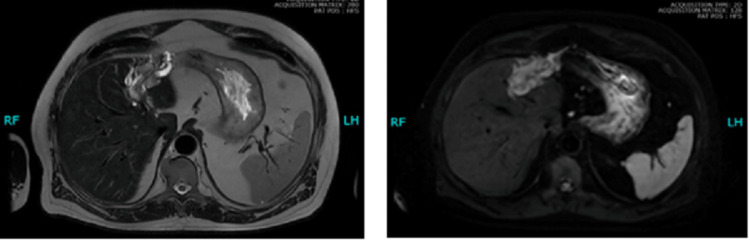
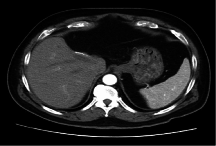
An intraductal papillary neoplasm involving the biliary tree is an unusual premalignant condition of epithelial origin, identified by its cystic dilatation of the biliary channels. Being a slow-growing tumor, surgery offers the best curative rate, especially in the setting of a low-grade disease. Here, we present a case of a localized, low-grade, intraductal papillary neoplasm of the bile duct (IPNB), residing in the liver, which was treated with resection of the liver lobe. The adjuvant treatment and prognosis highly depend upon the presence of dysplasia or a co-existent invasive malignancy. To the best of our knowledge, being a rare entity, this is the first case to be reported from Pakistan.
Keywords: bile duct; intraductal papillary neoplasm; intrahepatic; low grade; surgical resection.
Copyright © 2023, Samar et al.
Conflict of interest statement
The authors have declared that no competing interests exist.
Figures
Similar articles
-
Branch-type intraductal papillary neoplasm of the bile duct treated with laparoscopic anatomical resection: a case report.Surg Case Rep. 2020 May 15;6(1):103. doi: 10.1186/s40792-020-00864-3. Surg Case Rep. 2020. PMID: 32415464 Free PMC article.
-
Clinicopathological characterization of so-called "cholangiocarcinoma with intraductal papillary growth" with respect to "intraductal papillary neoplasm of bile duct (IPNB)".Int J Clin Exp Pathol. 2014 May 15;7(6):3112-22. eCollection 2014. Int J Clin Exp Pathol. 2014. PMID: 25031730 Free PMC article.
-
Recurrent intraductal papillary neoplasm of the bile duct due to intraductal dissemination: a case report and literature review.Surg Case Rep. 2021 Nov 5;7(1):238. doi: 10.1186/s40792-021-01318-0. Surg Case Rep. 2021. PMID: 34739634 Free PMC article.
-
Intraductal papillary neoplasm of the bile duct: review of updated clinicopathological and imaging characteristics.Br J Surg. 2023 Aug 11;110(9):1229-1240. doi: 10.1093/bjs/znad202. Br J Surg. 2023. PMID: 37463281 Review.
-
Intraductal Papillary Neoplasm of Bile Duct: Updated Clinicopathological Characteristics and Molecular and Genetic Alterations.J Clin Med. 2020 Dec 9;9(12):3991. doi: 10.3390/jcm9123991. J Clin Med. 2020. PMID: 33317146 Free PMC article. Review.
References
-
- Precancerous lesions of the biliary tree. Klöppel G, Adsay V, Konukiewitz B, Kleeff J, Schlitter AM, Esposito I. Best Pract Res Clin Gastroenterol. 2013;27:285–297. - PubMed
-
- Gallbladder cancer worldwide: geographical distribution and risk factors. Randi G, Franceschi S, La Vecchia C. Int J Cancer. 2006;118:1591–1602. - PubMed
-
- The 2019 WHO classification of tumours of the digestive system. Nagtegaal ID, Odze RD, Klimstra D, et al. http://of Tumours Editorial Board. Histopathology. 2020;76:182–188. - PMC - PubMed
-
- Intraductal papillary neoplasm of the bile duct presenting with hepatogastric fistula: a case report and literature review. Chan WH, Chen CM, Wang SY, et al. https://www.frontiersin.org/journals/oncology/articles/10.3389/fonc.2023.... Front Oncol. 2023;13:1193918. - PMC - PubMed
Publication types
LinkOut - more resources
Full Text Sources