

Suspected Enhanced S-Cone Syndrome: A Case Report
- PMID: 37719593
- PMCID: PMC10505071
- DOI: 10.7759/cureus.43660
Suspected Enhanced S-Cone Syndrome: A Case Report
Abstract
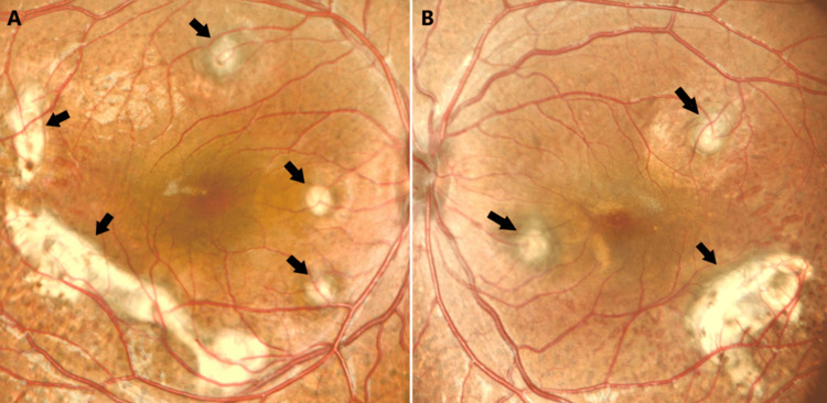
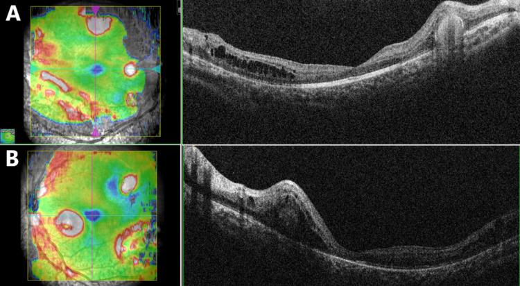
Enhanced S-cone syndrome (ESCS) is a rare type of retinal dystrophy disorder that is linked to NR2E3 gene mutation and NRL gene mutations less widely. The disease is characterized by increased S-cones number and marked degeneration in rods and M- and L-cone receptors. The patient suffers from night blindness from an early age. Examination of the fundus of the eye shows nummular pigmented lesions, but they are not specific to ESCS. The diagnosis can be confirmed with electroretinography. We report a case of a four-year-old girl suspected of having ESCS based on her clinical picture, fundus examination, and electroretinography.
Keywords: case report; diagnosis; electroretinography; enhanced s-cone syndrome; retinal dystrophy.
Copyright © 2023, Alnosair et al.
Conflict of interest statement
The authors have declared that no competing interests exist.
Figures
Similar articles
-
Homozygosity for a Recessive Loss-of-Function Mutation of the NRL Gene Is Associated With a Variant of Enhanced S-Cone Syndrome.Invest Ophthalmol Vis Sci. 2016 Oct 1;57(13):5361-5371. doi: 10.1167/iovs.16-19505. Invest Ophthalmol Vis Sci. 2016. PMID: 27732723
-
Clinical and molecular characterization of enhanced S-cone syndrome in children.JAMA Ophthalmol. 2014 Nov;132(11):1341-9. doi: 10.1001/jamaophthalmol.2014.2343. JAMA Ophthalmol. 2014. PMID: 25079116
-
A novel mutation (Cys83Tyr) in the second zinc finger of NR2E3 in enhanced S-cone syndrome.Graefes Arch Clin Exp Ophthalmol. 2011 Feb;249(2):201-8. doi: 10.1007/s00417-010-1482-y. Epub 2010 Aug 20. Graefes Arch Clin Exp Ophthalmol. 2011. PMID: 20725840
-
Enhanced S-Cone Syndrome (Goldmann-Favre Syndrome).Adv Exp Med Biol. 2018;1085:153-156. doi: 10.1007/978-3-319-95046-4_28. Adv Exp Med Biol. 2018. PMID: 30578501 Review.
-
NR2E3 mutations in enhanced S-cone sensitivity syndrome (ESCS), Goldmann-Favre syndrome (GFS), clumped pigmentary retinal degeneration (CPRD), and retinitis pigmentosa (RP).Hum Mutat. 2009 Nov;30(11):1475-85. doi: 10.1002/humu.21096. Hum Mutat. 2009. PMID: 19718767 Review.
References
-
- Retinochoroidal anastomosis associated with enhanced S-cone syndrome. Zerbib J, Blanco Garavito R, Gerber S, et al. Retin Cases Brief Rep. 2019;13:295–299. - PubMed
Publication types
LinkOut - more resources
Full Text Sources