

Idiopathic Intracranial Hypertension Secondary to Chronic Inflammatory Demyelinating Polyradiculoneuropathy
- PMID: 37719616
- PMCID: PMC10505048
- DOI: 10.7759/cureus.43648
Idiopathic Intracranial Hypertension Secondary to Chronic Inflammatory Demyelinating Polyradiculoneuropathy
Abstract
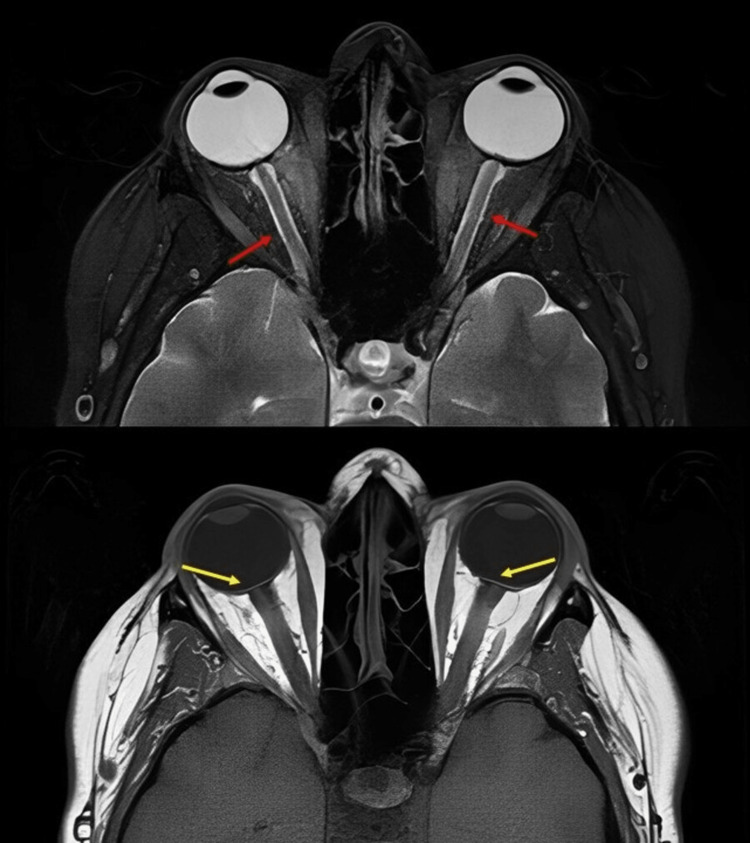
Chronic inflammatory demyelinating polyradiculoneuropathy (CIDP) is the most common immune-mediated inflammatory polyneuropathy, defined as progressive or relapsing symptoms for over two months with pathological or electrophysiological evidence of peripheral nerve demyelination. Papilledema is optic nerve head edema secondary to increased intracranial pressure or infiltrative/infectious etiologies. Regardless of the cause, visual loss is one of the feared manifestations due to optic nerve damage. We present a 50-year-old female patient with CIDP who developed papilledema that was secondary to increased intracranial pressure from high protein content in the cerebrospinal fluid (CSF) and elevated body mass index (BMI) secondary to prednisone use. Treatment with acetazolamide completely resolved the papilledema and headaches, and the patient was later maintained on mycophenolate, intravenous immunoglobulin (IVIG), rituximab, and prednisone. To the best of our knowledge, this is the first case that describes successful medical management of increased intracranial pressure in the setting of CIDP.
Keywords: acetazolamide; chronic inflammatory demyelinating polyradiculoneuropathy; hydrocephalus; idiopathic intracranial hypertension; papilledema.
Copyright © 2023, Gül et al.
Conflict of interest statement
The authors have declared that no competing interests exist.
Figures
References
-
- Epidemiology of inflammatory neurological and inflammatory neuromuscular diseases in Tottori Prefecture, Japan. Kusumi M, Nakashima K, Nakayama H, Takahashi K. Psychiatry Clin Neurosci. 1995;49:169–174. - PubMed
-
- Chronic inflammatory demyelinating polyneuropathy: update on diagnosis, immunopathogenesis and treatment. Lehmann HC, Burke D, Kuwabara S. J Neurol Neurosurg Psychiatry. 2019;90:981–987. - PubMed
Publication types
LinkOut - more resources
Full Text Sources