

What is the most predictive magnetic resonance imaging finding of rotator cuff tear concomitant with shoulder stiffness?
- PMID: 37719831
- PMCID: PMC10499857
- DOI: 10.1016/j.jseint.2023.05.001
What is the most predictive magnetic resonance imaging finding of rotator cuff tear concomitant with shoulder stiffness?
Abstract
Background: Common magnetic resonance imaging (MRI) findings in adhesive capsulitis are not often evident in rotator cuff tear concomitant with shoulder stiffness. This study aimed to determine the most predictive MRI finding of rotator cuff tear with shoulder stiffness to differentiate from that without stiffness.
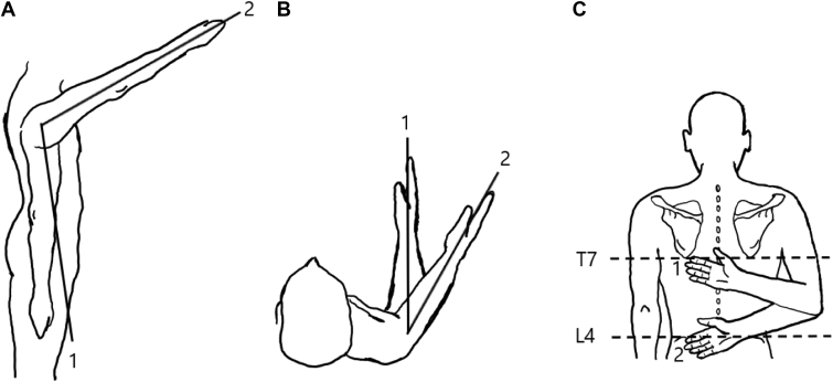
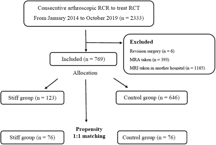
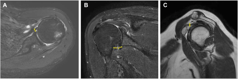
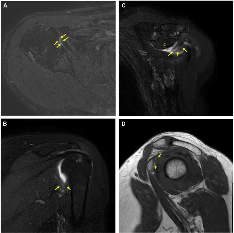
Materials and methods: The data of patients who underwent arthroscopic rotator cuff repair between January 2014 and October 2019 were retrospectively reviewed. Stiffness was defined as forward flexion <120°, external rotation at side <30°, and internal rotation at back <L3 in the active range of motion. Propensity score matching (1-to-1) was performed between the stiff and control groups by sex, age, and tear size, and 76 patients per group were matched. Anterior capsular thickness, maximal humeral/glenoid capsular thickness in the axillary recess, coracohumeral ligament thickness, the presence of hyperintensity in the anterior capsule and humeral/glenoid capsule in the axillary recess, and hyperintensity and obliteration of the subcoracoid fat triangle were evaluated.
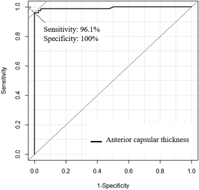
Results: Anterior capsular thickness, glenoid capsular thickness in the axillary recess, and anterior and axillary capsular hyperintensities were significantly more dominant in the stiff group (all P < .05) than in the control group. Anterior capsular thickness and anterior capsular abnormal hyperintensity could be used to differentiate between the stiff and control groups (P < .05). Anterior capsular thickness showed high diagnostic performance with an area under the receiver operating characteristic curve of 0.993. The cut-off value for stiffness was 3.07 mm (sensitivity, 96.1%; specificity, 100%).
Conclusion: Anterior capsular thickening and abnormal hyperintensity were the most predictive MRI findings for stiffness in patients with rotator cuff tear and stiffness to differentiate from patients with rotator cuff tear without stiffness.
Keywords: Adhesive capsulitis; Anterior capsular abnormal hyperintensity; Anterior capsular thickness; Magnetic resonance imaging; Rotator cuff tear; Stiff shoulder.
© 2023 The Author(s).
Figures
Similar articles
-
Correlations between clinical features and MRI findings in early adhesive capsulitis of the shoulder: a retrospective observational study.BMC Musculoskelet Disord. 2020 Aug 13;21(1):542. doi: 10.1186/s12891-020-03569-8. BMC Musculoskelet Disord. 2020. PMID: 32791997 Free PMC article.
-
Anterior capsular abnormality: another important MRI finding for the diagnosis of adhesive capsulitis of the shoulder.Skeletal Radiol. 2019 Apr;48(4):543-552. doi: 10.1007/s00256-018-3064-8. Epub 2018 Sep 11. Skeletal Radiol. 2019. PMID: 30206678
-
MRI Findings Predictive of Shoulder Stiffness in Patients With Full-Thickness Rotator Cuff Tears.AJR Am J Roentgenol. 2020 May;214(5):1146-1151. doi: 10.2214/AJR.19.21973. Epub 2020 Feb 18. AJR Am J Roentgenol. 2020. PMID: 32069080
-
Comparison of surgical outcomes between rotator cuff repair with and without rotator interval capsular release for rotator cuff tears to prevent and improve postoperative stiffness: a meta-analysis.Eur J Orthop Surg Traumatol. 2020 Oct;30(7):1263-1275. doi: 10.1007/s00590-020-02695-2. Epub 2020 May 18. Eur J Orthop Surg Traumatol. 2020. PMID: 32424473
-
Magnetic resonance imaging features for diagnosing adhesive capsulitis of the shoulder: a systematic review and meta-analysis.BMC Musculoskelet Disord. 2025 Apr 16;26(1):368. doi: 10.1186/s12891-025-08592-1. BMC Musculoskelet Disord. 2025. PMID: 40241075 Free PMC article.
Cited by
-
Establishment and validation of a screening and risk predication model for rotator cuff tear and shoulder stiffness.Ann Jt. 2025 Jul 30;10:24. doi: 10.21037/aoj-25-16. eCollection 2025. Ann Jt. 2025. PMID: 40791897 Free PMC article.
References
-
- Carrillon Y., Noel E., Fantino O., Perrin-Fayolle O., Tran-Minh V.A. Magnetic resonance imaging findings in idiopathic adhesive capsulitis of the shoulder. Rev Rhum Engl Ed. 1999;66:201–206. - PubMed
-
- Chen Y., Chen S., Qiao Y., Ge Y., Li H., Chen J., et al. A Long preoperative duration of Symptoms is associated with Worse Functional Outcomes after 1-stage arthroscopic treatment of rotator cuff tears with shoulder stiffness. Am J Sports Med. 2017;45:2336–2344. doi: 10.1177/0363546517707202. - DOI - PubMed
LinkOut - more resources
Full Text Sources