[Preprint]. 2023 Sep 6:rs.3.rs-3304297.
doi: 10.21203/rs.3.rs-3304297/v1.

Altered peripheral taste function in a mouse model of inflammatory bowel disease

Affiliations

Altered peripheral taste function in a mouse model of inflammatory bowel disease

Guangkuo Dong et al. Res Sq. .

Update in

Abstract

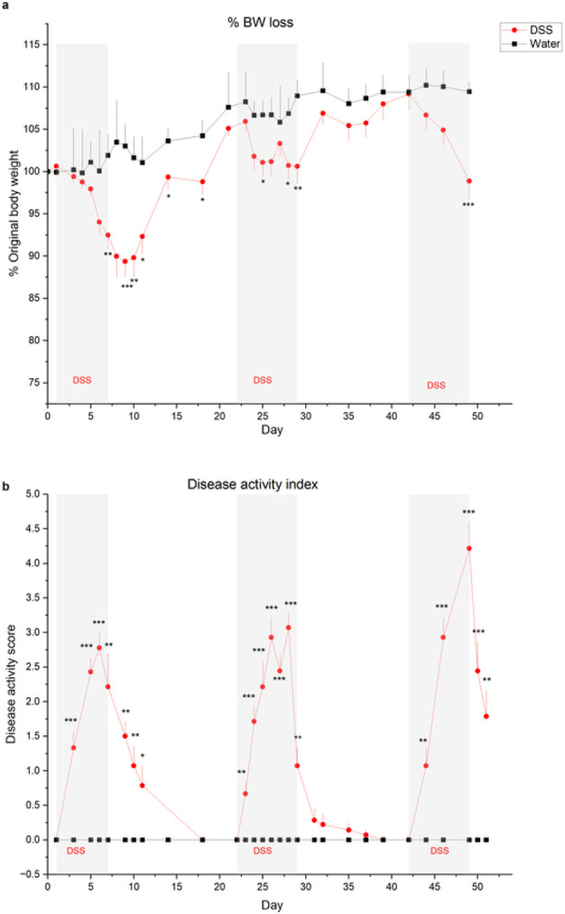
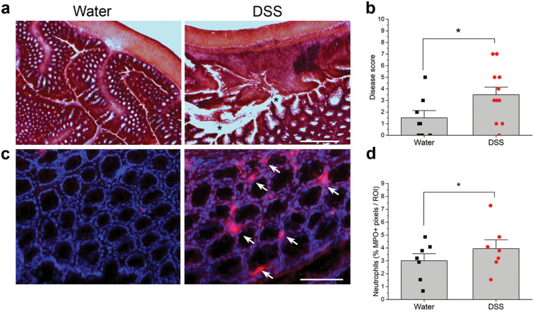
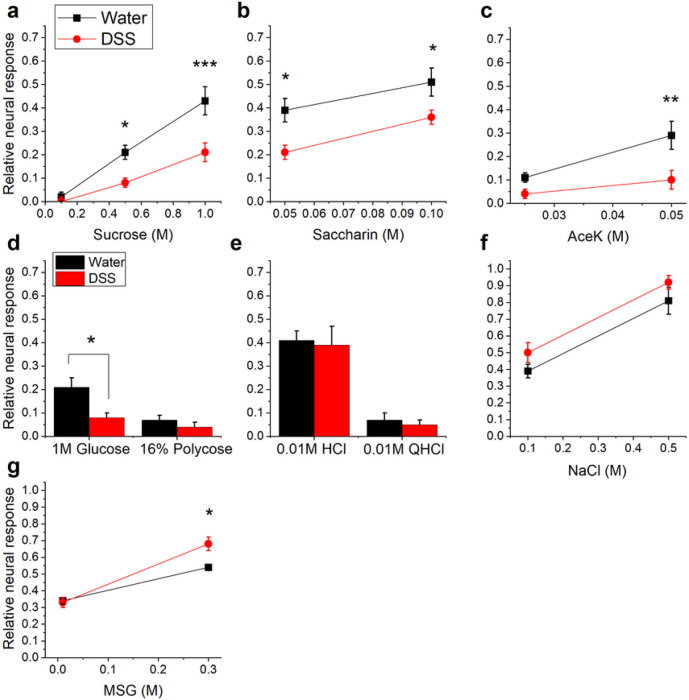
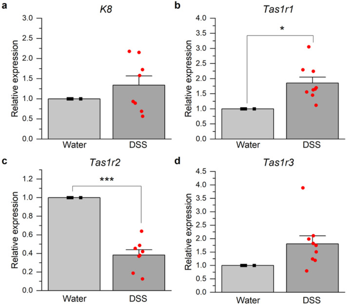
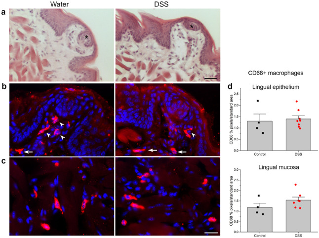
Increased sugar intake and taste dysfunction have been reported in patients with inflammatory bowel disease (IBD), a chronic disorder characterized by diarrhea, pain, weight loss and fatigue. It was previously unknown whether taste function changes in mouse models of IBD. Mice consumed dextran sodium sulfate (DSS) during three 7-day cycles to induce chronic colitis. DSS-treated mice displayed signs of disease, including significant weight loss, diarrhea, loss of colon architecture, and inflammation of the colon. After the last DSS cycle we assessed taste function by recording electrophysiological responses from the chorda tympani (CT) nerve, which transmits activity from lingual taste buds to the brain. DSS treatment significantly reduced neural taste responses to natural and artificial sweeteners. Responses to carbohydrate, salt, sour or bitter tastants were unaffected in mice with colitis, but umami responses were modestly elevated. DSS treatment modulated the expression of receptor subunits that transduce sweet and umami stimuli in oral taste buds as a substrate for functional changes. Dysregulated systemic cytokine responses, or dysbiosis that occurs during chronic colitis may be upstream from changes in oral taste buds. We demonstrate for the first time that colitis alters taste input to the brain, which could exacerbate malnutrition in IBD patients.

Keywords: Tas1r1; Tas1r2; chorda tympani nerve; dextran sulfate sodium (DSS)-induced colitis; electrophysiology; gut inflammation; sweet; umami.


Conflict of interest statement

Conflict of interest statement: The authors declare no competing financial interests.

Figures

Figure 1
Figure 1. 7-day cycles of 2% DSS in drinking water induce colitis.
a Mean (± SEM) body weight expressed as a percentage of original body weight over time. Mice lost significantly more body weight at several time points during or after DSS treatment (n=13) compared to water-treated controls (n=5). Grey shaded blocks indicate DSS treatment days. b Mean Disease Activity Index scores (± SEM) were significantly elevated on most days in DSS-treated mice compared to controls. Individual scores representing the magnitude of body weight loss, blood in stool, and diarrhea are summed to calculate the Disease Activity score as described in Methods. *p < 0.05; **p < 0.001, ***p < 0.0001. Actual p values are listed in Table 1.
Figure 2
Figure 2. DSS treatment elicited inflammation and loss of tissue structure in the colon.
a Hematoxylin and eosin staining reveals the loss of tissue architecture in the colon of mice treated with DSS. Black stars indicate severe tissue damage. b Disease scores reflecting inflammation and tissue breakdown (see Methods) were significantly higher in mice drinking DSS (n=6) compared to controls (n=4). cMyeloperoxidase (MPO)-expressing neutrophils (red) are more prominent in the colon of DSS-treated mice. Blue nuclei are stained with DAPI. d Neutrophil density was significantly higher in DSS-treated mice (n=4) compared to controls (n=4). *p< 0.05. Bar in A = 60 μm; B = 75 μm.
Figure 3
Figure 3. Chorda tympani nerve responses to taste stimuli.
Representative responses from a water-treated control or b DSS-treated mouse. Responses to 0.5M NH4Cl were robust in both groups, and were used as a standard stimulus to check the stability of the preparation. Responses to sucrose and artificial sweeteners are suppressed in the DSS-treated mouse. Dotted vertical lines indicate stimulus onset and rinse. Black bars under the first 0.5M NH4Cl response indicate 20 sec.
Figure 4
Figure 4. Taste responses to natural and artificial sweeteners are significantly reduced in mice with DSS-induced colitis.
Mean neural taste responses (±SEM) to taste stimuli, expressed relative to 0.5M NH4Cl responses, were recorded 56-61 days after the start of the experiment. Three cycles of DSS significantly inhibited responses to a0.5M and 1.0M sucrose, b both concentrations of saccharin, c the higher concentration of aceK, and dglucose. Neural response magnitudes to d the starch, polycose, e HCl, QHCl, and f NaCl were similar in DSS-treated (n = 15) and control (n = 8) groups. g There was a slight but significant increase in responses to the umami stimulus, 0.3M MSG, n mice with colitis. *p < 0.05; **p < 0.01; ***p < 0.0001.
Figure 5
Figure 5. DSS-induced colitis modulates the expression of lingual sweet and umami taste receptor genes.
Lingual mRNA expression in DSS-treated mice (n = 9) was expressed relative to values for water controls (n = 4). a The expression of the taste receptor cell marker gene, keratin (K)8, was similar between groups. b The umami taste receptor subunit, Tas1r1, was significantly increased in mice with colitis compared to controls. cExpression of the sweet taste receptor subunit, Tas1r2, was significantly reduced by DSS treatment. dExpression levels of Tas1r3, which forms heteromeric umami receptors with Tas1r1and sweet receptors with Tas1r2, were similar between groups. *p < 0.05; ***p < 0.0001.
Figure 6
Figure 6. Chronic DSS treatment did not induce lingual inflammation or epithelial breakdown.
a Hematoxylin and eosin stained sections of anterior tongue from control DSS-treated mice demonstrate similar, intact taste buds (black asterisks), surrounding epithelium, and lamina propria without significant inflammatory infiltrates. CD68+ macrophages are similarly dense in bfungiform papillae and c lamina propria and underlying mucosa of the anterior tongue from both groups. dCD68+ pixels in a standard-sized area of interest from the two tissue compartments were similar between treatment groups (p > 0.05). White arrowheads in B indicate macrophages within fungiform papillae, and arrows in the lamina propria. Bar in a = 30 μm; c = 20 μm.

References

    1. Hendrickson B.A., Gokhale R. & Cho J.H. Clinical Aspects and Pathophysiology of Inflammatory Bowel Disease. Clin. Microbiol. Rev. 15, 79–94 (2002). - PMC - PubMed
    1. Abraham C. & Cho J.H. Inflammatory Bowel Disease. N. Engl. J. Med. 361, 2066–2078 (2009). - PMC - PubMed
    1. Massironi S., et al. Nutritional deficiencies in inflammatory bowel disease: therapeutic approaches. Clin. Nutr. 32, 904–910 (2013). - PubMed
    1. Weisshof R. & Chermesh I. Micronutrient deficiencies in inflammatory bowel disease. Curr. Opin. Clin. Nutr. Metab. Care 18, 576–581 (2015). - PubMed
    1. Hebuterne X., Filippi J. & Schneider S.M. Nutrition in adult patients with inflammatory bowel disease. Curr. Drug Targets 15, 1030–1038 (2014). - PubMed

Publication types

LinkOut - more resources