[Preprint]. 2023 Sep 7:rs.3.rs-3311121.
doi: 10.21203/rs.3.rs-3311121/v1.

Reduced suprasellar cistern cerebrospinal fluid motion in patients with Parkinson's disease revealed by magnetic resonance imaging with dynamic cycling of diffusion weightings

Affiliations

Reduced suprasellar cistern cerebrospinal fluid motion in patients with Parkinson's disease revealed by magnetic resonance imaging with dynamic cycling of diffusion weightings

Gabriela Pierobon Mays et al. Res Sq. .

Update in

Abstract

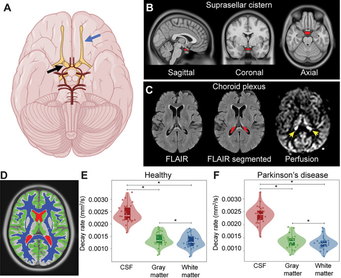
Background: Parkinson's disease is characterized by dopamine-responsive symptoms as well as aggregation and accumulation of a-synuclein protofibrils. New diagnostic methods assess a-synuclein aggregation characteristics from cerebrospinal fluid and recent pathophysiologic mechanisms suggest that cerebrospinal fluid circulation disruptions may precipitate a-synuclein retention. Here, we test the hypothesis that cerebrospinal fluid motion at the level of the suprasellar cistern is reduced in Parkinson's disease relative to healthy participants and this reduction relates to choroid plexus perfusion.

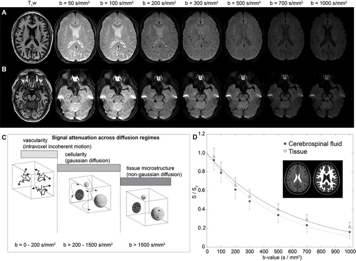
Methods: Diffusion weighted imaging (spatial resolution=1.8×1.8×4 mm) magnetic resonance imaging with cycling of diffusion weightings (b-values=0, 50, 100, 200, 300, 700, and 1000 s/mm2) over the approximate kinetic range of suprasellar cistern neurofluid motion was applied at 3-Tesla in Parkinson's disease (n=27; age=66±6.7 years) and healthy (n=32; age=68±8.9 years) participants. Wilcoxon rank-sum tests were applied to test the primary hypothesis that the decay rate of cerebrospinal fluid signal as a function of b-value, which reflects increasing fluid motion, is reduced in persons with versus without Parkinson's disease and inversely relates to choroid plexus activity assessed from perfusion-weighted magnetic resonance imaging (Spearman rank-order correlation; significance-criteria: p<0.05).

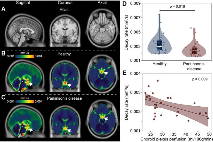
Results: Consistent with the primary hypothesis, decay rates were higher in healthy (D=0.00328±0.00123mm2/s) relative to Parkinson's disease (D=0.00256±0.0094mm2/s) participants (p=0.016). This finding was preserved after controlling for age and sex. An inverse correlation between choroid plexus perfusion and decay rate (p=0.011) was observed in Parkinson's disease participants.

Conclusions: Cerebrospinal fluid motion at the level of the suprasellar cistern is often reduced in adults with versus without Parkinson's disease and this reduction correlates on average with choroid plexus perfusion.

Keywords: DWI; Parkinson’s; Parkinson’s disease; cerebrospinal fluid; choroid plexus; cisterns; diffusion weighted imaging; glymphatic; neurofluid; suprasellar cistern; α-synuclein.


Figures

Figure 1
Figure 1. Diffusion weighted imaging (DWI) with dynamic cycling of low-to-intermediate b-values.
(A) Two slices at the level of the lateral ventricles (A) and suprasellar cistern (B) for a representative participant (age=77 years; sex=male) are shown. (C) The approximate regime of physiological sensitivity for increasing b-values, whereby low b-values < 200 s/mm2 have known sensitivity to vascular structures and intravoxel incoherent motion, intermediate b-values of approxiamtely 200 – 1500 s/mm2 are sensitive to cellularity in the regime of gaussian diffusion, and high b-values above 1500 s/mm2 are most sensitive to non-gaussian diffusion and tissue microstructure assessments. (D) Example decay curves as a function of low-to-intermediate b-value in the transition range of intravoxel incoherent motion and gaussian diffusion demonstrate differences in cerebrospinal fluid and tissue. Error bars across the region shown in the insert are depicted as one-sided for clarity.
Figure 2
Figure 2. Localization and segmentation of imaging regions.
(A) Suprasellar cistern location, which co-localizes with major cranial nerves including the olfactor (blue arrow) and optic (black arrow), along with circle of Willis (red vasculature). (B) Suprasellar cistern region of interest used in all participants overlaid on the standard 1 mm brain atlas. (C) Choroid plexus at the level of the lateral ventricles visible on FLuid Attenuated Inverstion Recovery (FLAIR) MRI, along with example of segmentation; the perfusion map is shown to right, which highlights (yellow arrows) the high choroid plexus perfusion signal which is comparable to gray matter perfusion signal. Given the high variability in choroid plexus anatomy, structures were segmented in native space for each participant using previously-reported machine learning routines (see Methods). (D) Example of tissue segmentation used to confirm abilities to evaluate differences in neurofluid and tissue motion. (E-F) Separately in each group, decay rates are significantly elevated in cerebrospinal fluid (CSF) relative to other tissue types (p<0.001), a well as in gray matter relative to white matter (p<0.001). The plots show boxplots overlaid on violin plots, with all participant data plotted as separate data points for completeness. See Table 1 for quantitative values. * p<0.001.
Figure 3
Figure 3. Group-averaged maps of decay rates in healthy participants and participants with Parkinson’s disease (PD).
Reduced supracellar flow is observed in PD relative to healthy participants (A-C). (D) Violin plots showing the distribution of suprasellar cistern decay rates in healthy vs. PD participants, suggesting reduced suprasellar cistern neurofluid motion in PD (p=0.016). The plots show boxplots overlaid on violin plots, with all participant data plotted as separate data points for completeness. (E) In PD participants only, reduced decay rates, indicative of slower neurofluid flow, are associated with higher choroid plexus perfusion values.
Figure 4
Figure 4. Case examples.
Case example of an age- and sex-matched healthy (A) and Parkinson’s disease (PD) (B) participant showing anatomical imaging at the level of the suprasellar cistern. For the same two participants, orthogonal depictions of the atlas and the the quantified decay rate maps are shown, scaled identically, which demonstrate reduced suprasellar cistern motion in the PD relative to healthy participant (white arrows: suprasellar cistern on quantified maps).

References

    1. Bloem BR, Okun MS, Klein C. Parkinson’s disease. Lancet. 2021;397(10291):2284–303. - PubMed
    1. McKnight CD, Trujillo P, Lopez AM, et al. Diffusion along perivascular spaces reveals evidence supportive of glymphatic function impairment in Parkinson disease. Parkinsonism Relat Disord. 2021;89:98–104. - PMC - PubMed
    1. Salman MM, Kitchen P, Iliff JJ, Bill RM. Aquaporin 4 and glymphatic flow have central roles in brain fluid homeostasis. Nat Rev Neurosci. 2021;22(10):650–1. - PubMed
    1. McKnight CD, Rouleau RM, Donahue MJ, Claassen DO. The Regulation of Cerebral Spinal Fluid Flow and Its Relevance to the Glymphatic System. Curr Neurol Neurosci Rep. 2020;20(12):58. - PMC - PubMed
    1. Ringstad G, Valnes LM, Dale AM et al. Brain-wide glymphatic enhancement and clearance in humans assessed with MRI. JCI Insight 2018;3(13). - PMC - PubMed

Publication types

LinkOut - more resources