. 2023 Aug 30;26(10):107792.
doi: 10.1016/j.isci.2023.107792. eCollection 2023 Oct 20.

Spinal TAOK2 contributes to neuropathic pain via cGAS-STING activation in rats

Affiliations

Spinal TAOK2 contributes to neuropathic pain via cGAS-STING activation in rats

Hui Zhang et al. iScience. .

Abstract

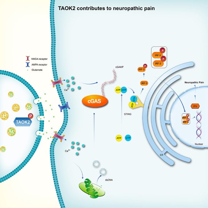
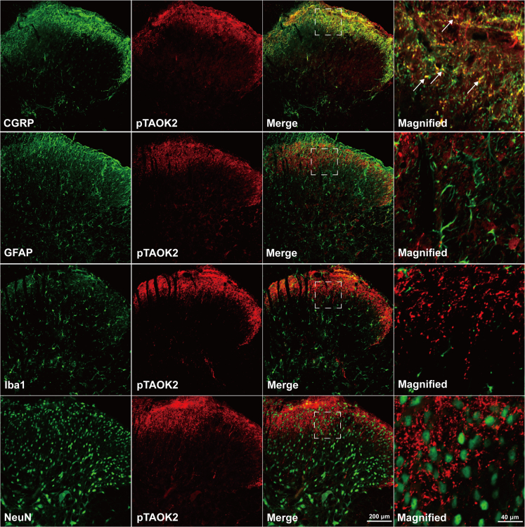
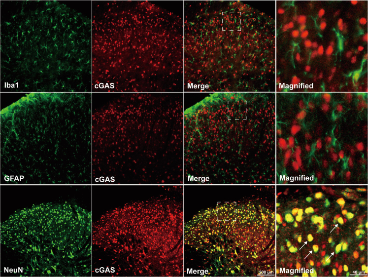
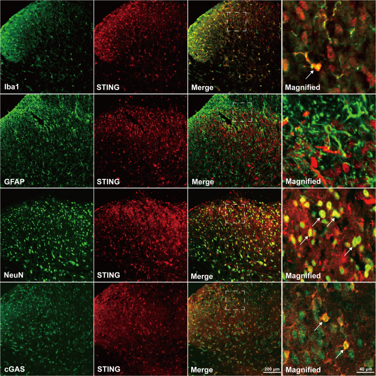
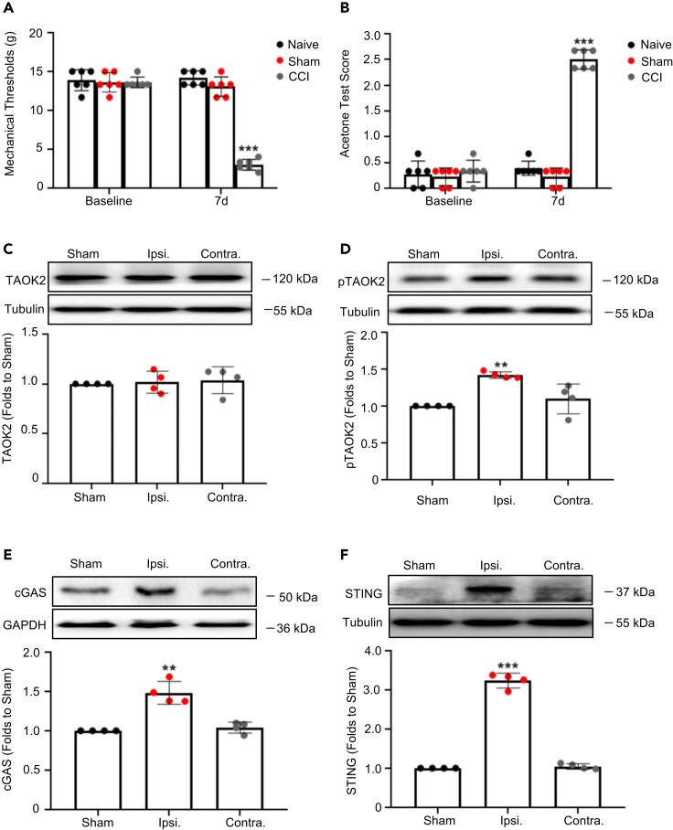
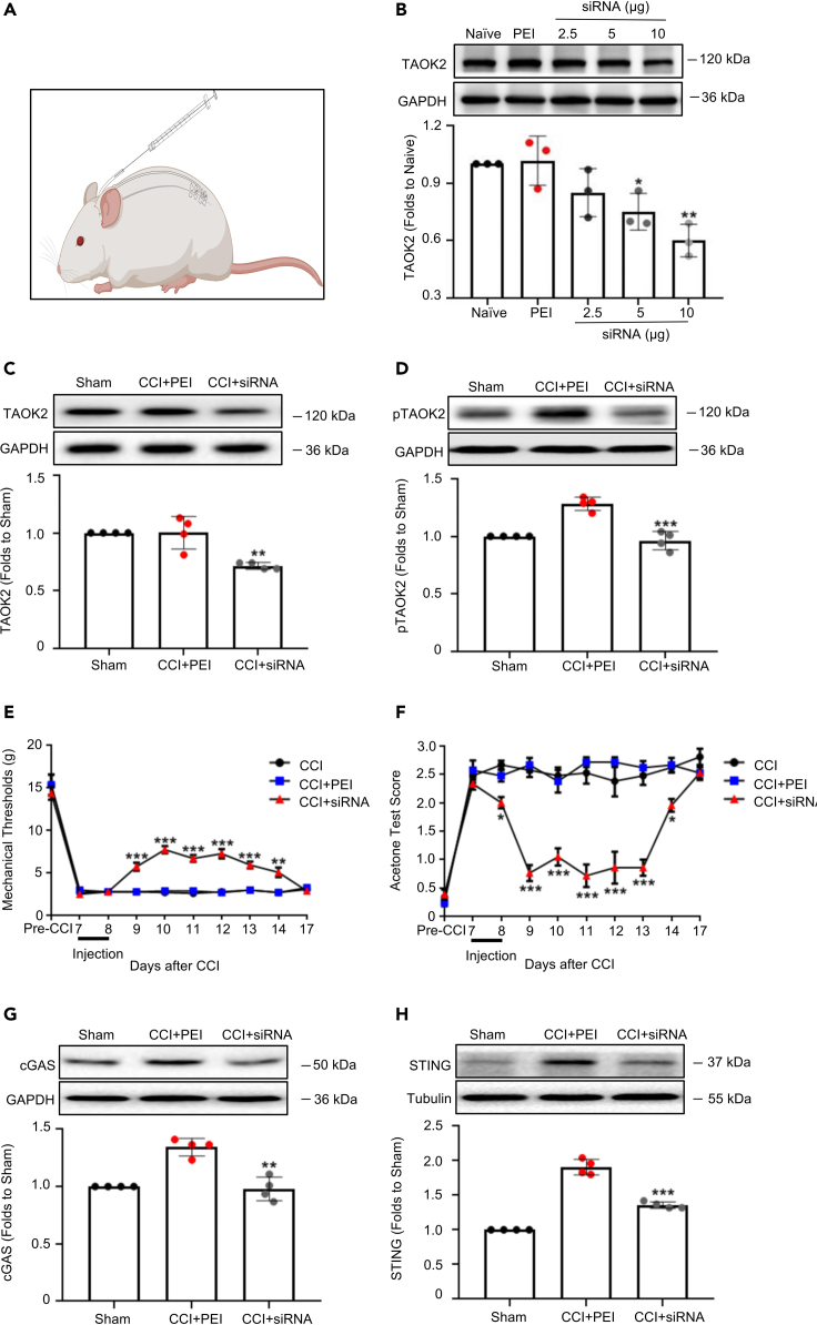
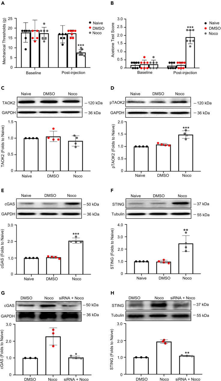
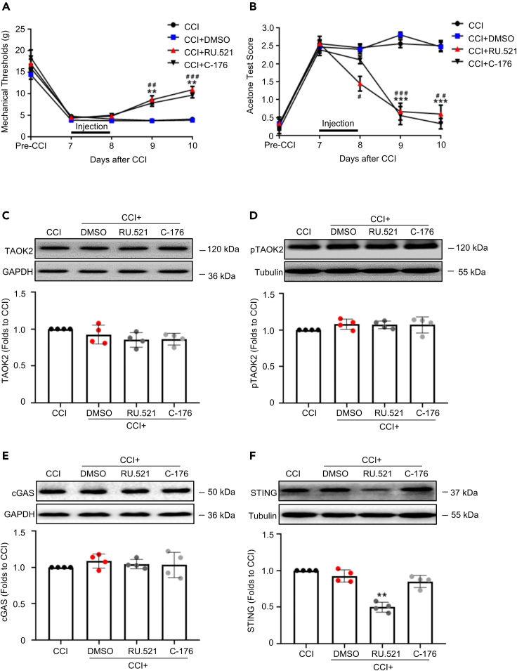
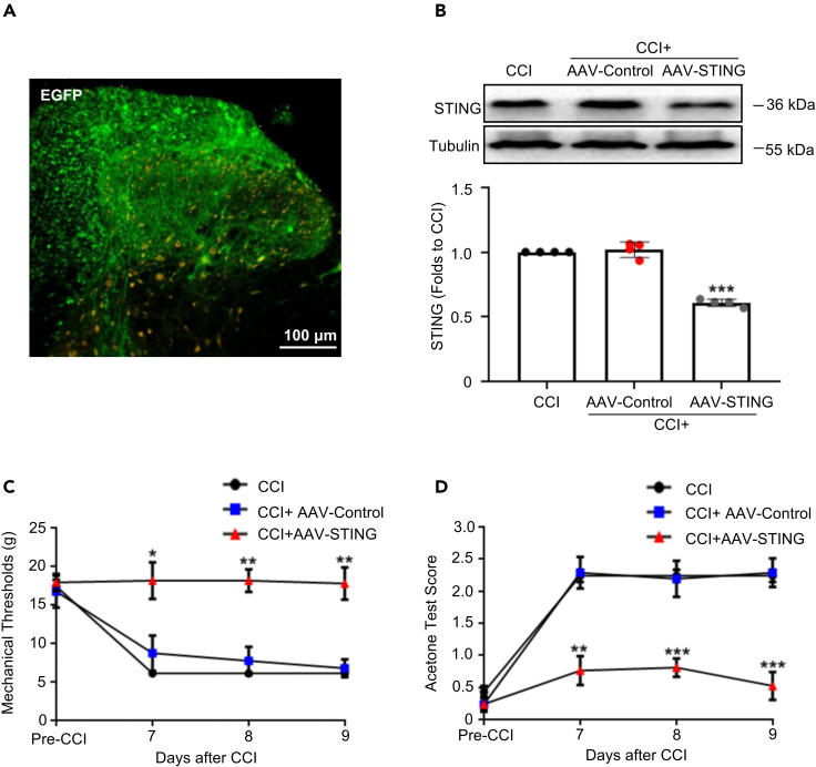
Thousand and one amino acid kinase 2 (TAOK2) is a member of the mammalian sterile 20 kinase family and is implicated in neurodevelopmental disorders; however, its role in neuropathic pain remains unknown. Here, we found that TAOK2 was enriched and activated after chronic constriction injury (CCI) in the rat spinal dorsal horn. Meanwhile, cyclic guanosine monophosphate-adenosine monophosphate synthase (cGAS)-stimulator of interferon genes (STING) signaling was also activated with hyperalgesia. Silencing TAOK2 reversed hyperalgesia and suppressed the activation of cGAS-STING signaling induced by CCI, while pharmacological activation of TAOK2 induced pain hypersensitivity and upregulation of cGAS-STING signaling in naive rats. Furthermore, pharmacological inhibition or gene silencing of cGAS-STING signaling attenuated CCI-induced hyperalgesia. Taken together, these data demonstrate that the activation of spinal TAOK2 contributes to CCI-induced hyperalgesia via cGAS-STING signaling activation, providing new molecular targets for the treatment of neuropathic pain.

Keywords: Cell biology; Molecular biology; Neuroscience.


Conflict of interest statement

The authors declare no competing interests.

Figures

None
Graphical abstract
Figure 1
Figure 1
The expression profile of TAOK2 in the spinal dorsal horn Double immunofluorescence staining shows colocalization of pTAOK2 with CGRP, but not with NeuN, GFAP, or Iba1. CGRP, calcitonin gene-related peptide; NeuN, neuron-specific nuclear protein; GFAP, glial fibrillary acidic protein; Iba1, ionized calcium-binding adaptor molecule-1. White arrows indicate colocalization.
Figure 2
Figure 2
Colocalization of cGAS with NeuN in the spinal dorsal horn Double immunofluorescence staining showed that cGAS colocalized with NeuN but not GFAP or Iba1. NeuN, neuron-specific nuclear protein; GFAP, glial fibrillary acidic protein; Iba1, ionized calcium-binding adaptor molecule-1. White arrows indicate colocalization.
Figure 3
Figure 3
Colocalization of STING with NeuN, Iba1, and cGAS in the spinal dorsal horn Double immunofluorescence staining showed that STING colocalized with NeuN and Iba1 but not GFAP. STING also colocalized with cGAS. NeuN, neuron-specific nuclear protein; GFAP, glial fibrillary acidic protein; Iba1, ionized calcium-binding adaptor molecule-1. White arrows indicate colocalization.
Figure 4
Figure 4
CCI-induced behavioral hyperalgesia and activation of TAOK2 and cGAS-STING signaling (A and B) The CCI group showed significantly lower MWT (A) and higher ATS (B) than did the sham group on the 7th day after surgery (∗∗∗p < 0.001, one-way ANOVA, n = 7). (C) Western blotting showed that, compared to that in the sham group, the total abundance of TAOK2 did not change in the CCI group. (D) Compared to that in the sham group, the phosphorylation level of TAOK2 was significantly increased in the ipsilateral spinal dorsal horn of the CCI group (∗∗p < 0.01, vs. sham, one-way ANOVA, n = 4). (E and F) Compared to that in the sham group, the expression of cGAS (E) and STING (F) was significantly upregulated in the ipsilateral spinal dorsal horn of the CCI group (∗∗p < 0.01, ∗∗∗p < 0.001, vs. Sham, one-way ANOVA, n = 4). Error bars represent the SEM.
Figure 5
Figure 5
Silencing TAOK2 reversed the behavioral hyperalgesia and suppressed the upregulation of cGAS-STING induced by CCI (A) A schematic diagram of the experiments. (B) Compared with that in the PEI group, the expression level of TAOK2 was significantly decreased in the siRNA (10 μg) group (∗∗p < 0.01, vs. PEI, one-way ANOVA, n = 3). (C and D) Compared to those in the CCI+PEI group, the total abundance of TAOK2 (C) and its phosphorylation level (D) were significantly decreased in the CCI+siRNA group (∗∗p < 0.01, ∗∗∗p < 0.001, vs. CCI+PEI, one-way ANOVA, n = 4). (E) Compared to that in the CCI+PEI group, the MWT significantly increased in the CCI+siRNA group from days 9 to 14 after CCI (∗∗p < 0.01, ∗∗∗p < 0.001, vs. CCI+PEI, two-way ANOVA, n = 7). (F) Compared to that in the CCI+PEI group, the ATS significantly decreased in the CCI+siRNA group from days 9 to 14 after CCI (∗p < 0.05, ∗∗∗p < 0.001, vs. CCI+PEI, two-way ANOVA, n = 7). (G and H) Compared to that in the CCI+PEI group, the expression of cGAS (G) and STING (H) was significantly downregulated in the CCI+siRNA group (∗∗p < 0.01, ∗∗∗p < 0.001, vs. CCI+PEI, one-way ANOVA, n = 4). Error bars represent the SEM.
Figure 6
Figure 6
Activation of TAOK2 induced behavioral hyperalgesia and activated cGAS-STING in naive rats (A and B) The nocodazole group showed significantly lower MWT (A) and higher ATS (B) after drug administration than did the DMSO group (∗∗∗p < 0.001, one-way ANOVA, n = 7). (C) The expression of TAOK2 showed no significant differences among the three groups. (D) Compared with that in the DMSO group, the phosphorylation level of TAOK2 significantly increased in the nocodazole group (∗∗∗p < 0.001, vs. DMSO, one-way ANOVA, n = 4). (E and F) Compared with that in the DMSO group, the expression of cGAS (E) and STING (F) was significantly upregulated in the nocodazole group (∗∗p < 0.01, ∗∗∗p < 0.001, vs. DMSO, one-way ANOVA, n = 4). (G and H) Moreover, compared to that in the nocodazole + PEI group, the expression of cGAS (G) and STING (H) was significantly decreased in the nocodazole + TAOK2-siRNA group (∗p < 0.05, ∗∗p < 0.01, vs. nocodazole + PEI, one-way ANOVA, n = 3). Error bars represent the SEM.
Figure 7
Figure 7
Inhibition of cGAS-STING reversed behavioral hyperalgesia induced by CCI (A) Compared with that in the CCI+DMSO group, the MWT significantly increased in the CCI+ RU.521 and CCI+C-176 groups on days 9 and 10 after CCI (∗∗p < 0.01, C-176 vs. DMSO; ##p < 0.01, ###p < 0.001, RU.521 vs. DMSO, two-way ANOVA, n = 7). (B) Compared with that in the CCI+DMSO group, the ATS significantly decreased in the CCI+ RU.521 and CCI+C-176 groups on days 8, 9, and 10 after CCI (∗∗∗p < 0.001, C-176 vs. DMSO; #p < 0.05, ##p < 0.01, ###p < 0.001, RU.521 vs. DMSO, two-way ANOVA, n = 7). (C and D) The expression of TAOK2 (C), phosphorylated TAOK2 (D), and cGAS (E) showed no significant differences in each group. (F) The expression of STING was significantly lower in the CCI+RU.521 group than in the CCI+DMSO group (∗∗p < 0.01, vs. DMSO, one-way ANOVA, n = 4). Error bars represent the SEM.
Figure 8
Figure 8
Knockdown of STING impeded the initiation of neuropathic pain after CCI (A) Enhanced green fluorescent protein was expressed in the spinal dorsal horn 3 weeks after transfection. (B) Compared to that in the CCI+AAV-Control group, the expression of STING significantly decreased in the CCI+AAV-STING group (∗∗∗p < 0.001, vs. CCI+AAV-Control, one-way ANOVA, n = 4). (C) Compared with that in the AAV-control group, the MWT significantly increased on days 7, 8, and 9 after CCI (∗p < 0.05, ∗∗p < 0.01, vs. CCI+AAV-Control, two-way ANOVA, n = 7). (D) Compared with that in the AAV-control group, the ATS significantly decreased on days 7, 8, and 9 after CCI (∗∗p < 0.01, ∗∗∗p < 0.001, vs. CCI+AAV-Control, two-way ANOVA, n = 7). Error bars represent the SEM. EGFP, enhanced green fluorescent protein.

Similar articles

Cited by

References

    1. Cohen S.P., Mao J. Neuropathic pain: mechanisms and their clinical implications. BMJ. 2014;348:f7656. doi: 10.1136/bmj.f7656. - DOI - PubMed
    1. Finnerup N.B., Haroutounian S., Kamerman P., Baron R., Bennett D.L.H., Bouhassira D., Cruccu G., Freeman R., Hansson P., Nurmikko T., et al. Neuropathic pain: an updated grading system for research and clinical practice. Pain. 2016;157:1599–1606. doi: 10.1097/j.pain.0000000000000492. - DOI - PMC - PubMed
    1. Cohen S.P., Vase L., Hooten W.M. Chronic pain: an update on burden, best practices, and new advances. Lancet. 2021;397:2082–2097. doi: 10.1016/S0140-6736(21)00393-7. - DOI - PubMed
    1. Ishikawa T., Murata K., Okuda H., Potapenko I., Hori K., Furuyama T., Yamamoto R., Ono M., Kato N., Fukazawa Y., Ozaki N. Pain-related neuronal ensembles in the primary somatosensory cortex contribute to hyperalgesia and anxiety. iScience. 2023;26 doi: 10.1016/j.isci.2023.106332. - DOI - PMC - PubMed
    1. Liang L.L., Yang J.L., Lü N., Gu X.Y., Zhang Y.Q., Zhao Z.Q. Synergetic analgesia of propentofylline and electroacupuncture by interrupting spinal glial function in rats. Neurochem. Res. 2010;35:1780–1786. doi: 10.1007/s11064-010-0244-x. - DOI - PubMed