. 2023 Aug 31:14:1109759.
doi: 10.3389/fimmu.2023.1109759. eCollection 2023.

Synthetic 5-amino-6-D-ribitylaminouracil paired with inflammatory stimuli facilitates MAIT cell expansion in vivo

Affiliations

Synthetic 5-amino-6-D-ribitylaminouracil paired with inflammatory stimuli facilitates MAIT cell expansion in vivo

Adam G Nelson et al. Front Immunol. .

Abstract

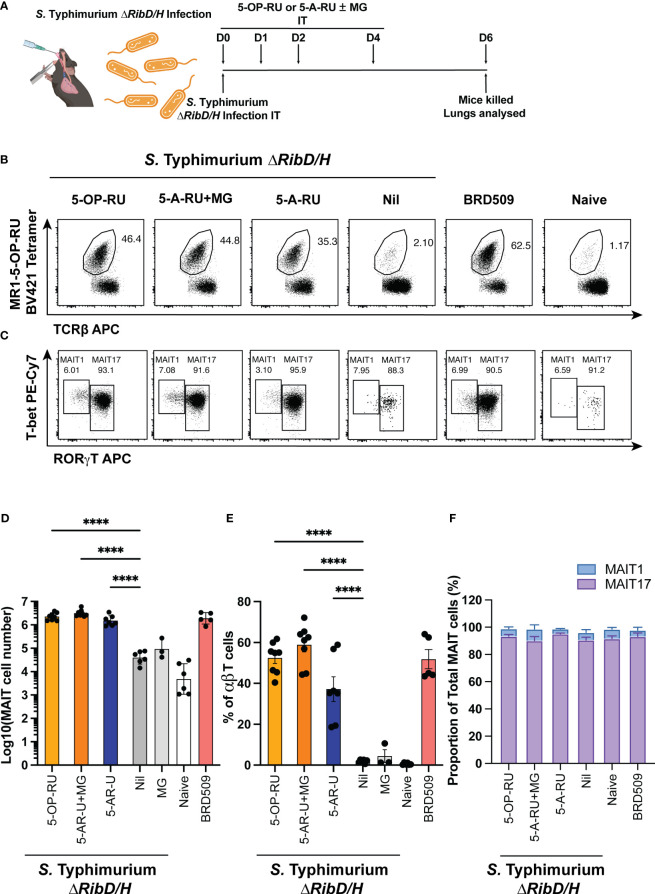
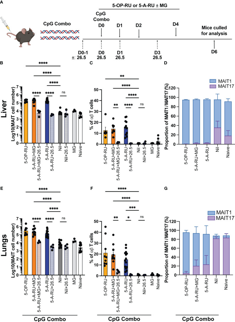
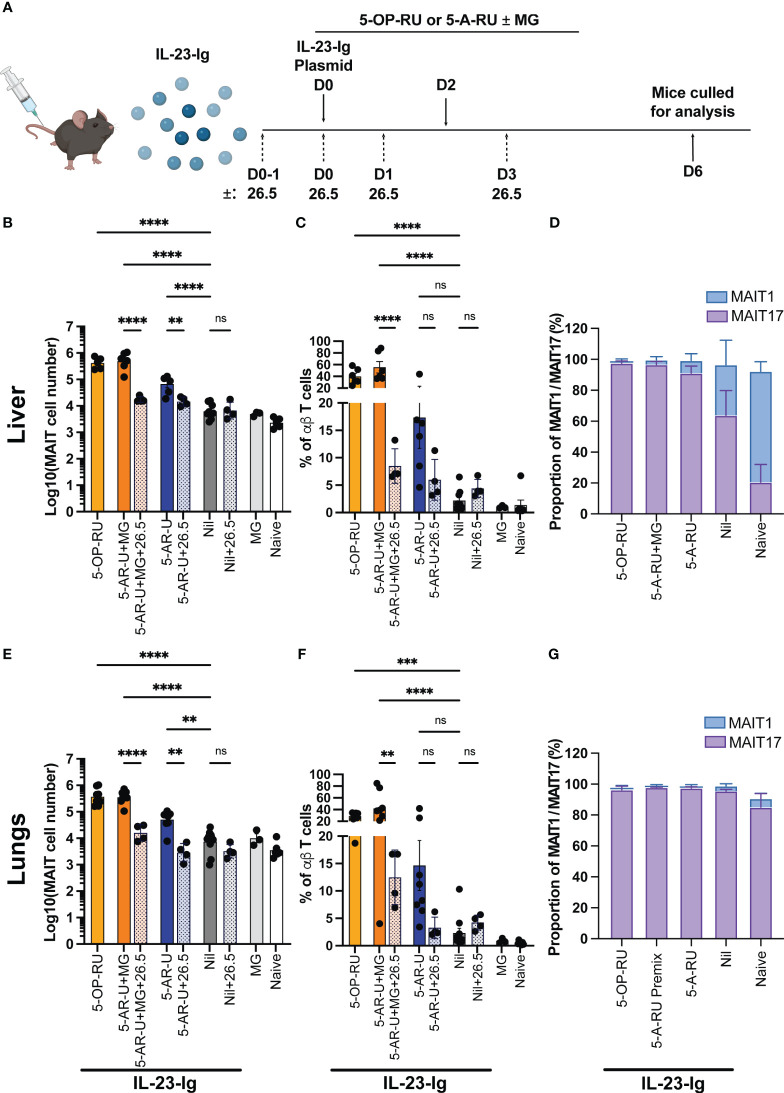
Introduction: Mucosal-associated invariant T (MAIT) cells are a population of innate-like T cells, which mediate host immunity to microbial infection by recognizing metabolite antigens derived from microbial riboflavin synthesis presented by the MHC-I-related protein 1 (MR1). Namely, the potent MAIT cell antigens, 5-(2-oxopropylideneamino)-6-D-ribitylaminouracil (5-OP-RU) and 5-(2-oxoethylideneamino)-6-D-ribitylaminouracil (5-OE-RU), form via the condensation of the riboflavin precursor 5-amino-6-D-ribitylaminouracil (5-A-RU) with the reactive carbonyl species (RCS) methylglyoxal (MG) and glyoxal (G), respectively. Although MAIT cells are abundant in humans, they are rare in mice, and increasing their abundance using expansion protocols with antigen and adjuvant has been shown to facilitate their study in mouse models of infection and disease.

Methods: Here, we outline three methods to increase the abundance of MAIT cells in C57BL/6 mice using a combination of inflammatory stimuli, 5-A-RU and MG.

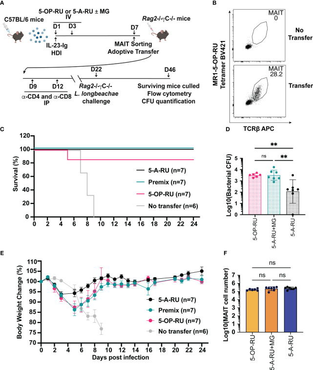
Results: Our data demonstrate that the administration of synthetic 5-A-RU in combination with one of three different inflammatory stimuli is sufficient to increase the frequency and absolute numbers of MAIT cells in C57BL/6 mice. The resultant boosted MAIT cells are functional and can provide protection against a lethal infection of Legionella longbeachae.

Conclusion: These results provide alternative methods for expanding MAIT cells with high doses of commercially available 5-A-RU (± MG) in the presence of various danger signals.

Keywords: 5-amino-6-D-ribitylaminouracil (5-A-RU); CpG; IL-23; MAIT cell boosting; MAIT cells; mouse model.


Conflict of interest statement

JM, DF, LK-N, JMc, SE, AC, and ZC are inventors of patents WO2014/005194 and WO2015/149130 describing MR1 tetramers and MR1 ligands. The remaining authors declare that the research was conducted in the absence of any commercial or financial relationships that could be construed as a potential conflict of interest.

Figures

Figure 1
Figure 1
MAIT cell accumulation in mice infected with riboflavin pathway impaired bacteria (S. Typhimurium ΔRibD/H) supplemented with synthetic 5-OP-RU or 5-A-RU±MG. (A) Schematic outlining infection of mice with S. Typhimurium ∆RibD/H, intratracheally (IT), and inoculation with four doses of either 5-OP-RU (50 pmol, 50 μL), 5-A-RU+MG (5-A-RU+MG: 32.5 nmol +110.5 nmol, 50 μL), 5-A-RU (5-A-RU: 32.5 nmol, 50 μL) or MG (110.5 nmol, 50 μL) on days 0 (D0), D1, D2 and D4, before harvest of lungs on D6. (B) Representative flow cytometry plots with gated MAIT cell frequency indicated and (C) representative flow cytometry plots with gated MAIT1 (T-bet high, RORγT low, left gate) and MAIT17 (T-bet high or low, RORγT high, right gate) frequencies. Bar graphs showing (D) absolute MAIT cell numbers, (E) MAIT cell frequency and (F) MAIT1 and MAIT17 as a proportion of total MAIT cells, from the lungs infected with 2 x 10⁷ CFU of S. Typhimurium ∆RibD/H and treated with four doses of either 5-OP-RU, 5-A-RU+MG 5-A-RU or MG IT on days 0, 1, 2 and 4; or infected with 2.5x10⁶ CFU S. Typhimurium BRD509 IT day 0, or naïve mice. Mice were killed and lungs were collected on day 6. Data show mean ± SEM and dots represent individual mice (n=3-8). Statistical significance is indicated by: **** (p<0.0001). One-way ANOVA with Tukey correction was performed on log-transformed data or percentage data. Data were pooled from two independent experiments.
Figure 2
Figure 2
MAIT cell accumulation in mice inoculated with TLR-9 agonist CpG combo and synthetic 5-OP-RU or 5-A-RU ± MG in an MR1-dependent manner. (A) Schematic outlining CpG combo vaccination strategy. Mice were intravenously (IV) administered CpG and either 5-OP-RU, 5-A-RU+MG, or 5-A-RU on D0, D1, D2 and D4. Anti-MR1 monoclonal antibody 26.5 was administered on D0-1, D0, D1 and D3. Mice were killed on D6 and organs harvested for analysis. Bar graphs showing absolute MAIT cell numbers, MAIT cell percentage of αβ T cell and MAIT1 and MAIT17 as a proportion of total MAIT cells for the: liver (B-D), or the lungs (E-G) of mice inoculated with 10 nmol of CpG combo and four doses of either 5-OP-RU (2 nmol, 200 μL), 5-A-RU+MG (5-A-RU+MG: 1.3 μmol +4.42 μmol, 200 μL), 5-A-RU (5-A-RU 1.3 μmol, 200 μL), MG (4.42 μmol, 200 μL) or PBS (200 μL) IV days 1, 2, 3, and 5 ± 4 doses of MR1 blocking monoclonal antibody 26.5 (250 mg, 200 μL). Data show mean ± SEM and dots represent individual mice (n=3-14). Statistical significance is indicated by ns (≥0.05) * (p<0.05), ** (p<0.01), *** (p<0.001), **** (p<0.0001). One-way ANOVA with Tukey correction was performed on log-transformed data or percentage data. Data were pooled from four independent experiments.
Figure 3
Figure 3
MAIT cell accumulation in mice inoculated with IL-23-Ig plasmid and synthetic 5-OP-RU or 5-A-RU±MG in an MR1-dependent manner (A) Schematic outlining IL-23-Ig vaccination strategy. Mice were administered IL-23-Ig plasmid DNA HDI and either 5-OP-RU, 5-A-RU+MG, or 5-A-RU D0, and D2, with or without MR1 blocking monoclonal antibody 26.5 administered D0-1, D0, D1 and D3. Mice were killed on D6 and organs harvested for analysis. Bar graphs showing absolute MAIT cell numbers, MAIT cell percentage of αβ T cell and MAIT1 and MAIT17 as a proportion of total MAIT cells for the: liver (B–D), or the lungs (E–G) of mice inoculated with two doses of either 5-OP-RU (200 pmol, 200 μL), 5-A-RU+MG (5-A-RU+MG: 130 nmol +442 nmol, 200 μL), 5-A-RU (5-A-RU 130 nmol, 200 μL), MG (442 nmol, 200 μL) or PBS (200 μL), IV days 0 and 2. Data show mean ± SEM and dots represent individual mice (n=3-6). Statistical significance is indicated by ns (≥0.05), ** (p<0.01), *** (p<0.001); **** (p<0.0001). One-way ANOVA with Tukey correction was performed on log-transformed data or percentage data. Mann Whitney U tests were performed between 5-A-RU and 5-A-RU+26.5 as well as 5-A-RU and nil groups in lung absolute number panel E. Data were pooled from three independent experiments.
Figure 4
Figure 4
MAIT cells derived from IL-23-Ig vaccination scheme protect Rag2-/-γc-/- mice from lethal challenge with L. longbeachae. (A) Schematic outlining vaccination schedule (all mice received 2 μg IL-23-Ig plasmid DNA via HDI and were inoculated with two doses of either 5-OP-RU (200 pmol, 200 μL), 5-A-RU+MG (5-A-RU+MG: 130 nmol +442 nmol, 200 μL) or 5-A-RU (5-A-RU 130 nmol, 200 μL)), adoptive transfer, conventional T cell depletion, and challenge with L. longbeachae. (B) Representative plots showing the MAIT cell profile of Rag2-/-γc-/- mice transferred with MAIT cells compared to a Rag2-/-γc-/- mouse with no transfer. (C) Survival curve and (D) Weight change (%) of Rag2-/-γc-/- mice transferred with 5-OP-RU, 5-A-RU+MG premix, 5-A-RU or without transfer, post-infection with L. longbeachae. (E) CFU of L. longbeachae and (F) MAIT cell numbers in the lungs of mice who survived bacterial challenge at day 24 post-infection. Data show mean ± SEM and dots represent individual mice (n=6-7). Statistical significance is indicated by ns (≥0.05) and ** (p<0.01). One-way ANOVA with Tukey correction was performed on log-transformed data or percentage data.

Similar articles

Cited by

References

    1. Eckle SB, Corbett AJ, Keller AN, Chen Z, Godfrey DI, Liu L, et al. Recognition of vitamin B precursors and byproducts by mucosal associated invariant T cells. J Biol Chem (2015) 290(51):30204–11. doi: 10.1074/jbc.R115.685990 - DOI - PMC - PubMed
    1. Corbett AJ, Eckle SBG, Birkinshaw RW, Liu L, Patel O, Mahony J, et al. T-cell activation by transitory neo-antigens derived from distinct microbial pathways. Nature (2014) 509(7500):361–5. doi: 10.1038/nature13160 - DOI - PubMed
    1. Mak JYW, Xu W, Reid RC, Corbett AJ, Meehan BS, Wang H, et al. Stabilizing short-lived Schiff base derivatives of 5-aminouracils that activate mucosal-associated invariant T cells. Nat Commun (2017) 8(1):14599. doi: 10.1038/ncomms14599 - DOI - PMC - PubMed
    1. Kjer-Nielsen L, Corbett AJ, Chen Z, Liu L, Mak JY, Godfrey DI, et al. An overview on the identification of MAIT cell antigens. Immunol Cell Biol (2018) 96(6):573–87. doi: 10.1111/imcb.12057 - DOI - PubMed
    1. Treiner E, Duban L, Bahram S, Radosavljevic M, Wanner V, Tilloy F, et al. Selection of evolutionarily conserved mucosal-associated invariant T cells by MR1. Nature (2003) 422(6928):164–9. doi: 10.1038/nature01433 - DOI - PubMed

Publication types