

Deep learning based clinico-radiological model for paediatric brain tumor detection and subtype prediction
- PMID: 37720352
- PMCID: PMC10501890
- DOI: 10.37349/etat.2023.00159
Deep learning based clinico-radiological model for paediatric brain tumor detection and subtype prediction
Abstract
Aim: Early diagnosis of paediatric brain tumors significantly improves the outcome. The aim is to study magnetic resonance imaging (MRI) features of paediatric brain tumors and to develop an automated segmentation (AS) tool which could segment and classify tumors using deep learning methods and compare with radiologist assessment.
Methods: This study included 94 cases, of which 75 were diagnosed cases of ependymoma, medulloblastoma, brainstem glioma, and pilocytic astrocytoma and 19 were normal MRI brain cases. The data was randomized into training data, 64 cases; test data, 21 cases and validation data, 9 cases to devise a deep learning algorithm to segment the paediatric brain tumor. The sensitivity, specificity, positive predictive value (PPV), negative predictive value (NPV), and accuracy of the deep learning model were compared with radiologist's findings. Performance evaluation of AS was done based on Dice score and Hausdorff95 distance.
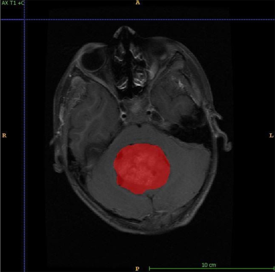
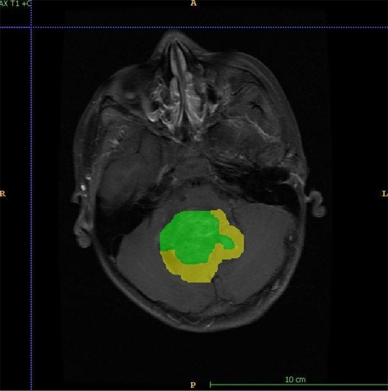
Results: Analysis of MRI semantic features was done with necrosis and haemorrhage as predicting features for ependymoma, diffusion restriction and cystic changes were predictors for medulloblastoma. The accuracy of detecting abnormalities was 90%, with a specificity of 100%. Further segmentation of the tumor into enhancing and non-enhancing components was done. The segmentation results for whole tumor (WT), enhancing tumor (ET), and non-enhancing tumor (NET) have been analyzed by Dice score and Hausdorff95 distance. The accuracy of prediction of all MRI features was compared with experienced radiologist's findings. Substantial agreement observed between the classification by model and the radiologist's given classification [K-0.695 (K is Cohen's kappa score for interrater reliability)].
Conclusions: The deep learning model had very high accuracy and specificity for predicting the magnetic resonance (MR) characteristics and close to 80% accuracy in predicting tumor type. This model can serve as a potential tool to make a timely and accurate diagnosis for radiologists not trained in neuroradiology.
Keywords: Deep learning model; artificial intelligence; brainstem glioma; ependymoma; medulloblastoma; paediatric brain tumors; pilocytic astrocytoma.
© The Author(s) 2023.
Conflict of interest statement
The authors declare that there is no conflict of interest.
Figures

References
-
- Lacayo A, Farmer PM. Brain tumors in children: a review. Ann Clin Lab Sci. 1991;21:26–35. - PubMed
LinkOut - more resources
Full Text Sources
Research Materials