

Cone beam computed tomography image guidance within a magnetic resonance imaging-only planning workflow
- PMID: 37720461
- PMCID: PMC10500022
- DOI: 10.1016/j.phro.2023.100472
Cone beam computed tomography image guidance within a magnetic resonance imaging-only planning workflow
Abstract
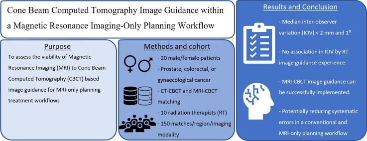
Background and purpose: Magnetic Resonance Imaging (MRI)-only planning workflows offer many advantages but raises challenges regarding image guidance. The study aimed to assess the viability of MRI to Cone Beam Computed Tomography (CBCT) based image guidance for MRI-only planning treatment workflows.
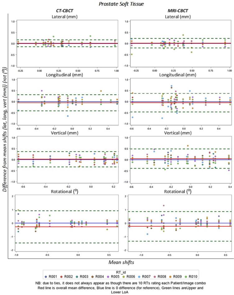
Materials and methods: An MRI matching training package was developed. Ten radiation therapists, with a range of clinical image guidance experience and experience with MRI, completed the training package prior to matching assessment. The matching assessment was performed on four match regions: prostate gold seed, prostate soft tissue, rectum/anal canal and gynaecological. Each match region consisted of five patients, with three CBCTs per patient, resulting in fifteen CBCTs for each match region. The ten radiation therapists performed the CBCT image matching to CT and to MRI for all regions and recorded the match values.
Results: The median inter-observer variation for MRI-CBCT matching and CT-CBCT matching for all regions were within 2 mm and 1 degree. There was no statistically significant association in the inter-observer variation in mean match values and radiation therapist image guidance experience levels. There was no statistically significant association in inter-observer variation in mean match values for MRI experience levels for prostate soft tissue and gynaecological match regions, while there was a statistically significant difference for prostate gold seed and rectum match regions.
Conclusion: The results of this study support the concept that with focussed training, an MRI to CBCT image guidance approach can be successfully implemented in a clinical planning workflow.
Keywords: Cone Beam Computed Tomography; Image-guided radiation therapy; MRI guided radiation therapy; MRI-only radiotherapy planning; Magnetic resonance imaging; Radiotherapy.
© 2023 The Authors.
Conflict of interest statement
The authors declare that they have no known competing financial interests or personal relationships that could have appeared to influence the work reported in this paper.
Figures
References
-
- Salembier C., Villeirs G., De Bari B., Hoskin P., Pieters B.R., Van Vulpen M., et al. ESTRO ACROP consensus guideline on CT- and MRI-based target volume delineation for primary radiation therapy of localized prostate cancer. Radiother Oncol. 2018;127:49–61. doi: 10.1016/j.radonc.2018.01.014. - DOI - PubMed
-
- Dowling J., O’Connor L., Acosta O., Raniga P., Crevoisierd R., Nunes J.C., et al. In: Biomedical Image Synthesis and Simulation: Methods and Applications. Burgos N., Svoboda D., editors. Academic Press; 2022. Image synthesis for MRI-only radiotherapy treatment planning. - DOI
LinkOut - more resources
Full Text Sources
