. 2023 Jun 1:27:100452.
doi: 10.1016/j.phro.2023.100452. eCollection 2023 Jul.

Automatically tracking brain metastases after stereotactic radiosurgery

Affiliations

Automatically tracking brain metastases after stereotactic radiosurgery

Dylan G Hsu et al. Phys Imaging Radiat Oncol. .

Abstract

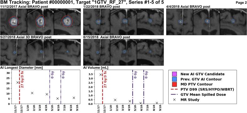
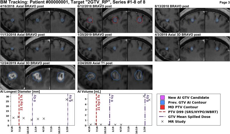
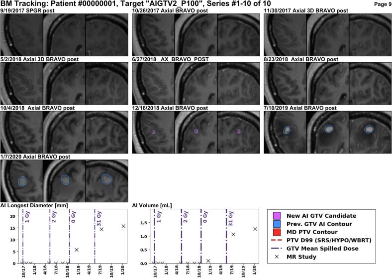
Background and purpose: Patients with brain metastases (BMs) are surviving longer and returning for multiple courses of stereotactic radiosurgery. BMs are monitored after radiation with follow-up magnetic resonance (MR) imaging every 2-3 months. This study investigated whether it is possible to automatically track BMs on longitudinal imaging and quantify the tumor response after radiotherapy.

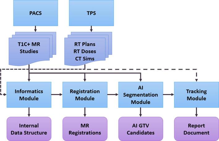
Methods: The METRO process (MEtastasis Tracking with Repeated Observations was developed to automatically process patient data and track BMs. A longitudinal intrapatient registration method for T1 MR post-Gd was conceived and validated on 20 patients. Detections and volumetric measurements of BMs were obtained from a deep learning model. BM tracking was validated on 32 separate patients by comparing results with manual measurements of BM response and radiologists' assessments of new BMs. Linear regression and residual analysis were used to assess accuracy in determining tumor response and size change.

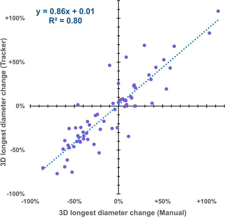
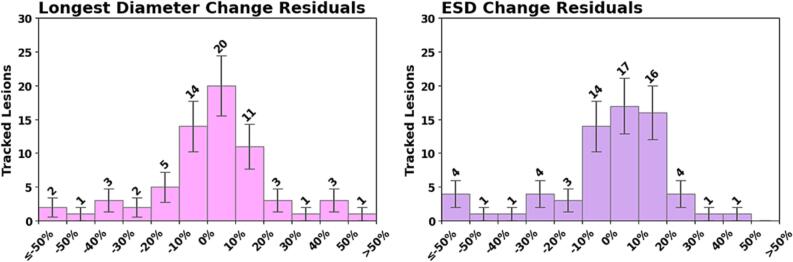
Results: A total of 123 irradiated BMs and 38 new BMs were successfully tracked. 66 irradiated BMs were visible on follow-up imaging 3-9 months after radiotherapy. Comparing their longest diameter changes measured manually vs. METRO, the Pearson correlation coefficient was 0.88 (p < 0.001); the mean residual error was -8 ± 17%. The mean registration error was 1.5 ± 0.2 mm.

Conclusions: Automatic, longitudinal tracking of BMs using deep learning methods is feasible. In particular, the software system METRO fulfills a need to automatically track and quantify volumetric changes of BMs prior to, and in response to, radiation therapy.

Keywords: Brain metastases; Deep learning; Image registration; Longitudinal tumor tracking; Stereotactic radiosurgery; T1 MR post-Gd.


Conflict of interest statement

The authors declare that they have no known competing financial interests or personal relationships that could have appeared to influence the work reported in this paper.

Figures

Fig. 1
Fig. 1
Schematic showing an overview of the METRO software. Planning and follow-up MR scans are obtained from the PACS. Treatment plans with calculated dose and CT scans are obtained from the TPS.
Fig. 2
Fig. 2
Linear regression of size response for treated BMs that were still visible on follow-up, as measured automatically by METRO versus manually. One outlier with diameter change greater than + 200% is suppressed.
Fig. 3
Fig. 3
Distributions of residual errors for the size changes measured by automatic METRO method for the BMs still visible at follow-up. The residual is the difference of the percent change measured with METRO minus that measured manually. Left: 3D longest diameter. Right: equivalent sphere diameter.
Fig. 4
Fig. 4
This 2 cm enhancing lesion in the right frontal lobe was treated with 27 Gy in three fractions using the MR scan from 11/12/2017 for planning. The planning target volume (PTV) is shown in red, and the longitudinal AI segmentations in blue. The lesion size is decreasing at all follow-up MR images with the last MR from 8/15/2018. The METRO report shows both the 3D longest diameter and the volume based on the AI segmentation at each MR. (For interpretation of the references to colour in this figure legend, the reader is referred to the web version of this article.)
Fig. 5
Fig. 5
A lesion in the right parietal lobe is targeted with single-fraction SRS using the MR from 6/12/2018 for treatment planning. There is not much size change in the first nine months after treatment, but a year and a half after treatment, at the MR scan from 12/24/2019, the lesion size is increasing. The increase in size must be evaluated for potential progression or necrosis.
Fig. 6
Fig. 6
This patient received three courses of SRS elsewhere in the brain, followed by WBRT (Rx 30 Gy). Then, the METRO workflow identified a new lesion in the right parietal lobe at the 7/10/2019 MR, which is increasing in size at subsequent follow-up on 1/7/2020.

References

    1. Lamba N., Wen P.Y., Aizer A.A. Epidemiology of brain metastases and leptomeningeal disease. Neuro Oncol. 2021;9:1447–1456. doi: 10.1093/neuonc/noab101. - DOI - PMC - PubMed
    1. Frisk G., Svensson T., Bäcklund L.M., Lidbrink E., Blomqvist P., Smedby K.E. Incidence and time trends of brain metastases admissions among breast cancer patients in Sweden. Br J Cancer. 2012;11:1850–1853. doi: 10.1038/bjc.2012.163. - DOI - PMC - PubMed
    1. Nayak L., Lee E.Q., Wen P.Y. Epidemiology of brain metastases. Curr Oncol Rep. 2012;1:48–54. doi: 10.1007/s11912-011-0203-y. - DOI - PubMed
    1. Steeg P.S., Camphausen K.A., Smith Q.R. Brain metastases as preventive and therapeutic targets. Nat Rev Cancer. 2011;5:352–363. doi: 10.1038/nrc3053. - DOI - PMC - PubMed
    1. Brown P.D., Ahluwalia M.S., Khan O.H., Asher A.L., Wefel J.S., Gondi V. Whole-Brain Radiotherapy for Brain Metastases: Evolution or Revolution? J Clin Oncol. 2018;5:483–491. doi: 10.1200/jco.2017.75.9589. - DOI - PMC - PubMed

LinkOut - more resources