

Advances in clinical examination of lacrimal gland
- PMID: 37720501
- PMCID: PMC10501785
- DOI: 10.3389/fmed.2023.1257209
Advances in clinical examination of lacrimal gland
Abstract
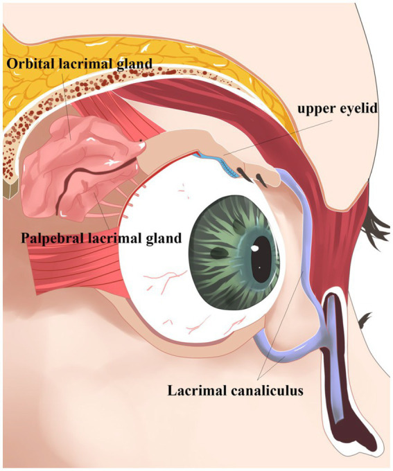
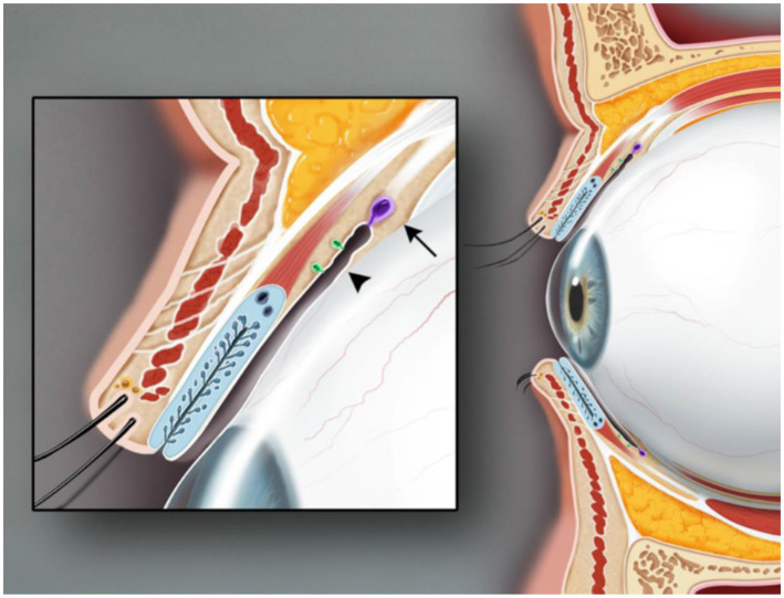
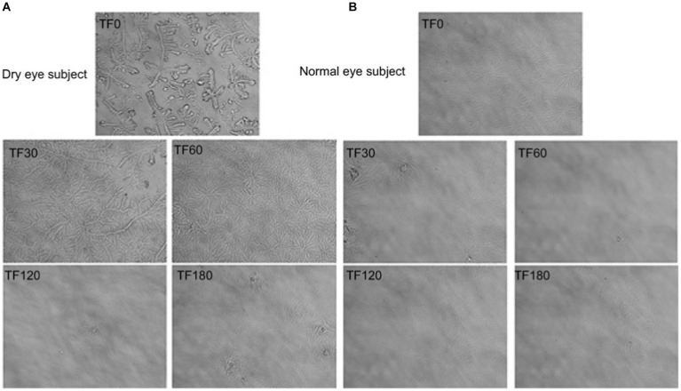
In humans, the lacrimal gland is located in the socket of the frontal bone above the outer orbital area. As an essential part of the eye surface, the gland is fixed to the orbital periosteum by connective tissue. The lacrimal gland passes through the outer tendon membrane, which divides the gland into larger orbital and minor eyelid glands. The lacrimal glands are the main contributors to tear film. They secrete electrolytes, proteins, and water to help nourish and protect the eye's surface. Furthermore, clinically, lacrimal glands are associated with a variety of inflammatory reactions and immune factors and are also vulnerable sites for tumors. Changes in tear gland morphology or secretory function affect tear film stability and tear secretion quality. Various technological devices have been developed and applied to lacrimal glands. This article systematically reviewed the clinical examination of the lacrimal gland to help inform personalized strategies for the diagnosis of lacrimal gland-related diseases.
Keywords: advance; clinical examination; function; lacrimal gland; morphology; review.
Copyright © 2023 Lin, Zhang, Shi, Wu and Ou.
Conflict of interest statement
The authors declare that the research was conducted in the absence of any commercial or financial relationships that could be construed as a potential conflict of interest.
Figures
Similar articles
-
Advances in lacrimal gland organoid development: Techniques and therapeutic applications.Biomed Pharmacother. 2025 Feb;183:117870. doi: 10.1016/j.biopha.2025.117870. Epub 2025 Jan 26. Biomed Pharmacother. 2025. PMID: 39870025 Review.
-
Lacrimal Gland Insufficiency in Aqueous Deficiency Dry Eye Disease: Recent Advances in Pathogenesis, Diagnosis, and Treatment.Semin Ophthalmol. 2022 Oct-Nov;37(7-8):801-812. doi: 10.1080/08820538.2022.2075706. Epub 2022 May 19. Semin Ophthalmol. 2022. PMID: 35587465 Review.
-
New insight into lacrimal gland function: Role of the duct epithelium in tear secretion.Ocul Surf. 2020 Oct;18(4):595-603. doi: 10.1016/j.jtos.2020.07.002. Epub 2020 Jul 21. Ocul Surf. 2020. PMID: 32707335 Review.
-
Age-related changes in morphology and secretory responses of male rat lacrimal gland.J Auton Nerv Syst. 1998 Apr 30;69(2-3):173-83. doi: 10.1016/s0165-1838(98)00026-5. J Auton Nerv Syst. 1998. PMID: 9696274
-
Is the main lacrimal gland indispensable? Contributions of the corneal and conjunctival epithelia.Surv Ophthalmol. 2016 Sep-Oct;61(5):616-27. doi: 10.1016/j.survophthal.2016.02.006. Epub 2016 Mar 9. Surv Ophthalmol. 2016. PMID: 26968256 Review.
Cited by
-
Epidemiology and tumor microenvironment of ocular surface and orbital tumors on growth and malignant transformation.Front Oncol. 2024 Oct 3;14:1388156. doi: 10.3389/fonc.2024.1388156. eCollection 2024. Front Oncol. 2024. PMID: 39421442 Free PMC article. Review.
-
Effect of a Nano-Sized Lipid-Based Eye Drop on Diabetic Dry Eye.Biomedicines. 2025 Mar 21;13(4):763. doi: 10.3390/biomedicines13040763. Biomedicines. 2025. PMID: 40299360 Free PMC article.
-
Wearable Electrochemical Glucose Sensors for Fluid Monitoring: Advances and Challenges in Non-Invasive and Minimally Invasive Technologies.Biosensors (Basel). 2025 May 12;15(5):309. doi: 10.3390/bios15050309. Biosensors (Basel). 2025. PMID: 40422047 Free PMC article. Review.
References
Publication types
LinkOut - more resources
Full Text Sources
Research Materials