. 2024 Mar;19(3):687-696.
doi: 10.4103/1673-5374.380905.

Activation of G-protein-coupled receptor 39 reduces neuropathic pain in a rat model

Affiliations

Activation of G-protein-coupled receptor 39 reduces neuropathic pain in a rat model

Longqing Zhang et al. Neural Regen Res. 2024 Mar.

Abstract

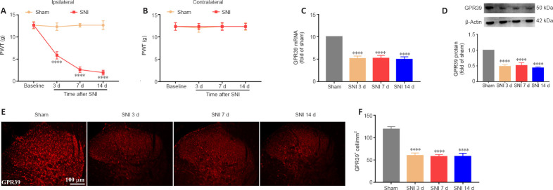
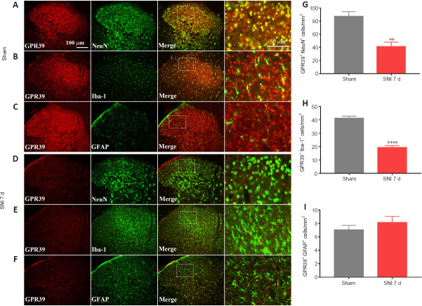
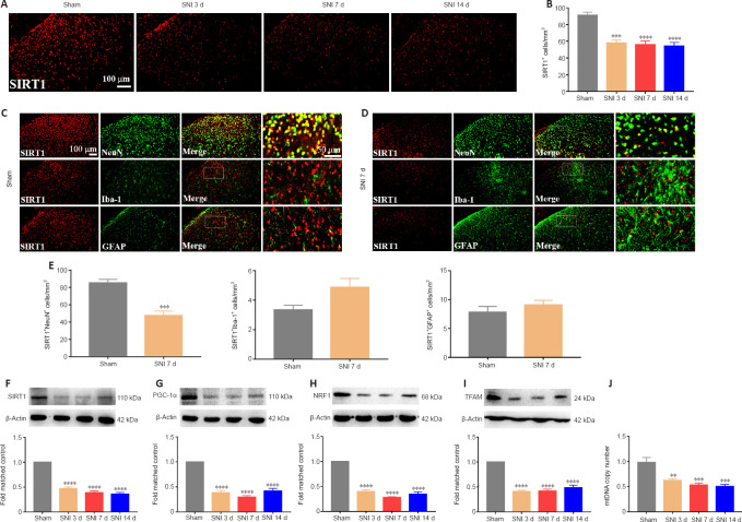
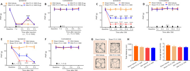
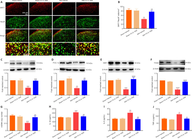
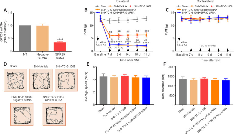
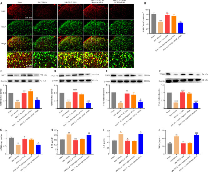
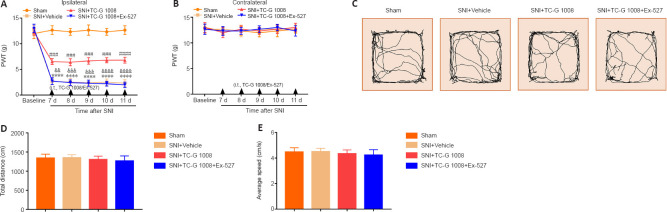
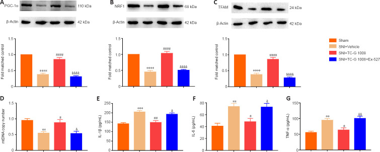
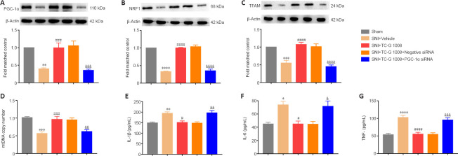
Activated G-protein-coupled receptor 39 (GPR39) has been shown to attenuate inflammation by interacting with sirtuin 1 (SIRT1) and peroxisome proliferator-activated receptor-γ coactivator 1α (PGC-1α). However, whether GPR39 attenuates neuropathic pain remains unclear. In this study, we established a Sprague-Dawley rat model of spared nerve injury-induced neuropathic pain and found that GPR39 expression was significantly decreased in neurons and microglia in the spinal dorsal horn compared with sham-operated rats. Intrathecal injection of TC-G 1008, a specific agonist of GPR39, significantly alleviated mechanical allodynia in the rats with spared nerve injury, improved spinal cord mitochondrial biogenesis, and alleviated neuroinflammation. These changes were abolished by GPR39 small interfering RNA (siRNA), Ex-527 (SIRT1 inhibitor), and PGC-1α siRNA. Taken together, these findings show that GPR39 activation ameliorates mechanical allodynia by activating the SIRT1/PGC-1α pathway in rats with spared nerve injury.

Keywords: G-protein-coupled receptor 39 (GPR39); mitochondrial transcription factor A (TFAM); neuroinflammation; neuropathic pain; nuclear respiratory factor 1 (NRF1); peroxisome proliferator-activated receptor-γcoactivator 1α(PGC-1α); sirtuin 1 (SIRT1); spinal cord.


Conflict of interest statement

None

Figures

Figure 1
Figure 1
SNI decreases PWT and lowers GPR39 expression in the spine. (A, B) SNI significantly reduced ipsilateral PWT, but not contralateral PWT, after nerve injury. (C, D) qPCR and WB results indicated that spinal GPR39 expression was down-regulated post-SNI. (E, F) IF staining results showed that GPR39 immunopositivity (red, CoraLite594) was decreased in the spinal dorsal horn of rats with SNI. Scale bars: 100 μm. All data are presented as the mean ± SEM (n = 5 rats/group). ****P < 0.0001, vs. sham group (A, B: two-way analysis of variance followed by Bonferroni’s post hoc test; C–F: one-way analysis of variance followed by Bonferroni’s post hoc test). SNI: Spared nerve injury; d: day; GPR39: G-protein-coupled receptor 39; qPCR: quantitative polymerase chain reaction; WB: western blotting; IF: immunofluorescence.
Figure 2
Figure 2
GPR39 localization in the spinal cord. (A–F) Representative immunostaining images of GPR39 (red, CoraLite594) expression in neurons (NeuN, green, CoraLite488), microglia (Iba-1, green, Fluorescein IsoThioCyanate), and astrocytes (GFAP, green, CoraLite488). GPR39 immunopositivity in neurons and microglia was decreased in the spinal dorsal horn of rats with SNI compared with sham rats. Scale bars: 100 μm, 50 μm (enlarged images). (G–I) Histograms demonstrating that GPR39 expression in neurons and microglia was decreased in rats with SNI. All data are presented as the mean ± SEM (n = 5 rats/group). **P < 0.01, ****P < 0.0001, vs. sham group (unpaired t-test). d: Day; GFAP: glial fibrillary acidic protein; GPR39: G-protein-coupled receptor 39; Iba-1: ionized calcium-binding adapter molecule 1; NeuN: neuronal nuclei; SNI: spared nerve injury.
Figure 3
Figure 3
Spared nerve injury inactivates the SIRT1/PGC-1α pathway and impairs mitochondrial biogenesis. (A, B) IF staining indicated that spinal SIRT1 (red, CoraLite594) expression was reduced post-SNI. Scale bars: 100 μm. (C, D) Representative immunostaining images of SIRT1 (red, CoraLite594) expression in neurons (NeuN, green, CoraLite488), microglia (Iba-1, green, Fluorescein IsoThioCyanate), and astrocytes (GFAP, green, CoraLite488). SIRT1 immunopositivity in neurons was decreased in the spinal dorsal horn of rats with SNI compared with sham rats. Scale bars: 100 μm, 50 μm (enlarged images). (E) SNI reduced SIRT1 expression in neurons. (F–I) SIRT1, PGC-1α, NRF1, and TFAM protein expression levels were decreased following SNI. (J) Mitochondrial biogenesis was impaired post-nerve injury. All data are presented as the mean ± SEM (n = 5 rats/group). **P < 0.01, ***P < 0.001, ****P < 0.0001, vs. sham group (A, B, F–J: one-way analysis of variance followed by Bonferroni’s post hoc test; C–E: unpaired t-test). d: Day; GFAP: glial fibrillary acidic protein; Iba-1: ionized calcium-binding adapter molecule 1; IF: immunofluorescence; mtDNA: mitochondrial DNA; NeuN: neuronal nuclei; NRF1: nuclear respiratory factor 1; PGC-1α: peroxisome proliferator-activated receptor-γ coactivator 1α; SIRT1: sirtuin 1; SNI: spared nerve injury; TFAM: mitochondrial transcription factor A.
Figure 4
Figure 4
Analgesic effect of TC-G 1008 on neuropathic pain in rats with SNI. (A, B) A single injection with TC-G 1008 (20 and 50 μg) reversed the decrease in ipsilateral PWT at 4 hours post-injection and did not affect the contralateral PWT. (C, D) Continuous intrathecal injection (i.t.) of TC-G 1008 (20 μg) improved SNI-induced mechanical allodynia and did not affect the contralateral PWT. (E, F) Early treatment of TC-G 1008 (20 μg) did not reduce the progresss of neuropathic pain. (G–I) Rat locomotor activity was not affected by TC-G 1008. All data are presented as the mean ± SEM (n = 5 rats/group). ****P < 0.0001, vs. sham + vehicle group; #P < 0.05, ##P < 0.01, ###P < 0.001, NRGR-19-687-g007.jpgP < 0.0001, vs. SNI + vehicle group (one- (G–I) or two-way (A–F) analysis of variance followed by Bonferroni’s post hoc test). PWT: Paw withdrawal threshold; SNI: spared nerve injury.
Figure 5
Figure 5
TC-G 1008 reverses neuropathic pain-induced impairment of mitochondrial biogenesis and neuroinflammation by activating the SIRT1/PGC-1α pathway. (A, B) TC-G 1008 elevated SIRT1 (red, CoraLite594) expression in neurons (NeuN, green, CoraLite488) post–nerve injury. Scale bars: 100 μm, 50 μm (enlarged images). (C–F) TC-G 1008 reversed the decrease in SIRT1/PGC-1α/NRF1/TFAM expression in the spinal cord induced by SNI. (G) TC-G 1008 reversed the reduction in mtDNA copy number caused by SNI. (H–J) TC-G 1008 lessened neuroinflammation in rats with nerve injury. All data are presented as the mean ± SEM (n = 5 rats/group). *P < 0.01, **P < 0.01, ***P < 0.001, ****P < 0.0001, vs. sham + vehicle group; #P < 0.05, ##P < 0.01, ###P < 0.001, ####P < 0.0001, vs. SNI + vehicle group (one-way analysis of variance followed by Bonferroni’s post hoc test). IL-1β: Interleukin-1β; IL-6: interleukin-6; mtDNA: mitochondrial DNA; NeuN: neuronal nuclei; NRF1: nuclear respiratory factor 1; SIRT1: sirtuin 1; PGC-1α: peroxisome proliferator-activated receptor-γ coactivator 1α; SNI: spared nerve injury; TFAM: mitochondrial transcription factor A; TNF-α: tumor necrosis factor-α.
Figure 6
Figure 6
TC-G 1008-induced improvement in mechanical allodynia is reversed by intrathecal injection with GPR39 siRNA. (A) GPR39 siRNA knocked down GPR39 expression (****P < 0.0001, vs. NT group). (B, C) GPR39 siRNA abrogated the analgesic effect of TC-G 1008 in rats with SNI (****P < 0.0001, vs. sham group; ##P < 0.01, ###P < 0.001, vs. SNI + vehicle group; &P < 0.05, &&P < 0.01, vs. SNI + TC-G 1008 group). (D–F) The OFT results indicated that GPR39 siRNA did not affect rat locomotor activity (P > 0.05). All data are presented as the mean ± SEM (n = 5 rats/group), and were analyzed by one- (A, D–F) or two-way (B and C) analysis of variance followed by Bonferroni’s post hoc test. d: Day; GRP39: G-protein-coupled receptor 39; i.t.: intrathecal injection; OFT: open field test; PWT: paw withdrawal threshold; siRNA: small interfering RNA; SNI: spared nerve injury.
Figure 7
Figure 7
Beneficial effects of TC-G 1008 on mitochondrial biogenesis and neuroinflammation are abrogated by intrathecal injection with GPR39 siRNA in rats with SNI. (A, B) TC-G 1008 up-regulated SIRT1 (red, CoraLite594) expression in neurons (NeuN, green, CoraLite488) in the spinal cord of rats with SNI, whereas GPR39 siRNA reversed this effect. Scale bars: 100 μm, 50 μm (enlarged images). (C–F) TC-G 1008 reversed the decrease in spinal SIRT1, PGC-1α, NRF1, and TFAM expression caused by SNI and administration of GPR39 siRNA reversed this effect. (G) GPR39 siRNA reversed the increase in mtDNA copy number induced by TC-G 1008 in rats with SNI. (H–J) GPR39 siRNA dampened the anti-inflammatory effect of TC-G 1008 in rats with SNI. All data are presented as the mean ± SEM (n = 5 rats/group). *P < 0.05, **P < 0.01, ***P < 0.001, ****P < 0.0001, vs. sham group; #P < 0.05, ##P < 0.01, ###P < 0.001, ####P < 0.0001, vs. SNI + vehicle group; &P < 0.05, &&P < 0.01, &&&P < 0.001, &&&&P < 0.0001, vs. SNI + TC-G 1008 group (one-way analysis of variance followed by Bonferroni’s post hoc test). GRP39: G-protein-coupled receptor 39; IL-1β: interleukin-1β; IL-6: interleukin-6; mtDNA: mitochondrial DNA; NeuN: neuronal nuclei; NRF1: nuclear respiratory factor 1; PGC-1α: peroxisome proliferator-activated receptor-γ coactivator 1α; siRNA: small interfering RNA; SIRT1: sirtuin 1; SNI: spared nerve injury; TFAM: mitochondrial transcription factor A; TNF-α: tumor necrosis factor-α.
Figure 8
Figure 8
A SIRT1 antagonist reverses the TC-G 1008-induced improvement in neuropathic pain in rats after nerve injury. (A, B) Ex-527 abolished the analgesic effect of TC-G 1008 in rats with SNI. (C–E) The OFT results showed that rat locomotor activity was not affected by Ex-527. All data are presented as the mean ± SEM (n = 5 rats/group). ****P < 0.0001, vs. sham group; ###P < 0.001, ####P < 0.0001, vs. SNI + vehicle group; &&P < 0.01, &&&P < 0.001, &&&&P < 0.0001, vs. SNI + TC-G 1008 group (A, B: two-way analysis of variance followed by Bonferroni’s post hoc test; C–E: one-way analysis of variance followed by Bonferroni’s post hoc test). d: Day; i.t.: intrathecal injection; OFT: open field test; PWT: paw withdrawal threshold; SNI: spared nerve injury.
Figure 9
Figure 9
Ex-527 abrogates the beneficial effects of TC-G 1008 on mitochondrial biogenesis and neuroinflammation in rats with SNI. (A–C) TC-G 1008 reversed the decrease in PGC-1α, NRF1, and TFAM expression in rats induced by nerve injury, but Ex-527 abrogated this effect. (D) Ex-527 reversed the increase in mtDNA copy number induced by TC-G 1008 in rats with SNI. (E–G) Ex-527 abrogated the anti-inflammatory effect of TC-G 1008 in rats with nerve injury. All data are presented as the mean ± SEM (n = 5 rats/group). **P < 0.01, ***P < 0.001, ****P < 0.0001, vs. sham group; #P < 0.05, ##P < 0.01, ####P < 0.0001, vs. SNI + vehicle group; &P < 0.05, &&P < 0.01, &&&&P < 0.0001, vs. SNI + TC-G 1008 group (one-way analysis of variance followed by Bonferroni’s post hoc test). IL-1β: Interleukin-1β; IL-6: interleukin-6; mtDNA: mitochondrial DNA; NRF1: nuclear respiratory factor 1; PGC-1α: peroxisome proliferator-activated receptor-γ coactivator 1α; SNI: spared nerve injury; TFAM: mitochondrial transcription factor A; TNF-α: tumor necrosis factor-α.
Figure 10
Figure 10
Beneficial effects of TC-G 1008 on mechanical allodynia are reversed by PGC-1α siRNA in rats with SNI. (A) PGC-1α siRNA knocked down PGC-1α expression (****P < 0.0001, vs. NT group, n = 5 rats/group). (B, C) PGC-1α siRNA abolished the analgesic effect of TC-G 1008 in rats with SNI (****P < 0.0001, vs. sham group; ##P < 0.01, ###P < 0.001, ####P < 0.0001, vs. SNI + vehicle group; &&P < 0.01, &&&P < 0.001, vs. SNI + TC-G 1008 group). (D–F) The OFT results indicated that rat locomotor activity was not affected by PGC-1α siRNA (P > 0.05). All data are presented as the mean ± SEM (n = 5 rats/group), and were analyzed by one (A, D–F) or two-way (B and C) analysis of variance followed by Bonferroni’s post hoc test. i.t.: Intrathecal injection; OFT: open field test; PGC-1α: peroxisome proliferator-activated receptor-γ coactivator 1α; PWT: paw withdrawal threshold; siRNA: small interfering RNA; SNI: spared nerve injury.
Figure 11
Figure 11
PGC-1α siRNA inhibits the beneficial effects of TC-G 1008 on mitochondrial biogenesis and neuroinflammation in rats with SNI. (A–C) The increase in PGC-1α, NRF1, and TFAM protein levels induced by TC-G 1008 was abolished by PGC-1α siRNA in rats with nerve injury. (D) PGC-1α siRNA reversed the increase in mtDNA copy number induced by TC-G 1008 in rats with SNI. (E–G) TC-G 1008-induced improvement in neuroinflammation in rats with SNI was abrogated by PGC-1α siRNA. All data are presented as the mean ± SEM (n = 5 rats/group). *P < 0.05, **P < 0.01, ***P < 0.001, ****P < 0.0001, vs. sham group; #P < 0.05, ###P < 0.001, ####P < 0.0001, vs. SNI + vehicle group; &P < 0.05, &&P < 0.01, &&&P < 0.001, &&&&P < 0.0001, vs. SNI + TC-G 1008 group (one-way analysis of variance followed by Bonferroni’s post hoc test). IL-1β: Interleukin-1β; IL-6: interleukin-6; mtDNA: mitochondrial DNA; NRF1: nuclear respiratory factor 1; PGC-1α: peroxisome proliferator-activated receptor-γ coactivator 1α; siRNA: small interfering RNA; SNI: spared nerve injury; TFAM: mitochondrial transcription factor A; TNF-α: tumor necrosis factor-α.

Similar articles

Cited by

References

    1. Bădescu L, Bădulescu O, Ciocoiu M, Bădescu M. Modulation of neuropathic pain in experimental diabetes mellitus. J Physiol Biochem. 2014;70:355–361. - PubMed
    1. Bordone L, Guarente L. Calorie restriction, SIRT1 and metabolism:understanding longevity. Nat Rev Mol Cell Biol. 2005;6:298–305. - PubMed
    1. Bouhassira D, Lantéri-Minet M, Attal N, Laurent B, Touboul C. Prevalence of chronic pain with neuropathic characteristics in the general population. Pain. 2008;136:380–387. - PubMed
    1. Chai X, Zhang W, Chang B, Feng X, Song J, Li L, Yu C, Zhao J, Si H. GPR39 agonist TC-G 1008 promotes osteoblast differentiation and mineralization in MC3T3-E1 cells. Artif Cells Nanomed Biotechnol. 2019;47:3569–3576. - PubMed
    1. Chang HC, Guarente L. SIRT1 and other sirtuins in metabolism. Trends Endocrinol Metab. 2014;25:138–145. - PMC - PubMed