

Integrated Molecular Characterization of Intraductal Papillary Mucinous Neoplasms: An NCI Cancer Moonshot Precancer Atlas Pilot Project
- PMID: 37721516
- PMCID: PMC10563795
- DOI: 10.1158/2767-9764.CRC-22-0419
Integrated Molecular Characterization of Intraductal Papillary Mucinous Neoplasms: An NCI Cancer Moonshot Precancer Atlas Pilot Project
Abstract
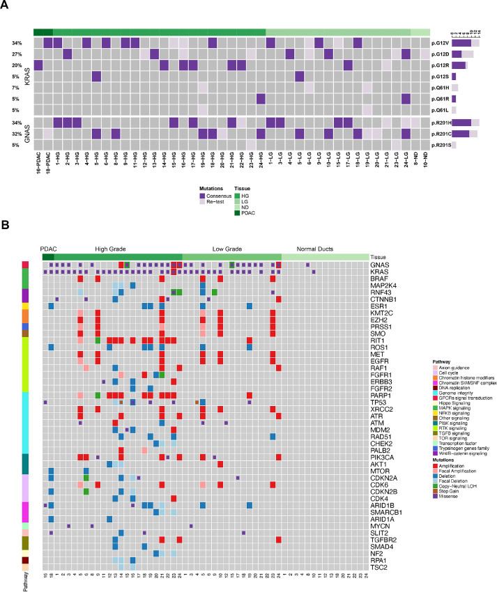
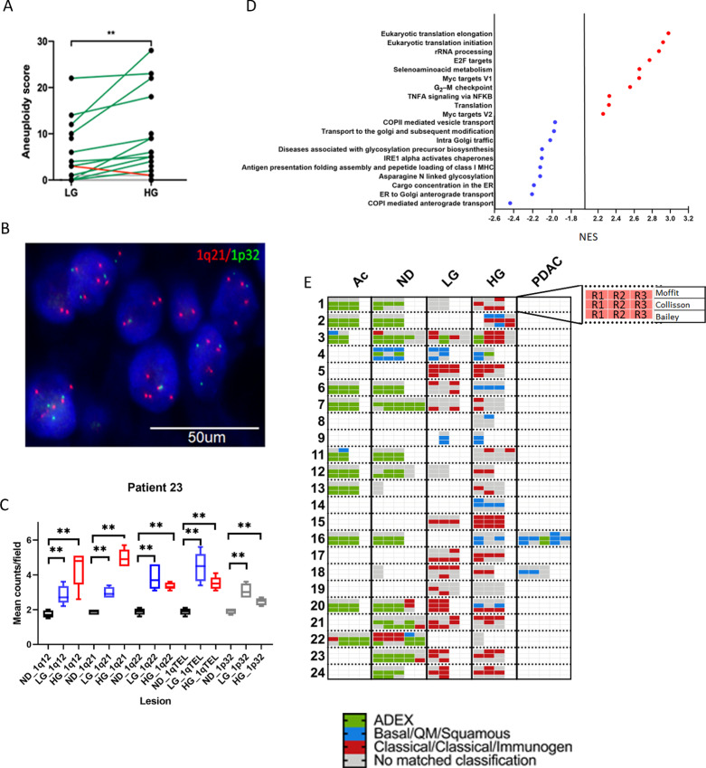
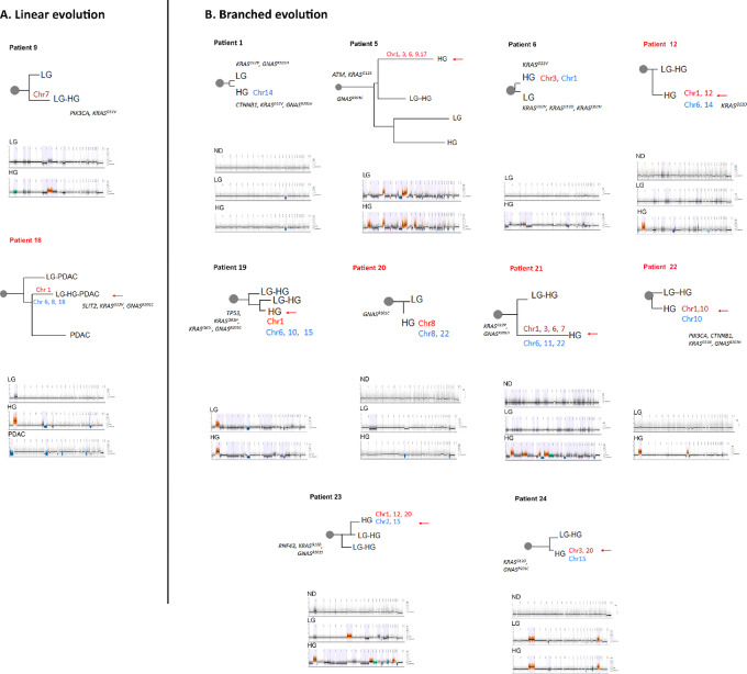
Intraductal papillary mucinous neoplasms (IPMN) are cystic precursor lesions to pancreatic ductal adenocarcinoma (PDAC). IPMNs undergo multistep progression from low-grade (LG) to high-grade (HG) dysplasia, culminating in invasive neoplasia. While patterns of IPMN progression have been analyzed using multiregion sequencing for somatic mutations, there is no integrated assessment of molecular events, including copy-number alterations (CNA) and transcriptional changes that accompany IPMN progression. We performed laser capture microdissection on surgically resected IPMNs of varying grades of histologic dysplasia obtained from 23 patients, followed by whole-exome and whole-transcriptome sequencing. Overall, HG IPMNs displayed a significantly greater aneuploidy score than LG lesions, with chromosome 1q amplification being associated with HG progression and with cases that harbored co-occurring PDAC. Furthermore, the combined assessment of single-nucleotide variants (SNV) and CNAs identified both linear and branched evolutionary trajectories, underscoring the heterogeneity in the progression of LG lesions to HG and PDAC. At the transcriptome level, upregulation of MYC-regulated targets and downregulation of transcripts associated with the MHC class I antigen presentation machinery as well as pathways related to glycosylation were a common feature of progression to HG. In addition, the established PDAC transcriptional subtypes (basal-like and classical) were readily apparent within IPMNs. Taken together, this work emphasizes the role of 1q copy-number amplification as a putative biomarker of high-risk IPMNs, underscores the importance of immune evasion even in noninvasive precursor lesions, and reinforces that evolutionary pathways in IPMNs are heterogenous, comprised of both SNV and CNA-driven events.
Significance: Integrated molecular analysis of genomic and transcriptomic alterations in the multistep progression of IPMNs, which are bona fide precursors of pancreatic cancer, identifies features associated with progression of low-risk lesions to high-risk lesions and cancer, which might enable patient stratification and cancer interception strategies.
© 2023 The Authors; Published by the American Association for Cancer Research.
Figures
References
-
- Rahib L, Smith BD, Aizenberg R, Rosenzweig AB, Fleshman JM, Matrisian LM. Projecting cancer incidence and deaths to 2030: the unexpected burden of thyroid, liver, and pancreas cancers in the United States. Cancer Res 2014;74:2913–21. - PubMed
-
- Kleeff J, Korc M, Apte M, La Vecchia C, Johnson CD, Biankin AV, et al. . Pancreatic cancer. Nat Rev Dis Primers 2016;2:16022. - PubMed
-
- Aziz H, Acher AW, Krishna SG, Cloyd JM, Pawlik TM. Comparison of society guidelines for the management and surveillance of pancreatic cysts: a review. JAMA Surg 2022;157:723–30. - PubMed
Publication types
MeSH terms
Grants and funding
- U54CA274371/NH/NIH HHS/United States
- U01 CA200468/CA/NCI NIH HHS/United States
- U01CA196406-03S1/NH/NIH HHS/United States
- U54 CA096297/CA/NCI NIH HHS/United States
- T32GM008806/NH/NIH HHS/United States
- U01 CA196406/CA/NCI NIH HHS/United States
- U54CA096297/NH/NIH HHS/United States
- U54 CA096300/CA/NCI NIH HHS/United States
- P30CA042014/NH/NIH HHS/United States
- U01CA200468/NH/NIH HHS/United States
- U01CA196403/NH/NIH HHS/United States
- P50CA221707/NH/NIH HHS/United States
- K22 CA258678/CA/NCI NIH HHS/United States
- P30 CA042014/CA/NCI NIH HHS/United States
- T32 GM008806/GM/NIGMS NIH HHS/United States
- T15LM011271/NH/NIH HHS/United States
- P50 CA221707/CA/NCI NIH HHS/United States
- U01 CA214263/CA/NCI NIH HHS/United States
- K22CA258678/NH/NIH HHS/United States
- T32CA009599/NH/NIH HHS/United States
- U54 CA274371/CA/NCI NIH HHS/United States
- UO1CA214263/NH/NIH HHS/United States
- T32CA217789/NH/NIH HHS/United States
- U54CA096300/NH/NIH HHS/United States
- T15 LM011271/LM/NLM NIH HHS/United States
- U01CA196406/NH/NIH HHS/United States
- U01 CA196403/CA/NCI NIH HHS/United States
- R21CA273974-01/NH/NIH HHS/United States
- R21 CA273974/CA/NCI NIH HHS/United States
- T32 CA009599/CA/NCI NIH HHS/United States
- T32 CA217789/CA/NCI NIH HHS/United States
LinkOut - more resources
Full Text Sources
Medical
Research Materials
