

Robot-assisted fluorescent sentinel lymph node identification in early-stage colon cancer
- PMID: 37721591
- PMCID: PMC10615938
- DOI: 10.1007/s00464-023-10394-2
Robot-assisted fluorescent sentinel lymph node identification in early-stage colon cancer
Abstract
Background: Patients with cT1-2 colon cancer (CC) have a 10-20% risk of lymph node metastases. Sentinel lymph node identification (SLNi) could improve staging and reduce morbidity in future organ-preserving CC surgery. This pilot study aimed to assess safety and feasibility of robot-assisted fluorescence-guided SLNi using submucosally injected indocyanine green (ICG) in patients with cT1-2N0M0 CC.
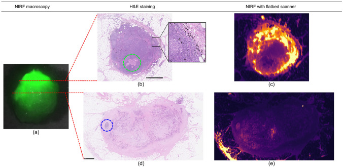
Methods: Ten consecutive patients with cT1-2N0M0 CC were included in this prospective feasibility study. Intraoperative submucosal, peritumoral injection of ICG was performed during a colonoscopy. Subsequently, the near-infrared fluorescence 'Firefly' mode of the da Vinci Xi robotic surgical system was used for SLNi. SLNs were marked with a suture, after which a segmental colectomy was performed. The SLN was postoperatively ultrastaged using serial slicing and immunohistochemistry, in addition to the standard pathological examination of the specimen. Colonoscopy time, detection time (time from ICG injection to first SLNi), and total SLNi time were measured (time from the start of colonoscopy to start of segmental resection). Intraoperative, postoperative, and pathological outcomes were registered.
Results: In all patients, at least one SLN was identified (mean 2.3 SLNs, SLN diameter range 1-13 mm). No tracer-related adverse events were noted. Median colonoscopy time was 12 min, detection time was 6 min, and total SLNi time was 30.5 min. Two patients had lymph node metastases present in the SLN, and there were no patients with false negative SLNs. No patient was upstaged due to ultrastaging of the SLN after an initial negative standard pathological examination. Half of the patients unexpectedly had pT3 tumours.
Conclusions: Robot-assisted fluorescence-guided SLNi using submucosally injected ICG in ten patients with cT1-2N0M0 CC was safe and feasible. SLNi was performed in an acceptable timespan and SLNs down to 1 mm were detected. All lymph node metastases would have been detected if SLN biopsy had been performed.
Keywords: Colon cancer; Image-guided surgery; Indocyanine green (ICG); Near-infrared fluorescence (NIRF); Robotic surgical procedures; Sentinel lymph node (SLN).
© 2023. The Author(s).
Conflict of interest statement
Daan J. Sikkenk and Esther C. J. Consten received grants from the ‘European Association for Endoscopic Surgery’ and ‘Stichting het Stichts Gastroenterologisch Genootschap’ for this study, but the funding had no role in the study. Esther C. J. Consten and Paul M. Verheijen are proctors for Intuitive Surgical. Daan J. Sikkenk, Andrea J. Sterkenburg, Thijs A. Burghgraef, Halil Akol, Matthijs P. Schwartz, René Arensman, Paul M. Verheijen, Wouter B. Nagengast and Esther C. J. Consten have no conflicts of interest or financial ties to disclose.
Figures

References
-
- Netherlands Cancer Registry. National data. https://iknl.nl/nkr-cijfers. Accessed 14 Nov 2022
-
- CBS (2022) Deaths; underlying cause of death (shortlist), sex, age. https://opendata.cbs.nl/statline/#/CBS/en/dataset/7052eng/table?ts=16684.... Accessed 14 Nov 2022
-
- Breekveldt ECH, Lansdorp-Vogelaar I, Toes-Zoutendijk E, Spaander MCW, van Vuuren AJ, van Kemenade FJ, Ramakers CRB, Dekker E, Nagtegaal ID, Krul MF, Kok NFM, Kuhlmann KFD, Vink GR, van Leerdam ME, Elferink MAG. Colorectal cancer incidence, mortality, tumour characteristics, and treatment before and after introduction of the faecal immunochemical testing-based screening programme in the Netherlands: a population-based study. Lancet Gastroenterol Hepatol. 2022;7:60–68. doi: 10.1016/S2468-1253(21)00368-X. - DOI - PubMed
-
- Backes Y, Elias SG, Bhoelan BS, Groen JN, van Bergeijk J, Seerden TCJ, Pullens HJM, Spanier BWM, Geesing JMJ, Kessels K, Kerkhof M, Siersema PD, de Vos tot Nederveen Cappel WH, van Lelyveld N, Wolfhagen FHJ, ter Borg F, Offerhaus GJA, Lacle MM, Moons LMG. The prognostic value of lymph node yield in the earliest stage of colorectal cancer: a multicenter cohort study. BMC Med. 2017;15:1–11. doi: 10.1186/s12916-017-0892-7. - DOI - PMC - PubMed
MeSH terms
Substances
LinkOut - more resources
Full Text Sources
