Review
. 2023 Nov;46(11):926-940.
doi: 10.1016/j.tins.2023.08.008. Epub 2023 Sep 16.

Microglia moonlighting after traumatic brain injury: aging and interferons influence chronic microglia reactivity

Affiliations
Review

Microglia moonlighting after traumatic brain injury: aging and interferons influence chronic microglia reactivity

Lynde M Wangler et al. Trends Neurosci. 2023 Nov.

Abstract

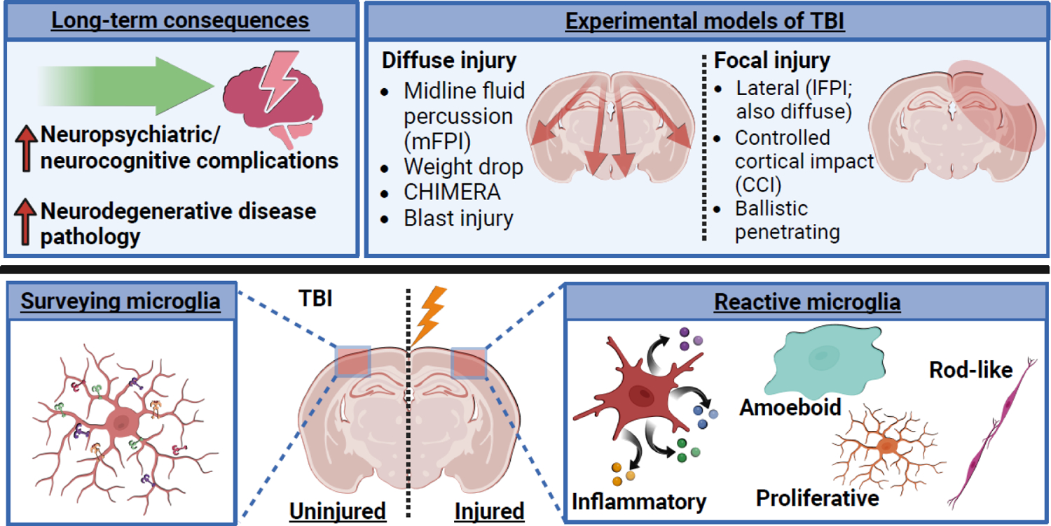
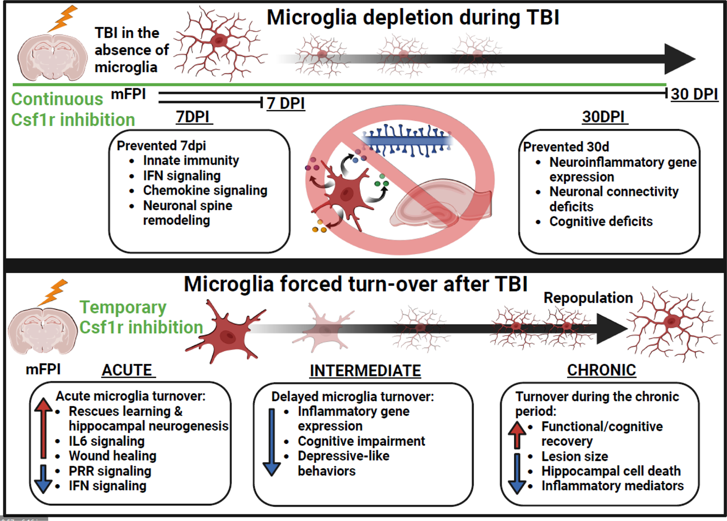
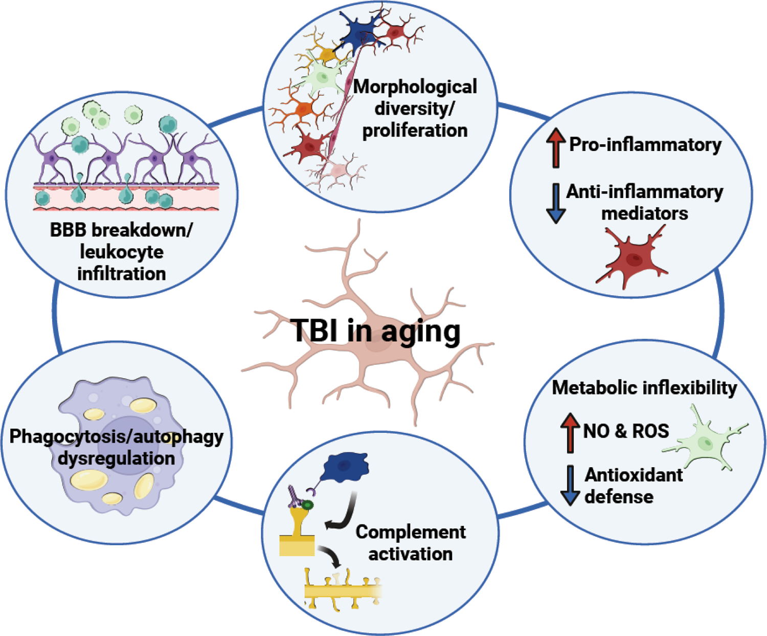
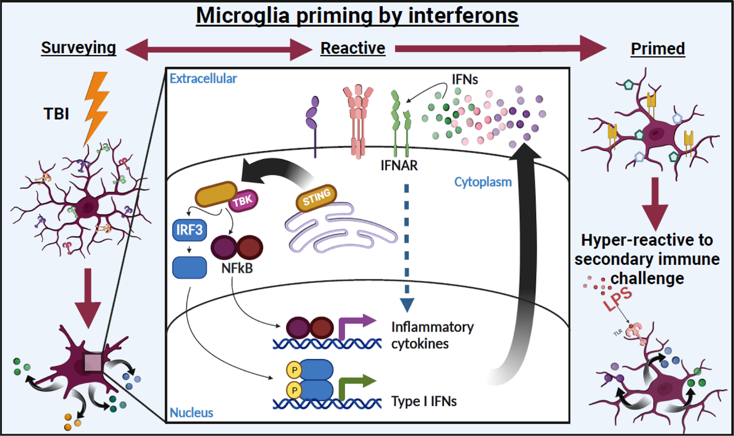
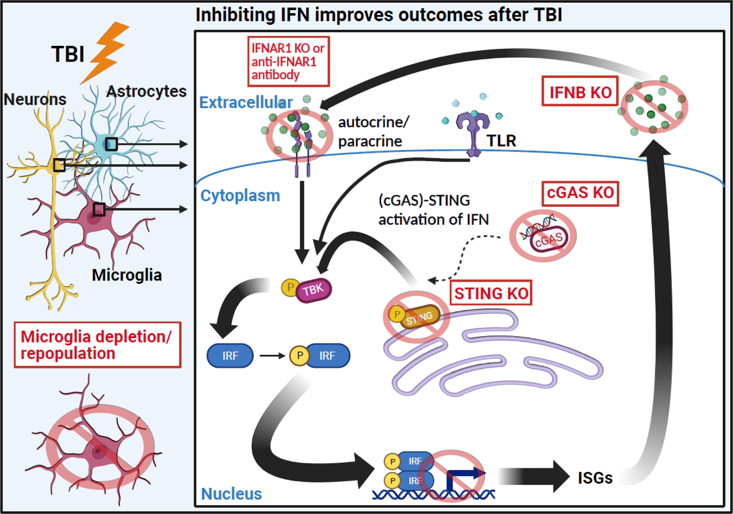
Most of the individuals who experience traumatic brain injury (TBI) develop neuropsychiatric and cognitive complications that negatively affect recovery and health span. Activation of multiple inflammatory pathways persists after TBI, but it is unclear how inflammation contributes to long-term behavioral and cognitive deficits. One outcome of TBI is microglial priming and subsequent hyper-reactivity to secondary stressors, injuries, or immune challenges that further augment complications. Additionally, microglia priming with aging contributes to exaggerated glial responses to TBI. One prominent inflammatory pathway, interferon (IFN) signaling, is increased after TBI and may contribute to microglial priming and subsequent reactivity. This review discusses the contributions of microglia to inflammatory processes after TBI, as well as the influence of aging and IFNs on microglia reactivity and chronic inflammation after TBI.

Keywords: microglia priming; neurodegeneration; neuroinflammation; neurotrauma.


Conflict of interest statement

Declaration of interests The authors declare no competing interests in relation to this work.

Figures

Figure 1.
Figure 1.. Experimental models for the study of microglia activation and neurological complications following traumatic brain injury.
Traumatic brain injury often leads to the development and persistence of neurological and neuropsychiatric complications, including cognitive decline, depression, anxiety, epilepsy, aggression, and neurodegenerative diseases [–20, 22, 23]. Preclinical researchers use several approaches to induce experimental TBIs to study the underlying pathophysiology. Microglia, the resident innate immune cells of the brain, are one well-documented cell type known to drive inflammation in the brain after TBI. Under steady-state conditions, microglia are actively surveying the micro-environment. Upon damage induced by TBI, microglia become reactive to drive several different processes. Microglia produce cytokines and chemokines (the signaling molecules of the immune system), become amoeboid and phagocytose debris, upregulate proliferation, and/or adopt rod-shaped morphology and align with damaged axons [39, 46]. Figure created using Biorender.com.
Figure 2.
Figure 2.. Microglia depletion and forced turn-over studies in animal models reveal stagespecific contributions of microglia in progression of pathology after TBI.
CSF1R antagonism results in rapid depopulation of microglia. Depletion of microglia throughout the time-course of TBI reverses inflammatory gene expression and neuronal spine remodeling, as well as cognitive deficits at a chronic timepoint, revealing the role of microglia in producing these effects after TBI [40, 46]. Forced turn-over or “repopulation” of microglia after TBI, by cessation of CSF1R antagonism, allows for assessment of applying microglia-specific intervention at later timepoints after injury. Acute (within 3 d), intermediate (5–14 d), or chronic (>30 d) repopulation of microglia after TBI generally improves inflammatory gene expression, cell death, cognitive impairment, depressive-like symptomology, and inflammatory responses to peripheral immune challenge[42, 48, 50, 51, 53]. These studies demonstrate that targeting microglia-mediated inflammation after TBI improves recovery. Figure created using Biorender.com.
Figure 3.
Figure 3.. Primed microglia promote amplified immune responses and worsened functional recovery in the aged brain following TBI.
Aged individuals are particularly vulnerable to TBI; those > 75 yr old have the highest incidence rates of TBI, as well as the most TBI-related hospitalizations and deaths. Preclinical research has defined several biological themes that drive these effects in aging, including amplified morphological restructuring of microglia; enhanced production of inflammatory mediators accompanied by deficient anti-inflammatory signaling; increased production of damaging ROS and NO and decreased antioxidant defense; enhanced complement activation and synapse loss; dysregulated phagocytosis and autophagy; and exacerbated blood brain barrier breakdown with peripheral cell infiltration. Figure created using Biorender.com.
Figure 4.
Figure 4.. Robust interferon activation primes microglia after TBI.
Microglia remain activated chronically after injury. Furthermore, while some microglia return to basal status, a subset of microglia adopt a “primed” profile characterized by increased reactivity to secondary stimuli (e.g., infection, stress, etc.). Microglia priming has been associated with increased inflammation and development of chronic cognitive and psychiatric complications following TBI. IFN activation has been associated with priming cells to respond to viral infection by increasing phagocytic and antigen presentation machinery similar to the profile of primed microglia. Figure created using Biorender.com.
Figure 5.
Figure 5.. Inhibiting interferon (IFN) signaling in animal models improves inflammation and functional recovery after TBI.
IFN gene expression is strongly induced by both focal and diffuse TBI in multiple cell types. Blocking the IFN response to TBI through various methods is generally beneficial to neuroinflammation and functional recovery. For instance, IFNAR (receptor) knockout or antibody inhibition decreases proinflammatory gene expression and lesion volume [96]. Direct knock-out of Ifnb also successfully ameliorates inflammatory gene expression [109]. Furthermore, knock-out of cGAS or STING, proteins that mediate the IFN response to TBI, improves neuronal pathology, inflammation, and functional recovery [98]. Figure created using Biorender.com.

Similar articles

Cited by

References

    1. Perry DC, et al., Association of traumatic brain injury with subsequent neurological and psychiatric disease: a meta-analysis. J Neurosurg, 2016. 124(2): p. 511–26. - PMC - PubMed
    1. Gualtieri T and Cox DR, The delayed neurobehavioural sequelae of traumatic brain injury. Brain Injury, 1991. 5(3): p. 219–232. - PubMed
    1. Bodnar CN, Morganti JM, and Bachstetter AD, Depression following a traumatic brain injury: uncovering cytokine dysregulation as a pathogenic mechanism. Neural Regen Res, 2018. 13(10): p. 1693–1704. - PMC - PubMed
    1. Millis SR, et al., Long-term neuropsychological outcome after traumatic brain injury. J Head Trauma Rehabil, 2001. 16(4): p. 343–55. - PubMed
    1. Salmond CH, et al., Changes over time in cognitive and structural profiles of head injury survivors. Neuropsychologia, 2006. 44(10): p. 1995–1998. - PubMed

Publication types