. 2023 Sep 18;14(1):5666.
doi: 10.1038/s41467-023-41381-9.

Pharmacological inhibition of TBK1/IKKε blunts immunopathology in a murine model of SARS-CoV-2 infection

Affiliations

Pharmacological inhibition of TBK1/IKKε blunts immunopathology in a murine model of SARS-CoV-2 infection

Tomalika R Ullah et al. Nat Commun. .

Abstract

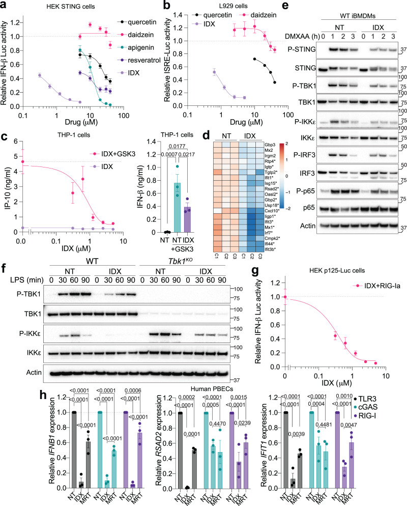
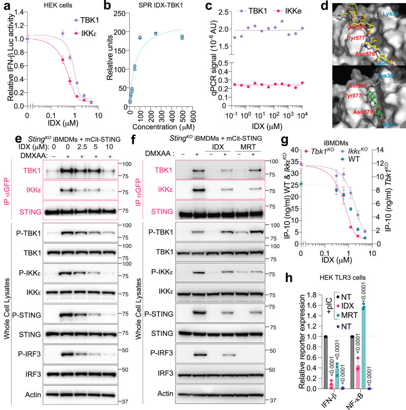
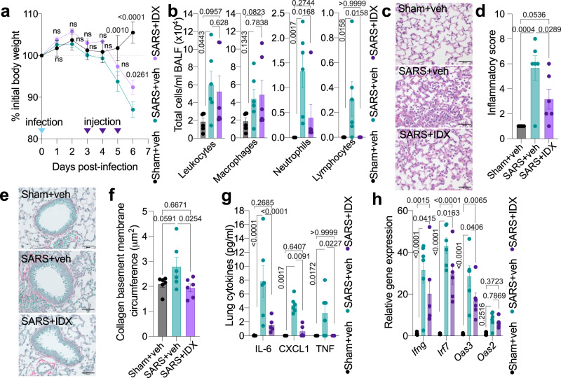
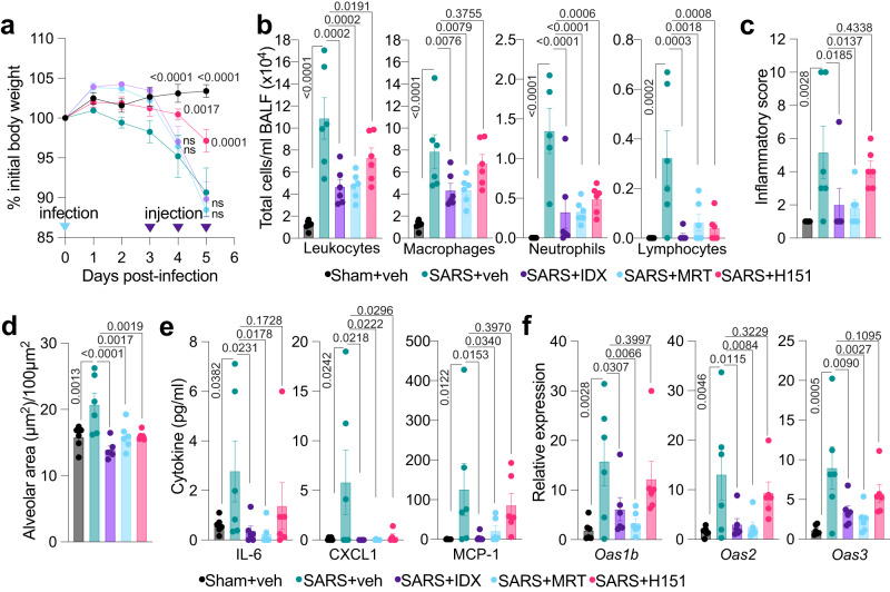
TANK-binding kinase 1 (TBK1) is a key signalling component in the production of type-I interferons, which have essential antiviral activities, including against SARS-CoV-2. TBK1, and its homologue IκB kinase-ε (IKKε), can also induce pro-inflammatory responses that contribute to pathogen clearance. While initially protective, sustained engagement of type-I interferons is associated with damaging hyper-inflammation found in severe COVID-19 patients. The contribution of TBK1/IKKε signalling to these responses is unknown. Here we find that the small molecule idronoxil inhibits TBK1/IKKε signalling through destabilisation of TBK1/IKKε protein complexes. Treatment with idronoxil, or the small molecule inhibitor MRT67307, suppresses TBK1/IKKε signalling and attenuates cellular and molecular lung inflammation in SARS-CoV-2-challenged mice. Our findings additionally demonstrate that engagement of STING is not the major driver of these inflammatory responses and establish a critical role for TBK1/IKKε signalling in SARS-CoV-2 hyper-inflammation.


Conflict of interest statement

Financial competing interests: O.F.L. and D.S.W. are employees of Noxopharm. G.E.K. owns equity in Noxopharm. M.P.G., J.Z., J.I.E., V.R.A., N.K. and P.M.H. receive funding from Noxopharm Ltd. to study the activity of IDX in inflammation. M.P.G. and P.M.H. received consulting fees from Noxopharm Ltd. Noxopharm Ltd. was involved in the conceptualisation, design, data collection, analysis and helped the preparation of the manuscript. M.P.G., J.Z., V.R.A., N.K., J.I.E. and P.M.H. do not personally own shares and/or equity in Noxopharm Ltd. G.E.K., O.F.L. and M.P.G. are named inventors of a patent (assignee: Noxopharm Ltd) relating to the use of IDX to treat inflammation associated with infection (WO/2021/195698, including patent US20210299085 granted). The remaining authors declare no competing interests.

Figures

Fig. 1
Fig. 1. Idronoxil inhibits STING, MAVS and TRIF driven responses.
a Interferon [IFN]-β luciferase expression in HEK293T mSTING cells overexpressing cGAS treated O/N with the indicated compounds (n = 3 independent experiments). b IFN-stimulated response element [ISRE]-Luciferase expression in L929 cells after 7 h stimulation with the mouse STING agonist DMXAA at 20 μg/ml and the indicated compounds (n = 3 independent experiment for all except Quercetin n = 2 independent experiments). c IFN-β (right) and IP-10 (left) protein levels in THP-1 monocytes after 6 h stimulation with the human STING agonist GSK3 at 100 nM and treatment with indicated idronoxil (IDX) doses. Right: 2.5 μM IDX was used (n = 3 independent experiments). d RNA sequencing analysis of the top 20 murine ISGs in RNA lysates from Trex1Q98X bone marrow-derived macrophages [BMDMs] treated O/N with 1.25 μM IDX (heat map of log2 expression relative to the average across all samples; significant genes are indicated with an asterisk) (n = 3 independent animals [C1–C3]). NT is non-treated. e Immunoblot of wild-type [WT] immortalised bone marrow-derived macrophages [iBMDMs] stimulated with 50 μg/ml DMXAA (STING) for the indicated times ±2.5 μM IDX (1 representative blot of 3 independent experiments shown). Molecular weight markers are shown in kDa. A doublet of STING bands appears upon STING activation. Phosphorylated proteins are indicated with [P]. f Immunoblot of wild-type and Tbk1KO iBMDMs stimulated with 200 ng/ml LPS (TLR4) ± 2.5 μM IDX treatment for the indicated time (1 representative blot of 3 independent experiments shown). Phosphorylated proteins are indicated with [P]. Molecular weight markers are shown in kDa. g IFN-β luciferase expression in p125HEK cells treated with indicated dose of IDX and stimulated O/N with 50 ng/ml of 3p-hpRNA (RIG-I agonist [RIG-Ia]) (n = 3 independent experiments). h RT-qPCR analyses of IFNB1/18S, RSAD2/18S and IFIT1/18S in RNA lysates from primary human bronchial epithelial cells [PBECs] from 3 independent donors treated with 5 μM IDX or 200 nM MRT, and stimulated 3 h with 1 μg/ml transfected polyI:C (MDA5/RIG-I), 25 μg/ml naked polyI:C (TLR3) or transfected with 2.5 μg/ml ISD70 (cGAS) (n = 3 independent donors). ac, g, h Data are mean ± s.e.m. c One-way or (h) two-way ANOVA with uncorrected Fisher’s LSD (with single pooled variance) multiple comparisons are shown. ac, g Non-linear regression analyses are shown. Exact p values are shown for all comparisons. Source data and detailed statistical analyses are provided as a Source Data file.
Fig. 2
Fig. 2. Idronoxil inhibits formation of TBK1 signalling complexes.
a Interferon [IFN]-β luciferase expression in HEK293T cells expressing the indicated constructs after O/N incubation (n = 3 independent experiments). b Surface plasmon resonance [SPR] analysis of idronoxil (IDX) binding to recombinant human TBK1 (representative of n = 3 independent experiments). c qPCR amplification signal of kinase concentration after KinomeScanTM assays, for the indicated IDX concentrations (averaged from n = 2 technical replicates). IKKe is IKKε. d In silico modelling of IDX binding (bottom—green) and STING C-terminal tail (top—yellow, PDB: 6nt9) to TBK1 dimer (the blue and red residues refer to the two different TBK1 units in the structure). Immunoblot of StingKO immortalised bone marrow-derived macrophages [iBMDMs] expressing mCitrine-STING, after 1 h stimulation with 50 μg/ml DMXAA (STING) and increasing doses of IDX (e), or 5 μM IDX or 100 nM MRT67307 [MRT] (f). e 1 representative blot of three independent experiments shown. f One representative blot of two independent experiments shown. Pink: pull-down of mCitrine-STING with anti-GFP antibody. Black: whole cell lysates. e, f Phosphorylated proteins are indicated with [P]. Molecular weight markers are shown in kDa. g IP-10 protein levels in wild-type (IC50 = 1.6 μM), Tbk1KO (IC50 = 0.75 μM), IkbkεKO [IkkεKO](IC50 = 2 μM) and wild-type [WT] iBMDM cells after O/N stimulation with 50 μg/ml DMXAA (STING) and increasing doses of IDX (n = 3 independent experiments). h HEK-TLR3 cells expressing NF-κB and IFN-β luciferase reporters were treated 6 h with 0.5 μg/ml poly(I:C) [pIC] (TLR3) in the presence of 5 μM IDX or 400 nM MRT (n = 3 independent experiments). a, g, h Data are mean ± s.e.m. ac, g Non-linear regression analyses are shown. h Two-way ANOVA with uncorrected Fisher’s LSD (with single pooled variance) multiple comparisons are shown. Exact p values are shown for all comparisons. Source data and detailed statistical analyses are provided as a Source Data file.
Fig. 3
Fig. 3. Idronoxil limits SARS-CoV-2-driven hyper-inflammation.
a Daily weights of K18-hACE2 mice after intranasal infection (day 0) with 103 PFU SARS-CoV-2 (n = 6 animals were examined per group in one experiment). Idronoxil [IDX]/vehicle injections were performed on days 3, 4, and 5, with mice culled on day 6 post-infection. Veh is vehicle, SARS is SARS-CoV-2 infected, and Sham is non-infected. b Bronchoalveolar lavage fluid [BALF] cell subsets were enumerated following Modified Giemsa staining to distinguish cell populations (n = 6 animals were examined per group in one experiment). c Histology of fixed lung lobes with Modified Giemsa staining (1 representative image of 6 per group) and (d) associated inflammatory scores (n = 6 animals were examined per group in one experiment). e Collagen deposition around the primary airways determined by Sirius Red/Fast Green staining (1 representative image of 6 per group) and (f) associated measurement of the collagen deposition around the primary airway basement circumference (n = 6 animals were examined per group in one experiment). c, e Scale bars of 50 μm are indicated. g Cytokine quantification in lung homogenates (n = 6 animals were examined per group in one experiment, however one outlier in the IDX + SARS group was removed in these analyses after ROUT outlier analysis). h mRNA transcript levels in lung homogenates. Expression of indicated genes is shown relative to Hprt (n = 6 animals were examined per group in one experiment, however qPCR amplification failed for one animal in the Sham + vehicle group). a, b, d, fh Data are mean ± s.e.m. a, g, h Two-way or (b, d, f) one-way ANOVA with uncorrected Fisher’s LSD (with single pooled variance) multiple comparisons are shown. Exact p values are shown for all comparisons except for (a) where “ns” is non-significant (p > 0.05). Source data and detailed statistical analyses are provided as a Source Data file.
Fig. 4
Fig. 4. TBK1 inhibition limits SARS-CoV-2-driven hyper-inflammation.
a Daily weights of K18-hACE2 mice after intranasal infection (day 0) with 103 PFU SARS-CoV-2. Idronoxil [IDX]/MRT67307 [MRT]/H151/vehicle injections were performed on days 3, 4, and 5, with mice culled on day 5 post-infection (n = 6 animals were examined per group in one experiment). Veh is vehicle, SARS is SARS-CoV-2 infected, and Sham is non-infected. b Bronchoalveolar lavage fluid [BALF] cell subsets were enumerated following Modified Giemsa staining to distinguish cell populations (n = 6 animals were examined per group in one experiment, however one outlier in the SARS + Vehicle group was removed in the Neutrophils analyses). c Histology of fixed lung lobes with associated inflammatory scores (n = 6 animals were examined per group in one experiment). d Alveolar thickness in the primary airways determined by histology (n = 6 animals were examined per group in one experiment). e Cytokine quantification in lung homogenates (n = 6 animals were examined per group in one experiment, however one outlier in each but the SARS + Vehicle group was removed in the CXCL1 analyses after ROUT outlier analysis). f mRNA transcript levels in lung homogenates. Expression of indicated genes is shown relative to Hprt (n = 6 animals were examined per group in one experiment). af Data are mean ± s.e.m. a Two-way or (bf) one-way ANOVA with uncorrected Fisher’s LSD (with single pooled variance) multiple comparisons are shown. Exact p values are shown for all comparisons except for (a) where the comparisons for days 0, 1, 2 and 3 are provided in Source Data File, and where “ns” is non-significant (p > 0.05). Source data and detailed statistical analyses are provided as a Source Data file.

References

    1. Liu, S. et al. Phosphorylation of innate immune adaptor proteins MAVS, STING, and TRIF induces IRF3 activation. Science34710.1126/science.aaa2630 (2015). - PubMed
    1. Hagan RS, Torres-Castillo J, Doerschuk CM. Myeloid TBK1 signaling contributes to the immune response to influenza. Am. J. Respir. Cell Mol. Biol. 2019;60:335–345. - PMC - PubMed
    1. Crow YJ, Manel N. Aicardi–Goutières syndrome and the type I interferonopathies. Nat. Rev. Immunol. 2015;15:429–440. - PubMed
    1. Hasan M, et al. Cutting edge: inhibiting TBK1 by compound II ameliorates autoimmune disease in mice. J. Immunol. 2015;195:4573–4577. - PMC - PubMed
    1. Louis C, et al. Therapeutic effects of a TANK‐binding kinase 1 inhibitor in germinal center–driven collagen‐induced arthritis. Arthritis Rheumatol. 2018;71:50–62. - PubMed

Publication types