. 2023 Sep 19;6(1):952.
doi: 10.1038/s42003-023-05298-9.

Loss of Grin2a causes a transient delay in the electrophysiological maturation of hippocampal parvalbumin interneurons

Affiliations

Loss of Grin2a causes a transient delay in the electrophysiological maturation of hippocampal parvalbumin interneurons

Chad R Camp et al. Commun Biol. .

Abstract

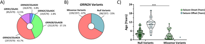
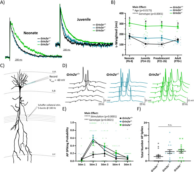
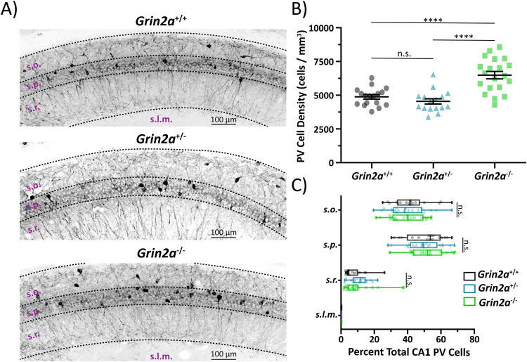
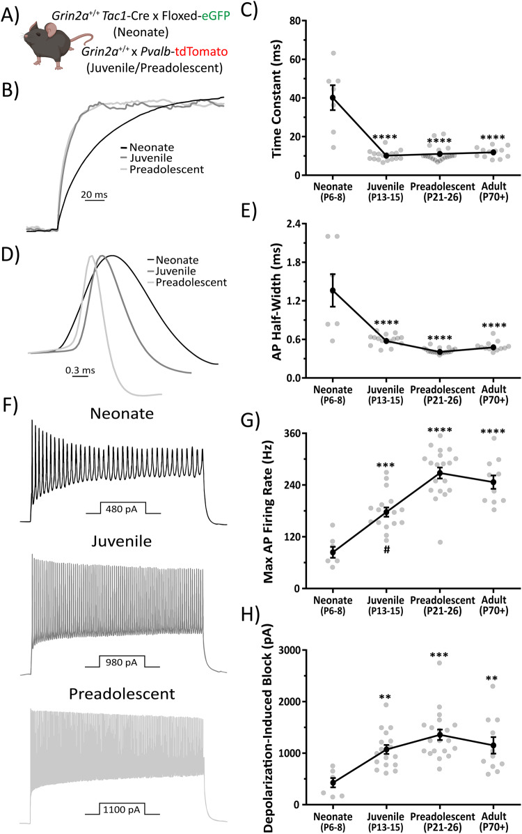
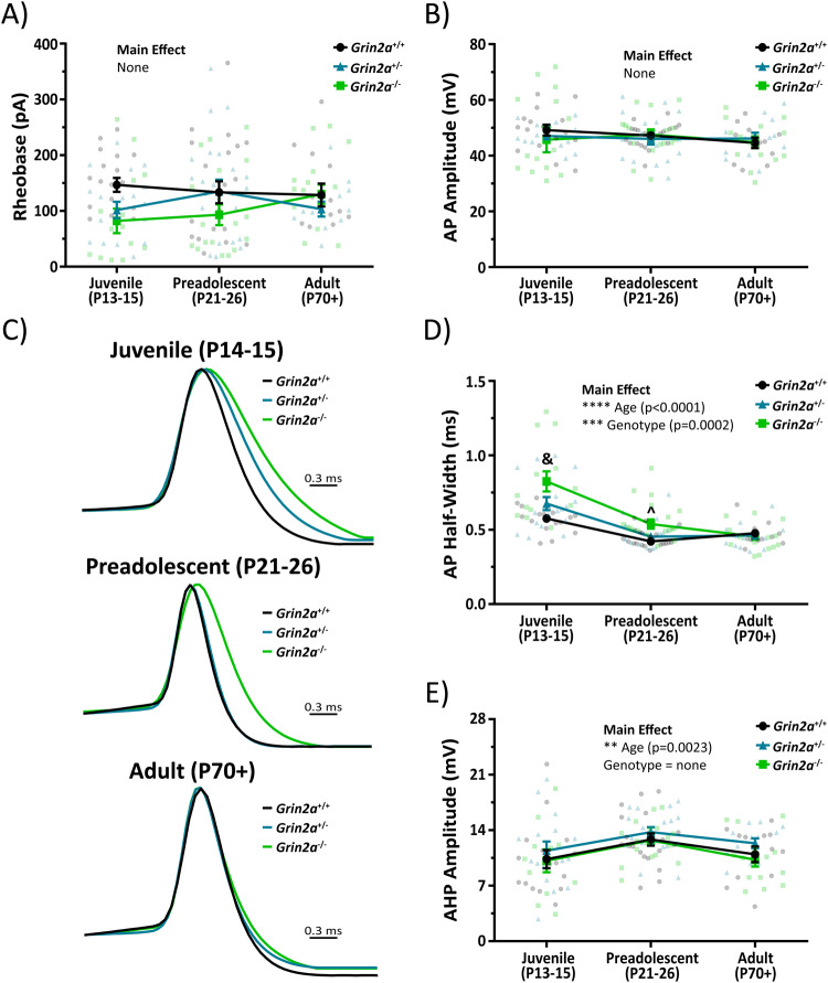
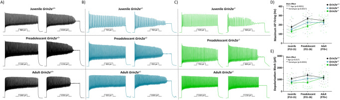
N-methyl-D-aspartate receptors (NMDARs) are ligand-gated ionotropic glutamate receptors that mediate a calcium-permeable component to fast excitatory neurotransmission. NMDARs are heterotetrameric assemblies of two obligate GluN1 subunits (GRIN1) and two GluN2 subunits (GRIN2A-GRIN2D). Sequencing data shows that 43% (297/679) of all currently known NMDAR disease-associated genetic variants are within the GRIN2A gene, which encodes the GluN2A subunit. Here, we show that unlike missense GRIN2A variants, individuals affected with disease-associated null GRIN2A variants demonstrate a transient period of seizure susceptibility that begins during infancy and diminishes near adolescence. We show increased circuit excitability and CA1 pyramidal cell output in juvenile mice of both Grin2a+/- and Grin2a-/- mice. These alterations in somatic spiking are not due to global upregulation of most Grin genes (including Grin2b). Deeper evaluation of the developing CA1 circuit led us to uncover age- and Grin2a gene dosing-dependent transient delays in the electrophysiological maturation programs of parvalbumin (PV) interneurons. We report that Grin2a+/+ mice reach PV cell electrophysiological maturation between the neonatal and juvenile neurodevelopmental timepoints, with Grin2a+/- mice not reaching PV cell electrophysiological maturation until preadolescence, and Grin2a-/- mice not reaching PV cell electrophysiological maturation until adulthood. Overall, these data may represent a molecular mechanism describing the transient nature of seizure susceptibility in disease-associated null GRIN2A patients.


Conflict of interest statement

H.Y. and S.F.T. are co-inventors of Emory-owned intellectual property. S.F.T. is a member of the SAB for Sage Therapeutics, Eumentis Therapeutics, the GRIN2B Foundation, the CureGRIN Foundation, and CombinedBrain. S.F.T. is a consultant for GRIN Therapeutics and Neurocrine. H.Y. is the PI on a research grant from Sage Therapeutics to Emory. S.F.T. is cofounder of NeurOp, Inc. and Agrithera. T.A.B. is a member of the SAB for GRIN2B Foundation, CureGRIN Foundation, GRIN Therapeutics, and Neurocrine; all remuneration has been made to his department. T.G.B. is PI on a research grant from Neumora to Emory University. J.R.L. is a consultant for GRIN Therapeutics and a member of the SAB for GRIN2B Foundation and CureGRIN Foundation. All other authors declare no competing interests.

Figures

Fig. 1
Fig. 1. Disease-associated null GRIN2A patients may display a transient seizure susceptibility not seen in disease-associated missense GRIN2A patients.
A Summary data adapted from Hansen et al. 2021 showing that most GRIN variants are found in the GRIN2A (44%; 297/679) gene. B Summary data adapted from Hansen et al. 2021 highlighting that a third (98/297) of all GRIN2A variants are null variants, which include nonsense variants, as well as chromosomal insertions, deletions, inversions, and translocations. C Disease-associated null GRIN2A variants display seizure onset susceptibility at a significantly older age than disease-associated missense GRIN2A variants (4.5 ± 0.2 years for null GRIN2A variants, n = 92 vs 3.1 ± 0.4 years for missense GRIN2A variants, n = 45; Mann-Whitney ranked sum, p = 0.0003). Currently, there are 20 disease-associated null GRIN2A patients with a previous history of seizures that were seizure- free at their last follow-up, with a mean seizure offset of 10.4 ± 0.8 years. These data are in stark contrast to disease-associated missense GRIN2A variants with available seizure offset data, as only one patient has been reported to be seizure-free at last follow-up (missense GRIN2A variant seizure onset age = 0.25 years with seizure offset at 1.7 years). For violin plots, solid middle line represents the median, with the dashed lines representing the 25th and 75th quantile. ***p < 0.001.
Fig. 2
Fig. 2. Juvenile CA1 circuit shows hyperexcitability in Grin2a+/− and Grin2a−/− mice.
A Evoked NMDAR-mediated excitatory postsynaptic currents (EPSCs) onto CA1 pyramidal cells from Grin2a+/+, Grin2a+/−, Grin2a−/− at various ages during development, with normalized representative traces shown for neonate and juvenile mice. B Two-way ANOVA showed significant main effects for both age (F3, 93 = 3.55; p = 0.0175) and genotype (F3, 93 = 71.4; p < 0.0001) of the tau-weighted describing the NMDAR-mediated EPSC decay time. At both neonate and adult timepoints, only Grin2a+/+ and Grin2a−/− mice have significantly different NMDAR-mediated EPSC decay times, whereas at the juvenile and preadolescent timepoints, all three genotypes have significantly different NMDAR-mediated EPSC decay times. Given that the juvenile stage was the earliest developmental window with separation at all three genotypes for the NMDAR-mediated EPSC decay time, we conducted further experiments at this age. C CA1 pyramidal cells from juvenile mice were current clamped at -60 mV and Schaffer collaterals were stimulated five times at 100 Hz for a total of 5 epochs. Stimulation intensity was set just below threshold to produce an action potential spike after a single Schaffer collateral stimulation. D Representative traces showing action potential spiking in response to successive Schaffer collateral stimulations. E Action potential spiking probability for each stimulus averaged over 5 epochs across all genotypes. Two-way ANOVA showed significant main effects for both genotype (F2, 225 = 6.351; p = 0.0021) and stimulation number (F4, 225 = 16.82; p < 0.0001), however, there was no interaction. F Total number of action potentials elicited over 5 epochs of 5-burst Schaffer collateral stimulation. One-way ANOVA showed there was no significant difference across the three genotypes (F = 2.997; p = 0.06). Data represented show mean ± SEM. AP = action potential; s.o. = stratum oriens; s.p. = stratum pyramidale; s.r. = stratum radiatum; n.s. = not significant. $ = Grin2a−/− significantly different than both Grin2a+/+ and Grin2a+/− at that age; # = all three genotypes significantly different than each other at that age.
Fig. 3
Fig. 3. Loss of Grin2a causes an increase in parvalbumin (PV) cell density in CA1.
A Representative images of CA1 hippocampal sections stained for PV in preadolescent mice. B CA1 PV cell density is significantly increased in Grin2a−/− mice compared to Grin2a+/+ mice (6488 ± 276 cells per mm3 in Grin2a−/− vs 4875 ± 162 cells per mm3 in Grin2a+/+; one-way ANOVA, p < 0.0001) and Grin2a+/− mice (6488 ± 276 cells per mm3 in Grin2a−/− vs 4542 ± 198 cells per mm3 in Grin2a+/−; one-way ANOVA, p < 0.0001). There is no difference in CA1 PV cell density between Grin2a+/+ and Grin2a+/− mice (one-way ANOVA, p = 0.58). C Despite an increase in cell density in Grin2a−/− mice, there is no difference in PV CA1 cellular lamination across all three genotypes. Data represented show mean ± SEM. s.o. = stratum oriens; s.p. = stratum pyramidale; s.r. = stratum radiatum; s.l.m. = stratum lacunosum moleculare; PV = parvalbumin; ****p < 0.0001; n.s. = not significant.
Fig. 4
Fig. 4. Loss of Grin2a does not alter cholecystokinin (CCK) cell density in CA1.
A Representative images of CA1 hippocampal sections stained for CCK in preadolescent mice. B CA1 CCK cell density is unchanged across all three genotypes (one-way ANOVA, p = 0.49). C There is no difference in CCK CA1 cellular lamination across all three genotypes. Data represented show mean ± SEM. s.o. = stratum oriens; s.p. = stratum pyramidale; s.r. = stratum radiatum; s.l.m. = stratum lacunosum moleculare; CCK = cholecystokinin; n.s. = not significant.
Fig. 5
Fig. 5. CA1 PV cells undergo electrophysiological maturation of passive and action potential firing properties.
A PV cells were visualized using either Pvalb-TdTomato or Tac1-Cre x floxed eGFP (see Supplemental Figs. S5 and S6). Mouse clipart from BioRender (permissions provided). B Representative, amplitude-normalized repolarization traces to highlight differences in membrane time constant following a −50 pA current injection at different developmental timepoints. C Membrane time constant is significantly prolonged in neonatal mice (F = 40.68, one-way ANOVA; p < 0.0001 for post-hoc multiple comparisons with every other developmental timepoint). D Action potential half-width is significantly prolonged in neonatal mice (F = 28, one-way ANOVA; p < 0.0001 for post-hoc multiple comparisons with every other developmental timepoint). E Representative, amplitude-normalized single action potential traces to highlight differences in half-width at different developmental timepoints. F Representative action potential trains elicited by various current injections depicted below each train to illustrate change in maximum action potential firing frequency during development. Traces shown are those just below threshold for depolarization-induced block of action potential firing. G Maximum action potential firing frequency is significantly decreased in neonatal mice (F = 28.3, one-way ANOVA; p < 0.001 for post-hoc multiple comparison test with juvenile mice, and p < 0.0001 for post-hoc multiple comparison test with preadolescent and adult mice). Juvenile mice also show a significantly decreased maximum action potential firing frequency compared to preadolescent mice (one-way ANOVA post-hoc multiple comparison test; p = 0.0039). H Current required for depolarization-induced block of action potential firing is significantly decreased in neonatal mice (F = 8.36, one-way ANOVA; p < 0.01 for post-hoc multiple comparison test with juvenile and adult mice, and p < 0.0001 for post-hoc multiple comparison test with preadolescent mice). Symbols are mean ± SEM. AP = action potential; depolarization block = Current required for depolarization-induced block of action potential firing. **p < 0.01; ***p < 0.001; ****p < 0.0001; # = juvenile mice significantly different than adult mice in one-way ANOVA post-hoc multiple comparison test.
Fig. 6
Fig. 6. The loss of Grin2a causes a transient change in passive electrophysiological properties in CA1 PV cells.
A Mouse model used to visualize PV cells in CA1 next to examples of a biocytin backfilled CA1 PV cell that was stained for TdTomato to indicate successful cellular identification. Mouse clipart from BioRender (permissions provided). B No change in resting membrane potential across all three genotypes at different developmental timepoints (two-way ANOVA). C No main effect of cellular capacitance across genotype (F2, 136 = 1.3; p = 0.28; two-way ANOVA) or age (F2, 136 = 0.32; p = 0.72; two-way ANOVA), however, there is a statistically significant interaction such that juvenile Grin2a+/− mice have a higher cellular capacitance than both Grin2a+/+ mice (150 ± 8.0 pF for Grin2a+/− vs 110 ± 6.5 pF for Grin2a+/+; p = 0.0061) and Grin2a−/− mice (150 ± 8.0 pF for Grin2a+/− vs 111 ± 11 pF for Grin2a−/−; p = 0.014). D Representative, amplitude-normalized repolarization traces to highlight differences in membrane time constant following a −50 pA current injection at different developmental timepoints. E Membrane time constant measurements show statistically significant main effects for both age (F2, 136 = 6.9; p = 0.0014; two-way ANOVA) and genotype (F2, 136 = 6.8; p = 0.0016; two-way ANOVA). There are also several statistically significant interactions such that both juvenile Grin2a+/− mice (20 ± 2.7 ms for Grin2a+/− vs 10 ± 0.6 ms for Grin2a+/+; p = 0.0011) and Grin2a−/− mice (22 ± 4.4 ms for Grin2a−/− vs 10 ± 0.6 ms for Grin2a+/+; p < 0.0001) displayed higher membrane time constants than Grin2a+/+ mice. In addition, preadolescent Grin2a−/− mice showed a higher membrane time constant than Grin2a+/+ mice (17 ± 1.9 ms for Grin2a−/− vs 11 ± 0.9 ms for Grin2a+/+; p = 0.048). F Input resistance measurements show statistically significant main effects for both age (F2, 133 = 5.0; p = 0.0082; two-way ANOVA) and genotype (F2, 133 = 12.2; p < 0.0001; two-way ANOVA). There are also several statistically significant interactions such that juvenile Grin2a−/− mice displayed higher input resistances than Grin2a+/+ mice (184 ± 23 MΩ for Grin2a−/− vs 96 ± 6.1 MΩ for Grin2a+/+; p < 0.0001) and Grin2a+/− mice (184 ± 23 MΩ for Grin2a−/− vs 128 ± 12 MΩ for Grin2a+/+; p = 0.0054). In addition, preadolescent Grin2a−/− mice showed a higher input resistance than Grin2a+/+ mice (149 ± 15 MΩ for Grin2a−/− vs 96 ± 7.9 MΩ for Grin2a+/+; p = 0.0031) and Grin2a+/− mice (149 ± 15 MΩ for Grin2a−/− vs 108 ± 9.3 MΩ for Grin2a+/+; p = 0.043). The sum of these data indicates an age- and gene-dependent transient delay in various passive electrical properties of CA1 PV cells. Symbols are mean ± SEM. RMP = resting membrane potential; # = Grin2a+/− significantly different than both Grin2a+/+ and Grin2a−/− at that age; $ = Grin2a+/+ significantly different than both Grin2a+/− and Grin2a−/− at that age; ^ = Grin2a−/− significantly different than Grin2a+/+ at that age; & = Grin2a−/− significantly different than both Grin2a+/+ and Grin2a+/− at that age; **p < 0.01; ***p < 0.001.
Fig. 7
Fig. 7. The loss of Grin2a causes a transient change in action potential waveform properties of CA1 PV cells.
There are no differences in A rheobase or B action potential amplitude regardless of age or genotype in CA1 PV cells. C Representative, amplitude-normalized single action potential traces to highlight differences in half-width at different developmental timepoints. D Action potential half-width measurements show statistically significant main effects for both age (F2, 134 = 47; p < 0.0001; two-way ANOVA) and genotype (F2, 134 = 9.3; p = 0.0002; two-way ANOVA). There are also several statistically significant interactions such that both juvenile Grin2a+/+ mice (0.58 ± 0.02 ms for Grin2a+/+ vs 0.82 ± 0.07 ms for Grin2a−/−; p < 0.0001) and Grin2a+/− mice (0.68 ± 0.04 ms for Grin2a+/− vs 0.82 ± 0.07 for Grin2a−/−; p < 0.005) displayed longer action potential half-widths than Grin2a−/− mice. In addition, preadolescent Grin2a−/− mice showed longer action potential half-widths than Grin2a+/+ mice (0.54 ± 0.03 ms for Grin2a−/− vs 0.42 ± 0.01 ms for Grin2a+/+; p = 0.0152). E Afterhyperpolarization amplitude of the action potential waveform showed a significant main effect for age (F2, 127 = 6.36; p = 0.0023; two-way ANOVA), but no main effect for genotype (F2, 127 = 2; p = 0.14; two-way ANOVA). Symbols are mean ± SEM. AP = action potential; AHP = afterhyperpolarization; ^ = Grin2a−/− significantly different than Grin2a+/+ at that age; & = Grin2a−/− significantly different than both Grin2a+/+ and Grin2a+/− at that age; **p < 0.01; ***p < 0.001; ****p < 0.0001.
Fig. 8
Fig. 8. The loss of Grin2a causes a transient change in action potential firing properties of CA1 PV cells.
Representative action potential trains elicited by current injections depicted below each train to illustrate the maximum action potential firing frequency and the current required for depolarization-induced block of action potential firing for A Grin2a+/+, B Grin2a+/−, and C Grin2a−/− mice during development. D Maximum action potential firing frequencies show a significant main effect for both age (F2, 135 = 29.8; p < 0.0001; two-way ANOVA) and genotype (F2, 135 = 6.7; p = 0.0017; two-way ANOVA). E Currents required to reach depolarization-induced block of action potential firing show a significant main effect for both age (F2, 135 = 4.2; p = 0.017; two-way ANOVA) and genotype (F2, 135 = 5.4; p = 0.006; two-way ANOVA). Symbols are mean ± SEM. AP = action potential; depolarization block = current required to reach depolarization-induced block of action potential firing; *p < 0.05; **p < 0.01; ***p < 0.001; ****p < 0.0001.

References

    1. Hansen KB, et al. Structure, function, and pharmacology of glutamate receptor ion channels. Pharmacol. Rev. 2021;73:298–487. - PMC - PubMed
    1. Collingridge G. Synaptic plasticity. The role of NMDA receptors in learning and memory. Nature. 1987;330:604–605. - PubMed
    1. Nakazawa K, McHugh TJ, Wilson MA, Tonegawa S. NMDA receptors, place cells and hippocampal spatial memory. Nat. Rev. Neurosci. 2004;5:361–372. - PubMed
    1. Watanabe D, et al. Ablation of cerebellar Golgi cells disrupts synaptic integration involving GABA inhibition and NMDA receptor activation in motor coordination. Cell. 1998;95:17–27. - PubMed
    1. Nash JE, Brotchie JM. A common signaling pathway for striatal NMDA and adenosine A2a receptors: implications for the treatment of Parkinson’s disease. J. Neurosci. 2000;20:7782–7789. - PMC - PubMed

Publication types

LinkOut - more resources