. 2023 Sep 18;15(1):73.
doi: 10.1186/s13073-023-01221-3.

Identification of novel genetic risk factors of dilated cardiomyopathy: from canine to human

Collaborators, Affiliations

Identification of novel genetic risk factors of dilated cardiomyopathy: from canine to human

Julia E Niskanen et al. Genome Med. .

Abstract

Background: Dilated cardiomyopathy (DCM) is a life-threatening heart disease and a common cause of heart failure due to systolic dysfunction and subsequent left or biventricular dilatation. A significant number of cases have a genetic etiology; however, as a complex disease, the exact genetic risk factors are largely unknown, and many patients remain without a molecular diagnosis.

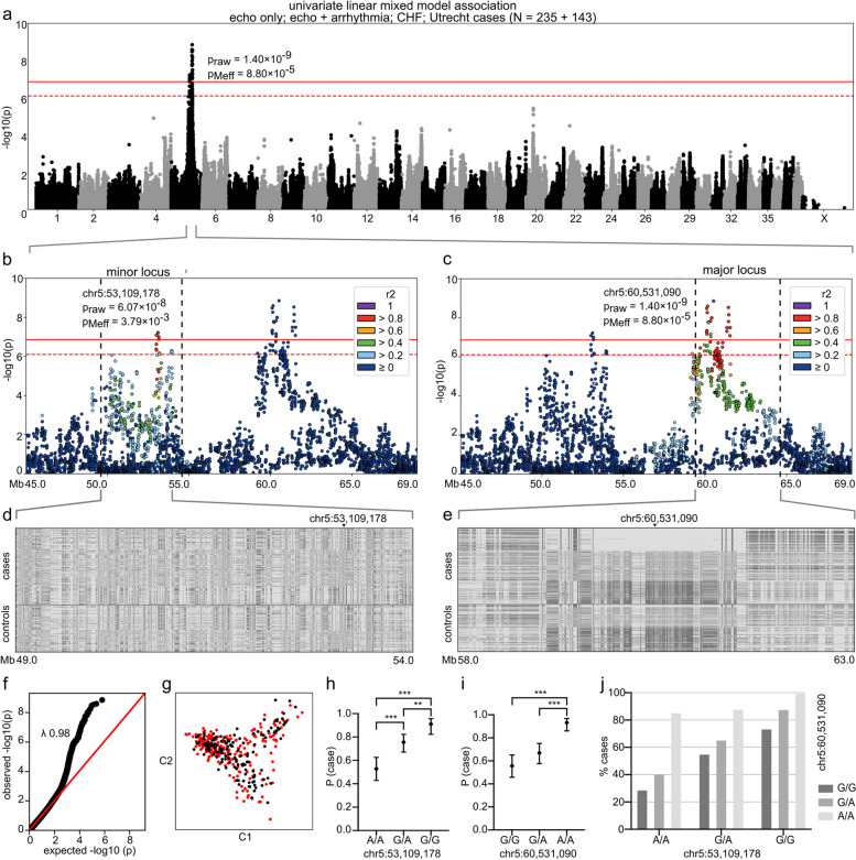
Methods: We performed GWAS followed by whole-genome, transcriptome, and immunohistochemical analyses in a spontaneously occurring canine model of DCM. Canine gene discovery was followed up in three human DCM cohorts.

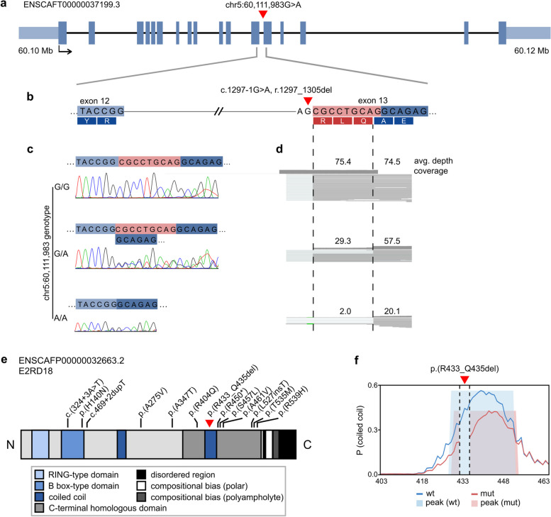
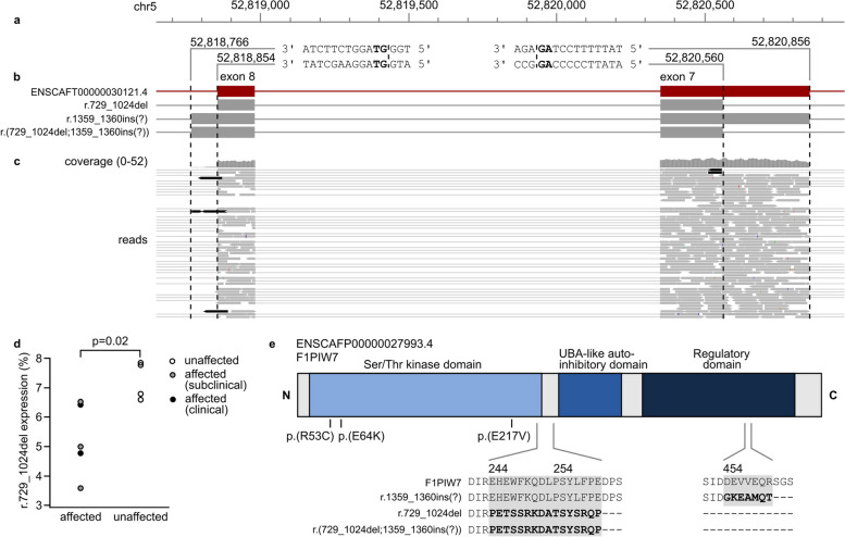
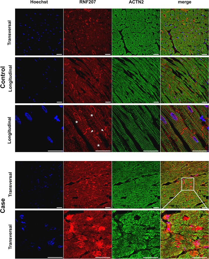
Results: Our results revealed two independent additive loci associated with the typical DCM phenotype comprising left ventricular systolic dysfunction and dilatation. We highlight two novel candidate genes, RNF207 and PRKAA2, known for their involvement in cardiac action potentials, energy homeostasis, and morphology. We further illustrate the distinct genetic etiologies underlying the typical DCM phenotype and ventricular premature contractions. Finally, we followed up on the canine discoveries in human DCM patients and discovered candidate variants in our two novel genes.

Conclusions: Collectively, our study yields insight into the molecular pathophysiology of DCM and provides a large animal model for preclinical studies.

Keywords: Arrhythmia; Cardiac; Cardiology; Companion animal; Complex trait; GWAS; Genetics; Transcriptomics.


Conflict of interest statement

HL has consulted, and JD is an employee of Wisdom Panel, Kinship, which offers canine DNA testing as a commercial service. The remaining authors declare that they have no competing interests.

Figures

Fig. 1
Fig. 1
GWAS with a univariate linear mixed model. The analysis included 235 cases from the “echo only,” “echo + arrhythmia,” and “CHF” subcohorts and Utrecht cohort and 143 controls from the “healthy” subcohort. In ac, Bonferroni-corrected and Meff significance thresholds are indicated with solid and dashed red lines, respectively. In d and e, the colors indicate the homozygous genotype for the case major allele (light gray), heterozygous genotype (middle gray), and homozygous genotype for the case minor allele (dark gray). In h and i, error bars indicate 95% confidence limits and asterisks ** and *** p-values < 0.01 and < 0.001, respectively. a A genome-wide significant two-locus signal occurs on chromosome 5: the major locus resides at 60 Mb and the minor locus at 53 Mb. The most significant SNP (praw = 1.40 × 10–9, pMeff = 8.80 × 10–5) is located at chr5:60,531,090. b A locus plot of chr5:45.0–69.0 Mb and SNP correlation structure of the index SNP chr5:53,109,178. c A locus plot of chr5:45.0–69.0 Mb and SNP correlation structure of the index SNP chr5:60,531,090. d Genotype plot of chr5:59–54 Mb. e Genotype plot of chr5:58–63 Mb. f Q-Q plot of the p-values (likelihood ratio test). g A multi-dimensional scaling (MDS) plot of the cases (red) and controls (black). h Probability of case status (P (case)) by genotype at chr5:53,109,178 (N = 372). i Probability of case status (P (case)) by genotype at chr5:60,531,090 (N = 372). j Frequency of case status by joint genotypes at ch5:53,109,178 and chr5:60,531,090 (N = 372)
Fig. 2
Fig. 2
Alternative splicing and potentially damaging human variants in RNF207. The position of the canine RNF207 splice variant (chr5:60,111,983G > A) is indicated with a red triangle. a Exon-intron structure of the canine RNF207 transcript (ENSCAFT00000037199.3). b The location of the variant at the splice acceptor site of exon 13 (ENSCAFT00000037199.3:c.1297-1G > A, r.1297_1305del). c Representative chromatograms of RNF207 cDNA in Dobermanns with different chr5:60,111,983 genotypes. The first nine bases of exon 13 are skipped in dogs with at least one copy of the A allele. d The average read depth and coverage at the variant site in the mRNA-seq data of the same Dobermanns as in c. Reads without the first nine bases of exon 13, indicated within the dashed lines, are observed in dogs with at least one copy of the A allele. Two reads that likely comprised pre-mRNA are observed at the deletion site in the A/A dog. e A schematic representation of the canine RNF207 protein (ENSCAFP00000032663.2, E2RD18). The p.(R433_Q435del) variant is indicated with a red triangle, and positions corresponding to protein-changing, potentially damaging variants in human cardiomyopathy patients with black lines. f Coiled-coil conformation of the wild type and p.(R433_Q435del) sequences at residues 403–463 as predicted by DeepCoil2. The coiled-coil domain may be affected by the loss of residues 433–435 in the altered protein. The propensity of coiled-coil conformation and detected peaks are indicated in blue and light blue for the wild-type sequence and in red and pink for the mutated sequence
Fig. 3
Fig. 3
Alternative splicing and potentially damaging human variants in PRKAA2. a Sequences of splice sites observed in canine RNA-seq data. The canonical GT and AG splice site sequences at the exon-intron junctions are indicated in bold. b Schematic representation of canine PRKAA2 transcripts and boundaries of exons 7 and 8: ENSCAFT00000030121.4, exon 7 deletion (chr5:52,820,560–52,820,856, r.729_1024del) mutant, intron 8 retention (chr5:52,818,766–52,818,853, c.1359-1360ins(?)) mutant, and a predicted transcript with both aberrant splice sites (r.(729_1024del;1359_1360ins(?))). c Alternate exon 7 and 8 splicing in the mRNA-seq reads of one affected Dobermann. The aberrant reads are indicated in black. d Proportional expression of the exon 7 mutant transcript detected with ddPCR in five DCM-affected and four unaffected Dobermanns. e Schematic representation of the canine PRKAA2 protein (ENSCAFP00000027993.4, F1PIW7) and the predicted amino acid sequences of the aberrant transcripts. The r.729_1024del variant is predicted to result in truncation upstream of the UBA-like autoinhibitory domain, and the c.1359-1360ins(?) transcript is predicted to contain a premature stop codon before the C-terminal regulatory domain involved in heterotrimerization. Positions corresponding to potentially damaging variants in human cardiomyopathy patients are indicated with black lines. ENSCAFT00000030121.4 was used as a reference sequence for the mutant transcripts, and domain annotation was obtained from CDD
Fig. 4
Fig. 4
Immunofluorescent staining of RNF207 in canine cardiac tissue. Healthy canine non-Dobermann cardiac tissue (top three rows) was stained for RNF207 to detect the differences in the expression or localization associated with disease and genotype. Additionally, tissue was stained for Hoechst in blue to show the nuclei and for actinin alpha 2 (ACTN2) in green to visualize the sarcomere structures. Localization is indicated from a transversal and longitudinal perspective. RNF207 is expressed in the cytoplasm of cardiomyocytes, intercalated disks (arrows), and perinuclearly (asterisks). In the cardiac tissue of the single DCM case homozygous for the RNF207c.1297-1G > A variant, a patchy expression resembling cellular mosaicism was detected (bottom row)

Similar articles

Cited by

  • Genome-wide association analysis provides insights into the molecular etiology of dilated cardiomyopathy.
    Zheng SL, Henry A, Cannie D, Lee M, Miller D, McGurk KA, Bond I, Xu X, Issa H, Francis C, De Marvao A, Theotokis PI, Buchan RJ, Speed D, Abner E, Adams L, Aragam KG, Ärnlöv J, Raja AA, Backman JD, Baksi J, Barton PJR, Biddinger KJ, Boersma E, Brandimarto J, Brunak S, Bundgaard H, Carey DJ, Charron P, Cook JP, Cook SA, Denaxas S, Deleuze JF, Doney AS, Elliott P, Erikstrup C, Esko T, Farber-Eger EH, Finan C, Garnier S, Ghouse J, Giedraitis V, Guðbjartsson DF, Haggerty CM, Halliday BP, Helgadottir A, Hemingway H, Hillege HL, Kardys I, Lind L, Lindgren CM, Lowery BD, Manisty C, Margulies KB, Moon JC, Mordi IR, Morley MP, Morris AD, Morris AP, Morton L, Noursadeghi M, Ostrowski SR, Owens AT, Palmer CNA, Pantazis A, Pedersen OBV, Prasad SK, Shekhar A, Smelser DT, Srinivasan S, Stefansson K, Sveinbjörnsson G, Syrris P, Tammesoo ML, Tayal U, Teder-Laving M, Thorgeirsson G, Thorsteinsdottir U, Tragante V, Trégouët DA, Treibel TA, Ullum H, Valdes AM, van Setten J, van Vugt M, Veluchamy A, Verschuren WMM, Villard E, Yang Y; COVIDsortium; DBDS Genomic Consortium; Estonian Biobank Research Team; HERMES Consortium; Asselbergs FW, Cappola TP, Dube MP, Dunn ME, Ellinor PT, Hingorani AD, Lang CC, … See abstract for full author list ➔ Zheng SL, et al. Nat Genet. 2024 Dec;56(12):2646-2658. doi: 10.1038/s41588-024-01952-y. Epub 2024 Nov 21. Nat Genet. 2024. PMID: 39572783 Free PMC article.
  • Association of the TTN, PDK4, and RNF207 mutations with dilated cardiomyopathy in Dobermanns from the United Kingdom.
    Dutton LC, Crosland A, Dukes-McEwan J, Connolly DJ. Dutton LC, et al. PLoS One. 2025 Mar 13;20(3):e0319932. doi: 10.1371/journal.pone.0319932. eCollection 2025. PLoS One. 2025. PMID: 40080479 Free PMC article.
  • AMPK Signalling Pathway: A Potential Strategy for the Treatment of Heart Failure with Chinese Medicine.
    Liu C, Guo X, Zhou Y, Wang H. Liu C, et al. J Inflamm Res. 2023 Nov 21;16:5451-5464. doi: 10.2147/JIR.S441597. eCollection 2023. J Inflamm Res. 2023. PMID: 38026240 Free PMC article. Review.
  • Advances in genetic diagnosis and therapy of hereditary heart disease: a bibliometric review from 2004 to 2024.
    Ma H, Wang Y, Jia Y, Xie L, Liu L, Zhang D, Ma X, Guo Y, Xu R. Ma H, et al. Front Med (Lausanne). 2025 Jan 8;11:1507313. doi: 10.3389/fmed.2024.1507313. eCollection 2024. Front Med (Lausanne). 2025. PMID: 39845823 Free PMC article. Review.
  • The DoGA consortium expression atlas of promoters and genes in 100 canine tissues.
    Hörtenhuber M, Hytönen MK, Mukarram AK, Arumilli M, Araujo CL, Quintero I, Syrjä P, Airas N, Kaukonen M, Kyöstilä K, Niskanen J, Jokinen TS, Mottaghitalab F, Takan I, Salokorpi N, Raman A, Stevens I, Iivanainen A, Yoshihara M, Gusev O, Bannasch D, Sukura A, Schoenebeck JJ; DoGA Consortium; Ezer S, Katayama S, Daub CO, Kere J, Lohi H. Hörtenhuber M, et al. Nat Commun. 2024 Oct 21;15(1):9082. doi: 10.1038/s41467-024-52798-1. Nat Commun. 2024. PMID: 39433728 Free PMC article.

References

    1. McNally EM, Mestroni L. Dilated cardiomyopathy: genetic determinants and mechanisms. Circ Res. 2017;121:731–748. doi: 10.1161/CIRCRESAHA.116.309396. - DOI - PMC - PubMed
    1. Jordan E, et al. Evidence-based assessment of genes in dilated cardiomyopathy. Circulation. 2021;144:7–19. doi: 10.1161/CIRCULATIONAHA.120.053033. - DOI - PMC - PubMed
    1. Tayal U, Prasad S, Cook SA. Genetics and genomics of dilated cardiomyopathy and systolic heart failure. Genome Med. 2017;9:20. doi: 10.1186/s13073-017-0410-8. - DOI - PMC - PubMed
    1. Esslinger U, et al. Exome-wide association study reveals novel susceptibility genes to sporadic dilated cardiomyopathy. PLoS One. 2017;12:e0172995. doi: 10.1371/journal.pone.0172995. - DOI - PMC - PubMed
    1. Aragam KG, et al. Phenotypic refinement of heart failure in a national biobank facilitates genetic discovery. Circulation. 2018 doi: 10.1161/CIRCULATIONAHA.118.035774. - DOI - PMC - PubMed

Publication types