

Choroid vascular changes in hyperopic anisometropia amblyopia using SS-OCTA
- PMID: 37723524
- PMCID: PMC10506216
- DOI: 10.1186/s12886-023-03121-x
Choroid vascular changes in hyperopic anisometropia amblyopia using SS-OCTA
Abstract
Purpose: To observe and understand the structural changes in choroidal vessels in eyes with hyperopic anisometropic amblyopia using swept-source optical coherence tomography angiography (SS-OCTA).
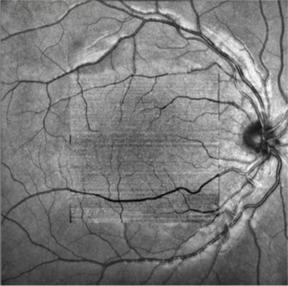
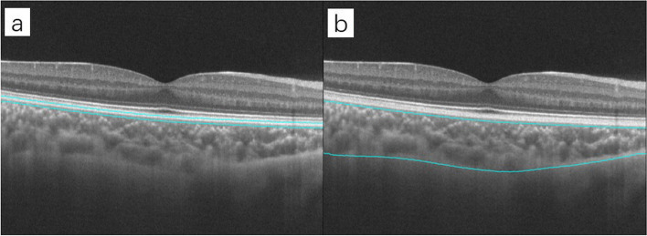
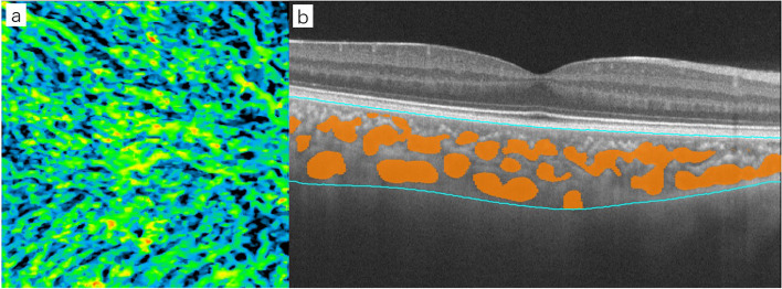
Methods: A total of 44 patients were enrolled in this study: 22 children with hyperopic anisometropic amblyopia and 22 age-matched controls. SS-OCTA was used to scan the 6*6 mm macular area of their eyes. The average choroidal thickness (CT) and choroidal capillary flow area (CC) in a 3 mm diameter area centered on the macular area were obtained. The choroidal vascularity volume (CVV) was automatically extracted and 3D reconstructed by inbuild software, and the three-dimensional choroidal vascularity index (3D-CVI) was calculated. The effect of amblyopia on the choroidal vessel structure was assessed using generalized linear estimating equations (GEEs) corrected for axial length, sex, age, and best-corrected visual acuity.
Results: The CC was greater in amblyopic eyes than in fellow eyes (P = 0.014) but was not significantly different from that in control eyes (P = 0.963). After correcting for sex, age, axial length, and visual acuity using GEEs, the mean CT in the amblyopic eyes was greater than that in the fellow eyes (P = 0.030) but was not significantly different from that in the control eyes (P = 0.160). The 3D-CVI in amblyopic eyes was higher than that in control eyes (P = 0.038) but was not significantly different from that in fellow eyes (P = 0.407). The three-dimensional choroidal vascularity volume (3D-CVV) was higher in amblyopic eyes than in fellow eyes (P = 0.046) and control eyes (P = 0.023).
Conclusions: We found that eyes with hyperopic anisometropic amblyopia demonstrated higher CT, CC and 3D-CVV values than the contralateral eyes after correction, while the 3D-CVI was unchanged. Compared with control eyes, amblyopic eyes had higher 3D-CVV and 3D-CVI values but similar CT and CC values. Amblyopic eyes may have different choroidal vascular structures from fellow and control eyes.
Keywords: Amblyopia; Anisometropia; Choroid; Choroidal capillary; Choroidal thickness; Choroidal vascularity index; Choroidal vascularity volume; Hyperopia; OCTA.
© 2023. BioMed Central Ltd., part of Springer Nature.
Conflict of interest statement
The authors declare no competing interests.
Figures

Similar articles
-
Choroidal vascular characteristics in anisometropic amblyopia: a comparative analysis.BMC Ophthalmol. 2025 May 21;25(1):301. doi: 10.1186/s12886-025-04143-3. BMC Ophthalmol. 2025. PMID: 40399854 Free PMC article.
-
Subfoveal Choroidal Thickness and Axial Length in Preschool Children with Hyperopic Anisometropic Amblyopia.Curr Eye Res. 2015 Sep;40(9):954-61. doi: 10.3109/02713683.2014.964418. Epub 2014 Oct 20. Curr Eye Res. 2015. PMID: 25330225
-
Assessment of the effects of myopic and hyperopic anisometropia on choroidal vascular structure in children using SS-OCTA.Ophthalmic Physiol Opt. 2024 May;44(3):525-536. doi: 10.1111/opo.13300. Epub 2024 Mar 8. Ophthalmic Physiol Opt. 2024. PMID: 38456753
-
Assessment of choroidal vascularity and choriocapillaris blood perfusion in Chinese preschool-age anisometropic hyperopic amblyopia children.Front Pediatr. 2022 Nov 17;10:1056888. doi: 10.3389/fped.2022.1056888. eCollection 2022. Front Pediatr. 2022. PMID: 36467467 Free PMC article.
-
[Macular thickness in unilateral amblyopia as measured by optical coherence tomography: a meta analysis].Zhonghua Yan Ke Za Zhi. 2014 Jul;50(7):504-10. Zhonghua Yan Ke Za Zhi. 2014. PMID: 25312459 Review. Chinese.
Cited by
-
Rapid myopization of the fellow eye in anisometropic amblyopia treated with 1% atropine: a case report.BMC Ophthalmol. 2025 Feb 17;25(1):77. doi: 10.1186/s12886-025-03910-6. BMC Ophthalmol. 2025. PMID: 39962389 Free PMC article.
-
Artificial Intelligence-Assisted Perfusion Density as Biomarker for Screening Diabetic Nephropathy.Transl Vis Sci Technol. 2024 Oct 1;13(10):19. doi: 10.1167/tvst.13.10.19. Transl Vis Sci Technol. 2024. PMID: 39388177 Free PMC article.
-
Choroidal vascular characteristics in anisometropic amblyopia: a comparative analysis.BMC Ophthalmol. 2025 May 21;25(1):301. doi: 10.1186/s12886-025-04143-3. BMC Ophthalmol. 2025. PMID: 40399854 Free PMC article.
-
Retinal and choroidal microvasculature and structural analysis in OCTA for refractive amblyopia diagnosis using machine learning.J Optom. 2025 Jul-Sep;18(3):100555. doi: 10.1016/j.optom.2025.100555. Epub 2025 May 6. J Optom. 2025. PMID: 40334350 Free PMC article.
References
-
- von Noorden GK. Amblyopia: a multidisciplinary approach. Proctor lecture Invest Ophthalmol Vis Sci. 1985;26(12):1704–1716. - PubMed
MeSH terms
LinkOut - more resources
Full Text Sources
Medical
