. 2023 Nov 2;15(11):mfad054.
doi: 10.1093/mtomcs/mfad054.

In vitro maturation of NiSOD reveals a role for cytoplasmic histidine in processing and metalation

Affiliations

In vitro maturation of NiSOD reveals a role for cytoplasmic histidine in processing and metalation

Priyanka Basak et al. Metallomics. .

Abstract

The importance of cellular low molecular weight ligands in metalloenzyme maturation is largely unexplored. Maturation of NiSOD requires post-translational N-terminal processing of the proenzyme, SodN, by its cognate protease, SodX. Here we provide evidence for the participation of L-histidine in the protease-dependent maturation of nickel-dependent superoxide dismutase (NiSOD) from Streptomyces coelicolor. In vitro studies using purified proteins cloned from S. coelicolor and overexpressed in E. coli support a model where a ternary complex formed between the substrate (SodN), the protease (SodX) and L-Histidine creates a novel Ni-binding site that is capable of the N-terminal processing of SodN and specifically incorporates Ni into the apo-NiSOD product. Thus, L-Histidine serves many of the functions associated with a metallochaperone or, conversely, eliminates the need for a metallochaperone in NiSOD maturation.

Keywords: in vitro maturation; low-molecular weight ligands; nickel; post-translational modification; protease (SodX); superoxide dismutase.


Conflict of interest statement

The authors declare no competing financial interests.

Figures

Graphical Abstract
Graphical Abstract
In vitro maturation of SodN.
Fig. 1
Fig. 1
The nickel active site in NiSOD (PDB ID: 1T6U). The inset shows the hexameric enzyme viewed down a threefold axis. Green spheres represent nickel atoms. The oxidized enzyme is 50:50 mixture of Ni(II) and Ni(III) where the latter is stabilized by an additional His1 coordination (dashed line).
Fig. 2
Fig. 2
Ni(II)-binding results for SodN and H15A-SodN using isothermal titration calorimetry (ITC). ITC thermograms (top) and binding isotherms (bottom) for titrations of (A) 0.15 mM of apo-SodN (blue) and (B) 0.15 mM apo H15A-SodN (cyan) with 2 mM Ni(II) in buffer containing 20 mM Tris, 200 mM NaCl, 1 mM TCEP, pH 8.0. One-site binding models were used to fit the isotherms in each case (solid black lines) to give binding stoichiometries of 1.03(2) and 0.47(2) Kd values of 21.1(1.7) μM and 20(2) μM, respectively.
Fig. 3
Fig. 3
Spectroscopic characterization of nickel complexes of NiSodN and NiSodN•His.(A) Overlay of the UV–vis spectra of 100 μM Ni(II)-SodN (blue), as-isolated recombinant NiSOD (orange), dithionite-reduced recombinant NiSOD (wine) plotted vs. ε and for XAS sample of NiSodN•His (green) plotted vs. absorbance. (B) Overlay of the normalized Ni K-edge XANES region of the XAS spectra of Ni-SodN (blue) and NiSodN•His (green) in 50 mM Tris, 200 mM NaBr, pH 8.0 compared with dithionite-reduced NiSOD (orange, Ref.16). Inset: Enlargement of the pre-edge region. (C) k3-weighted unfiltered EXAFS data for NiSodN (blue) and best-fit model from Table 2 (black). (D) Fourier-transformed EXAFS (k = 2–12.5 Å−1) data for NiSodN (blue) uncorrected for phase shifts and the best-fit model (black) from Table 2. (E) k3-weighted unfiltered EXAFS data for NiSodN•His (green) and best-fit model from Table 2 (black). (F) Fourier-transformed EXAFS (k = 2–12.5 Å−1) data for NiSodN•His (green) uncorrected for phase shifts and the best-fit model (black) from Table 2.
Fig. 4
Fig. 4
Homology model of SodX bound to the SodN peptide substrate. The SodX structure is shown in tan, and represented as a surface. The active site Ser28 nucleophile is colored magenta. The site of the glycan modification (residues 125–142) is shown in slate (backside). The inhibitor peptide bound to E. coli signal peptidase is shown in grey sticks, with heteroatoms colored, and likely corresponds to the position of the pro- peptide of SodN. The Ni-hook peptide (AHCDLPC; running vertically in the image in stick form, Ala—cyan, His—blue, Cys—yellow, CPL (Cys6-Pro5-Leu4)—green, with heteroatoms colored) was generated in PyMol and manually positioned to highlight the likely binding site with respect to Ser28.
Fig. 5
Fig. 5
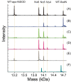
Deconvoluted ESI–MS spectra of proteolytic reaction mixtures containing SodN, SodX and free metals (Ni, Co, Zn). Reaction mixtures in assay buffer (50 mM Tris•HCl, pH 8.0) containing (A) 40 μM apo-SodN + 40 μM SodX (B) 40 μM SodN incubated with 80 μM NiCl2 (C) 40 μM SodX added to b (D) 40 μM SodX + 40 μM SodN pre-incubated with 80 μM CoCl2 (E) 40 μM SodX + 40 μM SodN pre-incubated with 80 μM ZnCl2. (F) 40 μM apo-H15A-SodN + 40 μM SodX.
Fig. 6
Fig. 6
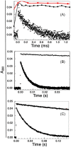
Activity assessment of NiSOD in cleavage and control experiments using pulse radiolysis. Kinetic traces showing the disappearance of pulse-radiolytically generated superoxide radical by monitoring optical absorbance at 260 nm (ε = 4000 M−1cm−1) for buffer (top trace in all panels), which represents the rate for the uncatalyzed bimolecular disproportionation of O2•−), NiSodN (panel A, •-•-•) and catalytic curves (bottom trace in all panels) for WT-NiSOD (panel A), metal-free cleavage reaction mixture (SodN + SodX) followed by Ni(II) addition (panel B) and cleavage reaction mixture (SodN + Ni + SodX) containing 0.01 mM L-histidine (panel C). The activity was assessed relative to Ni concentrations in each experiment.
Fig. 7
Fig. 7
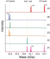
Deconvoluted ESI–MS traces of proteolysis reaction mixtures (40 μM SodX + 40 μM SodN) in assay buffer (50 mM Tris, 1 mM TCEP, pH 8.0) containing L-histidine. (A) with 0.1 mM L-histidine, (B) + 80 μM NiCl2 with 0.01 mM L-histidine, (C) + 80 μM NiCl2 with 0.10 mM L-histidine, (D) + 80 μM NiCl+ 1.0 mM L-histidine, (E) +80 μM NiCl2 with 10.0 mM L-histidine, (F) + 80 μM CoCl2 with 0.10 mM L-histidine.
Fig. 8
Fig. 8
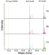
High-resolution deconvoluted ESI–MS traces of proteolytic reaction mixtures containing SodN, SodX, and Ni at different concentrations of D-histidine. Reaction mixtures containing 40 μM SodX and 40 μM SodN pre-incubated with 80 μM NiCl2 in assay buffer (50 mM Tris•HCl, 1 mM TCEP, pH 8.0) with (A) 0.010 mM D-histidine (B) 0.100 mM D-histidine (C) 1.0 mM D-histidine.
Scheme 1.
Scheme 1.
Scheme summarizing the in vitro N-terminal processing of SodN by SodX. Pathway A indicates that cleavage of SodN by SodX produces apo-NiSOD that can subsequently be nickelated to give active NiSOD. Pathway B indicates that in the presence of Ni(II) SodX does not cleave SodN to give NiSOD, likely due to the formation of a Ni(II) complex involving coordination of His15 at the cleavage site (see text). Pathway C indicates that the addition of L-His allows SodX cleavage of SodN in the presence of Ni(II), which occurs via a unique SodN•SodX•L-His ternary Ni complex (see text). Pathway D illustrates that cleavage in the absence of Ni(II) is inhibited by L-His.

Similar articles

Cited by

References

    1. Ackerman C. M., Lee S., Chang C. J., Analytical Methods for Imaging Metals in Biology: from Transition Metal Metabolism to Transition Metal Signaling, Anal. Chem., 2017, 89(1), 22–41. 10.1021/acs.analchem.6b04631 - DOI - PMC - PubMed
    1. Chandrangsu P., Rensing C., Helmann J. D., Metal Homeostasis and Resistance in Bacteria, Nat. Rev. Micro., 2017, 15(6), 338–350. 10.1038/nrmicro.2017.15 - DOI - PMC - PubMed
    1. Brawley H. N., Lindahl P. A., Low-Molecular-Mass Labile Metal Pools in Escherichia coli: Advances Using Chromatography and Mass Spectrometry, J. Biol. Inorg. Chem., 2021, 26(4), 479–494. 10.1007/s00775-021-01864-w - DOI - PMC - PubMed
    1. Callahan D. L., Baker A. J., Kolev S. D., Wedd A. G., Metal Ion Ligands in Hyperaccumulating Plants, J. Biol. Inorg. Chem., 2006, 11(1), 2–12. 10.1007/s00775-005-0056-7 - DOI - PubMed
    1. Montargès-Pelletier E., Chardot V., Echevarria G., Michot L. J., Bauer A., Morel J.-L., Identification of Nickel Chelators in Three Hyperaccumulating Plants: an X-Ray Spectroscopic Study, Phytochemistry, 2008, 69(8), 1695–1709. 10.1016/j.phytochem.2008.02.009 - DOI - PubMed

Publication types

LinkOut - more resources