. 2023 Oct;11(8):750-766.
doi: 10.1002/ueg2.12463. Epub 2023 Sep 18.

miR-10b-5p rescues leaky gut linked with gastrointestinal dysmotility and diabetes

Affiliations

miR-10b-5p rescues leaky gut linked with gastrointestinal dysmotility and diabetes

Hannah Zogg et al. United European Gastroenterol J. 2023 Oct.

Erratum in

Abstract

Background/aim: Diabetes has substantive co-occurrence with disorders of gut-brain interactions (DGBIs). The pathophysiological and molecular mechanisms linking diabetes and DGBIs are unclear. MicroRNAs (miRNAs) are key regulators of diabetes and gut dysmotility. We investigated whether impaired gut barrier function is regulated by a key miRNA, miR-10b-5p, linking diabetes and gut dysmotility.

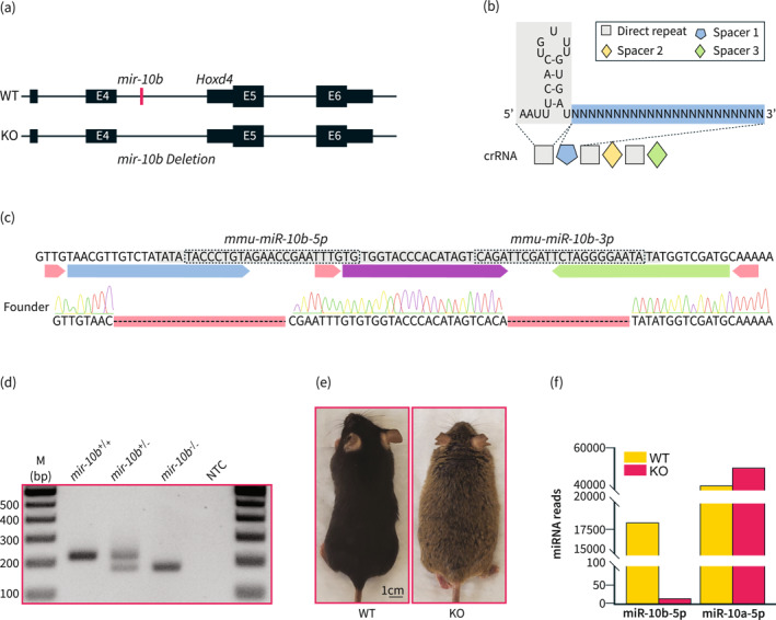
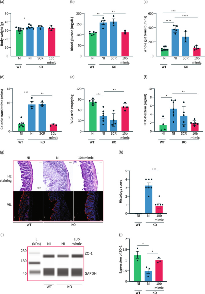
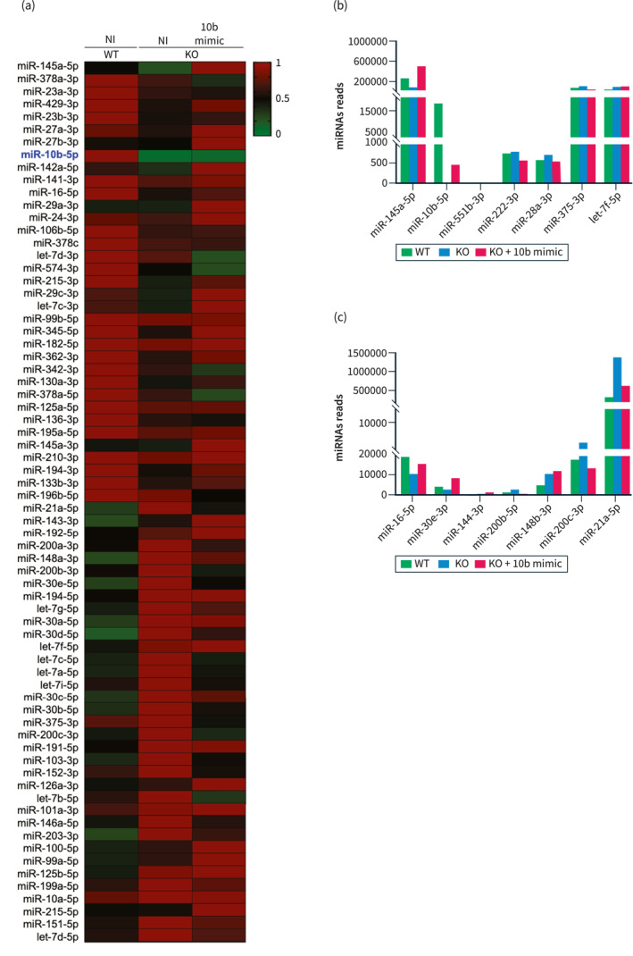
Methods: We created a new mouse line using the Mb3Cas12a/Mb3Cpf1 endonuclease to delete mir-10b globally. Loss of function studies in the mir-10b knockout (KO) mice were conducted to characterize diabetes, gut dysmotility, and gut barrier dysfunction phenotypes in these mice. Gain of function studies were conducted by injecting these mir-10b KO mice with a miR-10b-5p mimic. Further, we performed miRNA-sequencing analysis from colonic mucosa from mir-10b KO, wild type, and miR-10b-5p mimic injected mice to confirm (1) deficiency of miR-10b-5p in KO mice, and (2) restoration of miR-10b-5p after the mimic injection.

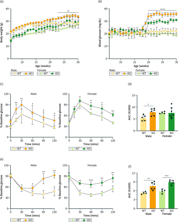
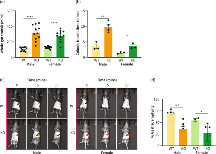
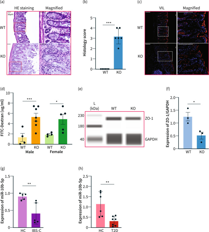
Results: Congenital loss of mir-10b in mice led to the development of hyperglycemia, gut dysmotility, and gut barrier dysfunction. Gut permeability was increased, but expression of the tight junction protein Zonula occludens-1 was reduced in the colon of mir-10b KO mice. Patients with diabetes or constipation- predominant irritable bowel syndrome, a known DGBI that is linked to leaky gut, had significantly reduced miR-10b-5p expression. Injection of a miR-10b-5p mimic in mir-10b KO mice rescued these molecular alterations and phenotypes.

Conclusions: Our study uncovered a potential pathophysiologic mechanism of gut barrier dysfunction that links both the diabetes and gut dysmotility phenotypes in mice lacking miR-10b-5p. Treatment with a miR-10b-5p mimic reversed the leaky gut, diabetic, and gut dysmotility phenotypes, highlighting the translational potential of the miR-10b-5p mimic.

Keywords: diabetic dysmotility; gastroparesis; intestinal barrier dysfunction; irritable bowel syndrome; microRNAs.


Conflict of interest statement

This author discloses the following: Seungil Ro and the University of Nevada Reno Office of Technology Transfer (serial no. 62/837,988, filed 24 April 2019) have published a PCT International Patent WO/2020/219872 entitled “Methods and compositions of miR‐10 mimics and targets thereof.” Seungil Ro is an employee and a member of the board of directors of RosVivo Therapeutics. Rajan Singh and Se Eun Ha are members of the board of directors of RosVivo Therapeutics. The remaining authors disclose no conflicts.

Figures

FIGURE 1
FIGURE 1
Generation of global mir‐10b knockout mice. (a) Schematic of the deletion of mir‐10b. mir‐10b is located within the intron of Hoxd4. (b) Schematic of the crRNA used to create the mir‐10b knockout (KO) mouse. (c) Sequencing results confirming the deletion of both miR‐10b‐5p and miR‐10b‐3p. (d) PCR confirmation of the mir‐10b KO mice. (e) Gross body image of 5‐month‐old male WT and 5‐month‐old male mir‐10b KO mice, which are from the same C57BL6J background. (f) miRNA reads of miR‐10b‐5p and miR‐10a‐5p in the colonic mucosa from WT and KO mice obtained by miRNA‐seq (n = 3). WT, wild type.
FIGURE 2
FIGURE 2
Knockout of mir‐10b leads to body weight gain and impaired glucose homeostasis. (a) Body weight comparison of both male and female mir‐10b knockout (KO) and WT mice. (b) Fasting blood glucose comparison of both male and female KO and WT mice. (c) Glucose tolerance tests of both male and female KO and WT mice. (d) AUC from (c). (e) Insulin tolerance tests of both male and female KO and WT mice. (f) AUC from (e). n = 4–7 per condition for each experiment. Error bar indicates mean ± SD, 2‐way analysis of variance (ANOVA). *p < 0.05; **p < 0.01; ***p < 0.001; ****p < 0.0001. WT, wild type.
FIGURE 3
FIGURE 3
mir‐10b knockout (KO) mice develop delayed gut transit. (a) Whole gut transit time comparison of both male and female mir‐10b KO and WT mice. (b) Colonic transit time comparison of both male and female KO and WT mice. (c) Gastric emptying images of both male and female KO and WT mice. (d) Quantification of percent gastric emptying after 30 min of both male and female KO and WT mice. n = 3–11 per condition for each experiment. Error bar indicates mean ± SD. *p < 0.05; **p < 0.01; ***p < 0.001; ****p < 0.0001. WT, wild type.
FIGURE 4
FIGURE 4
mir‐10b knockout (KO) mice develop the leaky gut phenotype. (a) H&E staining of colon cross sections from mir‐10b KO and WT 5‐month‐old male mice. Scale bars are 50 μm. (b) Histology score comparison of KO and WT 5‐month‐old male mice (n = 3) (c) Villin (red) and DAPI (blue) staining of colon cross sections from KO and WT mice. Scale bars are 100 μm. (d) Amount of FITC‐Dextran found in serum from both male and female KO and WT mice. (e), (f) Western blot and quantification of ZO‐1 expression from the colon of KO and WT male mice. (g), (h) miR‐10b‐5p expression in serum samples from patients with constipation‐predominant irritable bowel syndrome (IBS‐C) and type 2 diabetes (T2D), and healthy controls (HC) normalized by Snord15a expression (qPCR). n = 3–12 per condition for each experiment. Error bar indicates mean ± SD. *p < 0.05; ***p < 0.001.
FIGURE 5
FIGURE 5
Treatment with the miR‐10b mimic rescues the hyperglycemic, gut dysmotility, and leaky gut phenotypes. (a) Body weight comparison between WT non‐injected (NI) 5‐month‐old male mice, mir‐10b knockout (KO) 5‐month‐old male mice either injected with the scramble (SCR) RNA or the miR‐10b (10b) mimic, and NI KO 5‐month‐old male mice. (b) Blood glucose comparison between NI WT, NI KO, SCR KO, and 10b mimic injected KO mice. (c) Comparison of whole gut transit time between NI WT, NI KO, SCR KO, and 10b mimic injected KO mice. (d) Colonic transit time comparison between NI WT, NI KO, SCR KO, and 10b mimic injected KO mice. (e) Comparison of percent gastric emptying between NI WT, NI KO, SCR KO, and 10b mimic injected KO mice. (f) Amount of FITC‐Dextran found in serum from NI WT, NI KO, and 10b mimic injected KO mice. (g) Top: H&E staining of colon cross sections from WT, KO, and mimic injected KO mice. Scale bars are 50 μm. Bottom: Villin (red) and DAPI (blue) staining of colon cross sections from WT, KO, and mimic injected KO mice. Scale bars are 100 μm. (h) Histology score comparison NI WT, NI KO, and 10b mimic injected KO mice (n = 3) (i), (j) Western blot and quantification of ZO‐1 expression from the colon of WT, KO, and mimic injected KO mice. n = 3‐7 per condition for each experiment. Error bar indicates mean ± SD, 1‐way ANOVA. *p < 0.05; **p < 0.01; ***p < 0.001; ****p < 0.0001.
FIGURE 6
FIGURE 6
Dysregulated and restored miRNAs in the leaky gut of mir‐10b knockout (KO) male mice. (a) Heat map showing the normalized expression of differentially expressed miRNAs in the colonic mucosa from mir‐10b WT and KO 5‐month‐old mice injected with miR‐10b‐5p mimic or given no injection (NI), obtained by miRNA‐seq analysis. A heat map colors range from dark green to dark red, representing low and high expressions, respectively. (b) Dysregulated and restored miRNAs in the leaky gut of the mir‐10b KO mice and in the pathogenesis of gut dysmotility. (c) Dysregulated and restored miRNAs in the leaky gut of the mir‐10b KO mice and in the pathogenesis of intestinal barrier dysfunction.

Similar articles

Cited by

References

    1. Ogurtsova K, Guariguata L, Barengo NC, Ruiz PLD, Sacre JW, Karuranga S, et al. IDF diabetes Atlas: global estimates of undiagnosed diabetes in adults for 2021. Diabetes Res Clin Pract. 2022;183:109118. 10.1016/j.diabres.2021.109118 - DOI - PubMed
    1. Singh R, Zogg H, Wei L, Bartlett A, Ghoshal UC, Rajender S, et al. Gut microbial dysbiosis in the pathogenesis of gastrointestinal dysmotility and metabolic disorders. J Neurogastroenterol Motil. 2021;27(1):19–34. 10.5056/jnm20149 - DOI - PMC - PubMed
    1. Singh R, Zogg H, Ghoshal UC, Ro S. Current treatment options and therapeutic insights for gastrointestinal dysmotility and functional gastrointestinal disorders. Front Pharmacol. 2022;13:808195. 10.3389/fphar.2022.808195 - DOI - PMC - PubMed
    1. Camilleri M, Kuo B, Nguyen L, Vaughn VM, Petrey J, Greer K, et al. ACG clinical guideline: gastroparesis. Am J Gastroenterol. 2022;117(8):1197–1220. 10.14309/ajg.0000000000001874 - DOI - PMC - PubMed
    1. Chen RY, Kung VL, Das S, Hossain MS, Hibberd MC, Guruge J, et al. Duodenal microbiota in stunted undernourished children with enteropathy. N Engl J Med. 2020;383(4):321–333. 10.1056/NEJMoa1916004 - DOI - PMC - PubMed

Publication types