. 2021 Oct 21;1(1):47-77.
doi: 10.1515/mr-2021-0007. eCollection 2021 Oct.

Sympatho-adrenergic mechanisms in heart failure: new insights into pathophysiology

Affiliations

Sympatho-adrenergic mechanisms in heart failure: new insights into pathophysiology

Xiaojun Du. Med Rev (2021). .

Abstract

The sympathetic nervous system is activated in the setting of heart failure (HF) to compensate for hemodynamic instability. However, acute sympathetic surge or sustained high neuronal firing rates activates β-adrenergic receptor (βAR) signaling contributing to myocardial remodeling, dysfunction and electrical instability. Thus, sympatho-βAR activation is regarded as a hallmark of HF and forms pathophysiological basis for β-blocking therapy. Building upon earlier research findings, studies conducted in the recent decades have significantly advanced our understanding on the sympatho-adrenergic mechanism in HF, which forms the focus of this article. This review notes recent research progress regarding the roles of cardiac β2AR or α1AR in the failing heart, significance of β1AR-autoantibodies, and βAR signaling through G-protein independent signaling pathways. Sympatho-βAR regulation of immune cells or fibroblasts is specifically discussed. On the neuronal aspects, knowledge is assembled on the remodeling of sympathetic nerves of the failing heart, regulation by presynaptic α2AR of NE release, and findings on device-based neuromodulation of the sympathetic nervous system. The review ends with highlighting areas where significant knowledge gaps exist but hold promise for new breakthroughs.

Keywords: catecholamines; heart failure; sympathetic nervous system; β-adrenergic receptor; β-blocking therapy.


Conflict of interest statement

Competing interests: The author has no conflict of interest to declare.

Figures

Figure 1:
Figure 1:
Cardiac sympatho-β-adrenergic signaling leads to compensatory as well as adverse consequences. Surges or sustained hyperactivity of sympathetic nervous drive to the heart might be compensatory for hemodynamic stability. Yet sympatho-β-adrenergic overactivation acts as a double-edged sword to increase occurrence of detrimental events, most notably myocardial remodeling and β1AR downregulation/desensitization, which may further amplify central and efferent sympathetic activity. AR: adrenergic receptor.
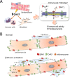
Figure 2:
Figure 2:
T-tubule localized β2AR signalosomes and redistribution of β2AR in the failing cardiomyocyte. A. Cardiomyocytes are rich in t-tubular structures, where sarcolemma membrane invaginate into the internal space of the cell body. T-tubules contain abundant lipid microdomains and caveolae and are rich in ion channels, e.g. L-type calcium channels, important for membrane potential and excitation-contraction (EC) coupling and mobilization of sarcoplasmic reticulum (SR) Ca2+ pool. T-tubules are the location of cluster of signaling proteins including G-proteins, adenylyl cyclase (AC), G-protein receptor kinase (GRK), A-kinase anchoring proteins (AKAP), protein kinase A (PKA), protein phosphatase 2A, and phosphodiesterases (PDE), forming highly efficient signalosomes. β2AR are also highly enriched in the caveolae/lipid microdomain of t-tubules where it induces local cAMP-PKA and Ca2+ signals. In comparison, β1AR are largely distributed throughout sarcolemma membrane and mediate a global cAMP-PKA and intracellular Ca2+ signals. B. In the failing myocardium, there is significant loss of t-tubular network and hence partial disintegration of signaling clusters. Hence, β2ARs undergo redistribution from t-tubules to sarcolemma membrane, which renders β2AR signaling upon ligand binding similar to that of β1AR. AR: adrenergic receptor.
Figure 3:
Figure 3:
Diagram depicting βAR signaling through the Hippo pathway in the heart. There is strong evidence for coupling of β-adrenergic and Hippo signaling pathways in the myocardium. βAR stimulation by genetic or pharmacological means effectively activates Hippo signaling featured by activation of upstream kinases Mst1 (mammalian strile-20 like kinase1) and Lats (large tumour suppressor homolog), and enhanced inhibitory Ser127-phosphorylation of YAP (yes-associated protein) and its paralogue TAZ (transcription coactivator with PDZ-binding motif). The latter results in cytoplasmic retention of YAP with increased binding to scaffold protein 14-3-3 and subsequent degradation via the ubiqutine/proteasome system. As a consequence, the activity of YAP as transcription co-activator or co-repressor, is turned off leading to altered expression of numerous YAP-target genes. Studies in vitro also suggest βAR/PKA-mediated direct Lats activation bypassing Mst (broken line). MOB1: Mps one binder kinase activator like 1; Sa: salvador homologue; TEAD: TEA-domain transcription factor. ⊗ Indicates epigenetic factors.
Figure 4:
Figure 4:
Sympathetic innervation of bone marrow and immune organs and βAR-mediated regulation of hematopoietic cells and immune cells. Sympathetic nerves, which innervate bones, can penetrate into the bone marrow compartment, where hematopoietic stem cells and progenitor cells (HSC) reside as specific niches. Catecholamines released by sympathetic varicosities activate hematopoiesis by stimulating β3AR in HSCs thereby promoting bone marrow cell differentiation, proliferation and egression of cells into blood circulation (a). After departing bone marrow, circulating immune cells relocate in immune organs like the spleen or lymph notes (b), where cells proliferate and mature and, upon sympathetic drive, depart immune organs into circulation (c). Circulating immune cells including monocytes (mono), lymphocytes (lym) and neutrophils (neu) then infiltrate the diseased myocardium (d) and promote regional inflammation. Immune cells are equipped mainly with β2ARs that regulate innate or acquired immunity, lymphocyte homing, immune cell maturation and expression of cytokines/chemokines. mϕ: macrophages; plate: platelet.
Figure 5:
Figure 5:
Cardiac phenotype of mice with dual deletion of β1AR and β2AR (β1/2-KO) under conditions of chronic pressure overload. Following a 12-week period of pressure overload, wild-type (WT) control mice displayed significant cardiac interstitial fibrosis (A), hypertrophy (B) and signs of HF (e.g. pulmonary congestion, LV dilatation and dysfunction, atrial thrombus formation). All these changes, together with upregulation of pro-fibrotic and inflammatory genes in the stressed myocardium, were abolished in dual β1/2-KO mice. These findings underscore an essential role of βAR signaling in mediating fibrosis and hypertrophy induced by pressure overload [148]. SH, sham-operation; TAC, transverse aorta constriction. Figure adopted from Kiriazis et al. with permission [148].
Figure 6:
Figure 6:
Schematic diagrams depicting βAR-mediated heterogeneous cell interactions promoting cardiac fibrosis. (A) Stimulation of βAR in cardiomyocytes induces expression and release of galectin-3 (Gal-3), which in turn activates expression of intermediate-conductance Ca2+-activated K+ (KCa3.1) channels in fibroblasts or immune cells. Activity of KCa3.1 channels leads to membrane hyperpolarization and subsequent Ca2+ influx via transient receptor potential (TRP) cation channels by which fibroblasts or inflammatory cells are activated [150]. (B) βAR activation in cardiomyocytes suppressed Cx43 expression and shifted Cx43 localization from interdisc to lateral sides of cells. Cx43 expression in fibroblasts is upregulated via direct activation of β2AR/PKA signaling cascade [142]. Besides, interleukin-18 (IL-18) released via βAR-mediated formation of inflammasomes in cardiomyocytes further upregulates Cx43 expression in fibroblasts [142]. These changes would favour the probability of cardiomyocyte-fibroblast coupling via Gap-junctions with consequent electrophysiological instability [, 152, 154], or activation of coupled fibroblasts [153].
Figure 7:
Figure 7:
Remodeling of cardiac sympathetic nerves in the failing myocardium. Most likely due to chronic sympathetic hyperactivity, cardiac sympathetic nerves exhibit functional and phenotypic changes. Changes in sympathetic neurons consist of reduced expression of tyrosine hydroxylase (TH, a limiting enzyme of NE synthesis) and NE transporter (NET), and yet expression of choline acetyltransferase (ChAT) is induced in certain neurons. These changes, defined as neurotransmitter switching, are characteristic of neuronal “rejuvenation”. The mechanism involves alterations of retrograde signaling of cardiomyocytes on neurons, notably upregulation of interleukin-6 (IL-6) family cytokines (e.g. leukaemia inhibitory factor, LIF) and down-regulation of nerve growth factor (NGF). LIF and NGF bind to respective receptors on the presynaptic membrane, undergo reverse-transportation to reach neuronal bodies, where they induce phenotypic changes. At varicosities, there are overt reduction in NE reuptake but increased NE breakdown by monoamine oxidase (MAO), which, together with excessive release and reduced biosynthesis, result in partial NE depletion. ACh: acetylcholine; DOPEG: 3,4-dihydroxyphenylethyl glycol; LIF-R: LIF receptor; TrkA: NGF-activated tyrosine kinase receptor; VMAT: vesicular monoamine transporter.
Figure 8:
Figure 8:
Neuromodulation with suppression of cardiovascular sympathetic nervous activity by device-based interventions. Increasing number of preclinical studies have shown that SNS activity could be suppressed through non-pharmacological device-based procedures, including thoracic spinal cord stimulation (TSCS), vagus nerve stimulation (VNS), carotid body denervation (CBD) or renal sympathetic denervation (RSD). Currently preclinical studies indicate that these interventions similarly induce central resetting, reduce cardiac sympathetic outflow and plasma NE levels, leading to cardioprotective actions. However, clinical studies on HF patients thus far could only replicate parts of these findings from large animal models with reasons to be explored.
Figure 9:
Figure 9:
Genetic activation of either βAR or Hippo signaling pathway induces similar alterations in cardiotranscriptome by RNA sequencing. Data were from adult transgenic (TG) mouse models with cardiac overexpression of β2AR (β2-TG) or mammalian sterile-20-like kinase 1 (Mst1-TG). In both models, RNA sequencing of the left ventricular myocardium revealed profound downregulation of numerous gene-sets of mitochondrial dynamics or metabolism, together with upregulation of fibrotic gene-sets. Data are based on published study [107] and our unpublished findings. BCAA: branched chain amino acids; ECM: extracellular matrix; NADH: nicotinamide adenine dinucleotide.

Similar articles

Cited by

References

    1. Armour JA, Murphy DA, Yuan BX, Macdonald S, Hopkins DA. Gross and microscopic anatomy of the human intrinsic cardiac nervous system. Anat Rec. 1997;247:289–98. doi: 10.1002/(sici)1097-0185(199702)247:2<289::aid-ar15>3.0.co;2-l. - DOI - PubMed
    1. Cohn JN, Levine TB, Olivari MT, Garberg V, Lura D, Francis GS, et al. Plasma norepinephrine as a guide to prognosis in patients with chronic congestive heart failure. N Engl J Med. 1984;311:819–23. doi: 10.1056/nejm198409273111303. - DOI - PubMed
    1. Hartupee J, Mann DL. Neurohormonal activation in heart failure with reduced ejection fraction. Nat Rev Cardiol. 2017;14:30–8. doi: 10.1038/nrcardio.2016.163. - DOI - PMC - PubMed
    1. Kaye DM, Lambert GW, Lefkovits J, Morris M, Jennings G, Esler MD. Neurochemical evidence of cardiac sympathetic activation and increased central nervous system norepinephrine turnover in severe congestive heart failure. J Am Coll Cardiol. 1994;23:570–8. doi: 10.1016/0735-1097(94)90738-2. - DOI - PubMed
    1. Parati G, Esler M. The human sympathetic nervous system: its relevance in hypertension and heart failure. Eur Heart J. 2012;33:1058–66. doi: 10.1093/eurheartj/ehs041. - DOI - PubMed

LinkOut - more resources