Infarcts of a Cardioembolic Source Mimicking Lacunar Infarcts: Case Series With Clinical and Radiological Correlation
- PMID: 37724216
- PMCID: PMC10505089
- DOI: 10.7759/cureus.43665
Infarcts of a Cardioembolic Source Mimicking Lacunar Infarcts: Case Series With Clinical and Radiological Correlation
Abstract
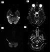
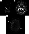
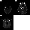
Lacunar strokes are the hallmark of cerebral small vessel disease. There are several well-established mechanisms for the pathogenesis of lacunar stroke, but the cardioembolic mechanism is not well-established. Three cases of acute ischemic stroke following elective cardiac and cerebral catheterization are reported. These cases had typical lacunar-looking infarcts on neuroimaging despite strong evidence of an embolic source with temporal correlation. Awareness of such findings and pathogenesis may help investigational workup and management of these patients.
Keywords: clinical correlation; embolic source; heart catheterization complications; lacunar infarcts; radiological correlation.
Copyright © 2023, Harazeen et al.
Conflict of interest statement
The authors have declared that no competing interests exist.
Figures


Similar articles
-
Clinical Characteristics and Outcome of Patients with Lacunar Infarcts and Concurrent Embolic Ischemic Lesions.Clin Neuroradiol. 2020 Sep;30(3):511-516. doi: 10.1007/s00062-019-00800-5. Epub 2019 Jun 3. Clin Neuroradiol. 2020. PMID: 31161343 Clinical Trial.
-
Silent brain infarcts in 755 consecutive patients with a first-ever supratentorial ischemic stroke. Relationship with index-stroke subtype, vascular risk factors, and mortality.Stroke. 1994 Dec;25(12):2384-90. doi: 10.1161/01.str.25.12.2384. Stroke. 1994. PMID: 7974577
-
Lacunar Stroke.2024 Mar 10. In: StatPearls [Internet]. Treasure Island (FL): StatPearls Publishing; 2025 Jan–. 2024 Mar 10. In: StatPearls [Internet]. Treasure Island (FL): StatPearls Publishing; 2025 Jan–. PMID: 33085363 Free Books & Documents.
-
Acute cardioembolic cerebral infarction: answers to clinical questions.Curr Cardiol Rev. 2012 Feb;8(1):54-67. doi: 10.2174/157340312801215791. Curr Cardiol Rev. 2012. PMID: 22845816 Free PMC article. Review.
-
Is investigating for carotid artery disease warranted in non-cortical lacunar infarction?Stroke. 2011 Jan;42(1):217-20. doi: 10.1161/STROKEAHA.110.600064. Epub 2010 Nov 24. Stroke. 2011. PMID: 21106953 Review.
References
Publication types
LinkOut - more resources
Full Text Sources
