

Effects of autophagy inhibitor 3-methyladenine on a diabetic mice model
- PMID: 37724274
- PMCID: PMC10475630
- DOI: 10.18240/ijo.2023.09.12
Effects of autophagy inhibitor 3-methyladenine on a diabetic mice model
Abstract
Aim: To investigate the role of autophagy inhibitor 3-methyladenine (3-MA) on a diabetic mice model (DM) and the potential mechanism.
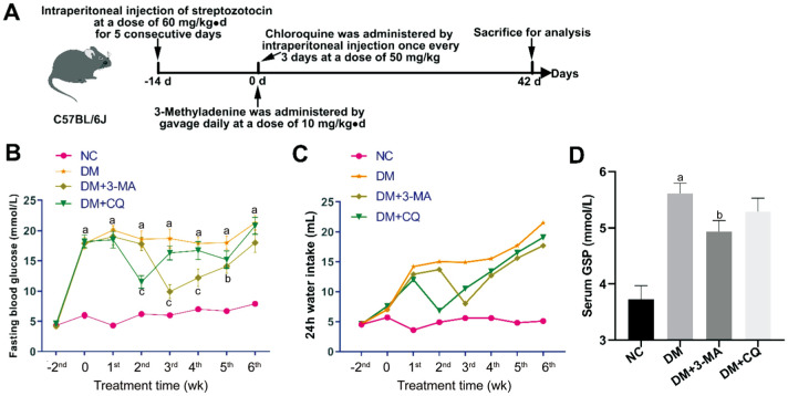
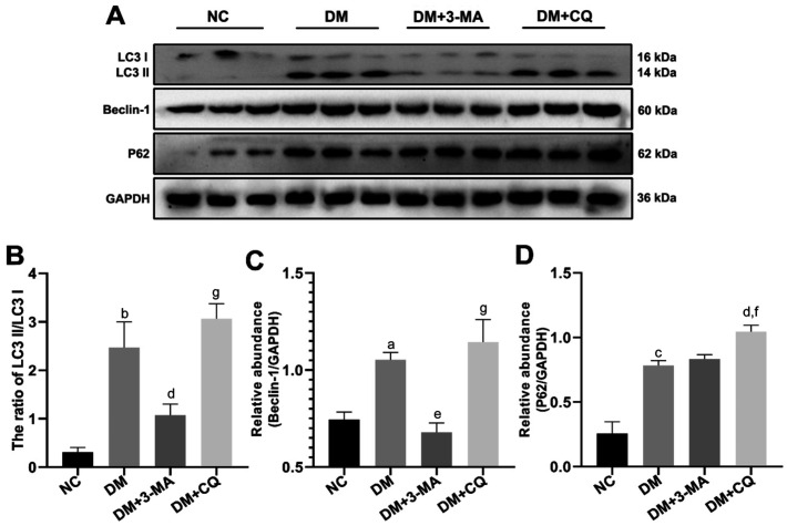
Methods: Male C57BL/6J mice were randomly divided into a normal control group (NC group) and an DM group. DM were induced by multiple low-dose intraperitoneal injection of streptozotocin (STZ) 60 mg/kg·d for 5 consecutive days. DM mice were randomly subdivided into untreated group (DM group), 3-MA (10 mg/kg·d by gavage) treated group (DM+3-MA group) and chloroquine (CQ; 50 mg/kg by intraperitoneal injection) treated group (DM+CQ group). The fasting blood glucose (FBG) levels were recorded every week. At the end of experiment, retinal samples were collected. The expression levels of pro-apoptotic proteins cleaved caspase-3, cleaved poly ADP-ribose polymerase 1 (PARP1) and Bax, anti-apoptotic protein Bcl-2, fibrosis-associated proteins Fibronectin and type 1 collagen α1 chain (COL1A1), vascular endothelial growth factor (VEGF), inflammatory factors interleukin (IL)-1β and tumor necrosis factor (TNF)-α, as well as autophagy related proteins LC3, Beclin-1 and P62 were determined by Western blotting. The oxidative stress indicators 8-hydroxydeoxyguanosine (8-OHdG) and malondialdehyde (MDA) were detected by commercial kits.
Results: Both 3-MA and CQ had short-term hypoglycemic effect on FBG and reduced the expression of VEGF and inflammatory factors IL-1β and TNF-α in DM mice. 3-MA also significantly alleviated oxidative stress indicators 8-OHdG and MDA, decreased the expression of fibrosis-related proteins Fibronectin and COL1A1, pro-apoptotic proteins cleaved caspase-3, cleaved PARP1, as well as the ratio of Bax/Bcl-2. CQ had no significant impact on the oxidative stress indicators, fibrosis, and apoptosis related proteins. The results of Western blotting for autophagy related proteins showed that the ratio of LC3 II/LC3 I and the expression of Beclin-1 in the retina of DM mice were decreased by 3-MA treatment, and the expression of P62 was further increased by CQ treatment.
Conclusion: 3-MA has anti-apoptotic and anti-fibrotic effects on the retina of DM mice, and can attenuate retinal oxidative stress, VEGF expression and the production of inflammatory factors in the retina of DM mice. The underlying mechanism of the above effects of 3-MA may be related to its inhibition of early autophagy and hypoglycemic effect.
Keywords: 3-methyladenine; apoptosis; autophagy; diabetic mellitus; fibrosis; mice.
International Journal of Ophthalmology Press.
Figures


Similar articles
-
Autophagy inhibitor 3-methyladenine attenuates renal injury in streptozotocin-induced diabetic mice.Iran J Basic Med Sci. 2024;27(7):793-800. doi: 10.22038/IJBMS.2024.71378.15518. Iran J Basic Med Sci. 2024. PMID: 38800022 Free PMC article.
-
[Biological role and related mechanism of autophagy in acute lung injury of hemorrhagic shock mice].Zhonghua Wei Zhong Bing Ji Jiu Yi Xue. 2024 Aug;36(8):848-852. doi: 10.3760/cma.j.cn121430-20231007-00844. Zhonghua Wei Zhong Bing Ji Jiu Yi Xue. 2024. PMID: 39238409 Chinese.
-
[Effect of electroacupuncture on myocardial inflammatory injury and apoptosis in mice with acute myocardial ischemia based on VEGF-C/VEGFR-3 pathway].Zhongguo Zhen Jiu. 2022 Nov 12;42(11):1269-77. doi: 10.13703/j.0255-2930.20211226-k0003. Zhongguo Zhen Jiu. 2022. PMID: 36397225 Chinese.
-
[Electroacupuncture promotes gastrointestinal motility by activating autophagy of Cajal interstitial cells via downregulating PI3K/Akt/mTOR signaling pathway in stomach of diabetic gastro-paresis rats].Zhen Ci Yan Jiu. 2022 Dec 25;47(12):1060-7. doi: 10.13702/j.1000-0607.20211241. Zhen Ci Yan Jiu. 2022. PMID: 36571220 Chinese.
-
[Chloroquine Enhances BIIB021-induced Apoptosis in Chronic Myeloid Leukemia Cells Bearing T315I Mutation].Zhongguo Shi Yan Xue Ye Xue Za Zhi. 2022 Aug;30(4):1005-1010. doi: 10.19746/j.cnki.issn.1009-2137.2022.04.005. Zhongguo Shi Yan Xue Ye Xue Za Zhi. 2022. PMID: 35981354 Chinese.
Cited by
-
Regulation role of miR-204 on SIRT1/VEGF in metabolic memory induced by high glucose in human retinal pigment epithelial cells.Int J Ophthalmol. 2024 Jul 18;17(7):1232-1237. doi: 10.18240/ijo.2024.07.06. eCollection 2024. Int J Ophthalmol. 2024. PMID: 39026923 Free PMC article.
-
Autophagy inhibitor 3-methyladenine attenuates renal injury in streptozotocin-induced diabetic mice.Iran J Basic Med Sci. 2024;27(7):793-800. doi: 10.22038/IJBMS.2024.71378.15518. Iran J Basic Med Sci. 2024. PMID: 38800022 Free PMC article.
-
Lacidipine, thiamine pyrophosphate and their combination on the ocular ischemic syndrome induced by bilateral common carotid artery ligation.Int J Ophthalmol. 2024 May 18;17(5):815-821. doi: 10.18240/ijo.2024.05.04. eCollection 2024. Int J Ophthalmol. 2024. PMID: 38766328 Free PMC article.
-
Potential of autophagy in subretinal fibrosis in neovascular age-related macular degeneration.Cell Mol Biol Lett. 2025 Apr 30;30(1):54. doi: 10.1186/s11658-025-00732-8. Cell Mol Biol Lett. 2025. PMID: 40307700 Free PMC article. Review.
-
RTA408 alleviates retinal ganglion cells damage in mouse glaucoma by inhibiting excessive autophagy.PLoS One. 2024 Nov 11;19(11):e0313446. doi: 10.1371/journal.pone.0313446. eCollection 2024. PLoS One. 2024. Retraction in: PLoS One. 2025 Jul 14;20(7):e0327995. doi: 10.1371/journal.pone.0327995. PMID: 39527591 Free PMC article. Retracted.
References
-
- Debele GR, Kanfe SG, Weldesenbet AB, Ayana GM, Jifar WW, Raru TB. Incidence of diabetic retinopathy and its predictors among newly diagnosed type 1 and type 2 diabetic patients: a retrospective follow-up study at tertiary health-care setting of Ethiopia. Diabetes Metab Syndr Obes. 2021;14:1305–1313. - PMC - PubMed
-
- Wong TY, Sabanayagam C. Strategies to tackle the global burden of diabetic retinopathy: from epidemiology to artificial intelligence. Ophthalmologica. 2020;243(1):9–20. - PubMed
LinkOut - more resources
Full Text Sources
Research Materials
Miscellaneous