. 2023 Nov 1;325(5):L675-L688.
doi: 10.1152/ajplung.00281.2022. Epub 2023 Sep 19.

The hemodynamic interplay between pulmonary ischemia-reperfusion injury and right ventricular function in lung transplantation: a translational porcine model

Affiliations

The hemodynamic interplay between pulmonary ischemia-reperfusion injury and right ventricular function in lung transplantation: a translational porcine model

Michaela Orlitová et al. Am J Physiol Lung Cell Mol Physiol. .

Abstract

Lung transplantation (LTx) is a challenging procedure. Following the process of ischemia-reperfusion injury, the transplanted pulmonary graft might become severely damaged, resulting in primary graft dysfunction. In addition, during the intraoperative window, the right ventricle (RV) is at risk of acute failure. The interaction of right ventricular function with lung injury is, however, poorly understood. We aimed to address this interaction in a translational porcine model of pulmonary ischemia-reperfusion injury. Advanced pulmonary and hemodynamic assessment was used, including right ventricular pressure-volume loop analysis. The acute model was based on clamping and unclamping of the left lung hilus, respecting the different hemodynamic phases of a clinical lung transplantation. We found that forcing entire right ventricular cardiac output through a lung suffering from ischemia-reperfusion injury increased afterload (pulmonary vascular resistance from baseline to end experiment P < 0.0001) and induced right ventricular failure (RVF) in 5/9 animals. Notably, we identified different compensation patterns in failing versus nonfailing ventricles (arterial elastance P = 0.0008; stroke volume P < 0.0001). Furthermore, increased vascular pressure and flow produced by the right ventricle resulted in higher pulmonary injury, as measured by ex vivo CT density (correlation: pressure r = 0.8; flow r = 0.85). Finally, RV ischemia as measured by troponin-T was negatively correlated with pulmonary injury (r = -0.76); however, troponin-T values did not determine RVF in all animals. In conclusion, we demonstrate a delicate balance between development of pulmonary ischemia-reperfusion injury and right ventricular function during lung transplantation. Furthermore, we provide a physiological basis for potential benefit of extracorporeal life support technology.NEW & NOTEWORTHY In contrast to the abundant literature of mechanical pulmonary artery clamping to increase right ventricular afterload, we developed a model adding a biological factor of pulmonary ischemia-reperfusion injury. We did not only focus on the right ventricular behavior, but also on the interaction with the injured lung. We are the first to describe this interaction while addressing the hemodynamic intraoperative phases of clinical lung transplantation.

Keywords: lung transplantation; porcine model; pulmonary ischemia-reperfusion injury; right ventricular function.


Conflict of interest statement

No conflicts of interest, financial or otherwise, are declared by the authors.

Figures

None
Graphical abstract
Figure 1.
Figure 1.
A: open chest experimental setup demonstrating the instrumentation. B: clinical lung transplantation (LTx) setting vs. experimental protocol: overview of sampling and parameter recording. AO, aorta; H, heart; LA, left atrium; LL – left lung; PA, pulmonary artery.1—LA pressure catheter; 2—PA flow probe; 3—PA pressure catheter; 4—vascular clamp on left PA and left main bronchus; 5—left PA flow probe; 6—vessel loop around Right PA enabling its closure during 6H; BL, baseline; 1H, 1 hour; 2H, 2 hours; 3H, 3 hours; 4H, 4 hours; 5H, 5 hours; 6H, 6 hours; RL CLA, right PA clamping; Hemodyn., hemodynamic parameters, Resp., respiratory parameters; PV, pressure-volume; W/D, wet-to-dry ratio; BAL, bronchoalveolar lavage; CT, computed tomography.
Figure 2.
Figure 2.
Lung injury assessment. A: wet-to-dry (W/D) ratio reflecting left lung (LL) injury. B: physiological parameters: left pulmonary vein (LPV) Po2 measured at 100% fraction of inspired O2 at baseline (BL) and during reperfusion; lung compliance at baseline and during reperfusion, both reflecting LL injury. C: computed tomography (CT) scan, example of increased density in clamping experimental group (CLA) compared with control experimental group (CON). D: severity of LL injury quantified by CT-measured parenchymal density. E: example of LL histology slide scored as 2 (CON) and 7 (CLA). F: histological lung injury severity score confirming severe LL injury. *P < 0.05; **P < 0.01; ***P < 0.001; ****P < 0.0001. Data presented as individual values and means ±SD; t test (A, D, and F) and two-way ANOVA (B) was used; CON (n = 7), CLA (n = 9). HU, Hounsfield units; RL, right lung; LPV, left pulmonary vein; Po2, partial pressure of oxygen; 4H, 4 hours; 5H, 5 hours; ½ 6H, halfway through 6H; 6H, 6 hours; CON (blue circle), control group; CLA (orange circle), clamping group; gray line (in B) indicates change of hemodynamic phase.
Figure 3.
Figure 3.
Pulmonary hemodynamics and afterload. A: HR, heart rate increased at 6H. B: mPAP, mean pulmonary artery pressure increased both as a response to left lung (LL) (1H) and right lung (RL) clamping (6H). C: RV CO, right ventricular cardiac output decreased throughout the experiment. D: LPA flow, left pulmonary artery flow increased after RL clamping. E: PVR, pulmonary vascular resistance increased in a same trend as mPAP. F: Ea, arterial elastance increased at 6H as a response to mechanical clamping and pulmonary injury; BL, baseline; 1H, 1 Hour; 2H, 2 Hours; 3H, 3 Hours; 4H, 4 Hours; 5H, 5 Hours; 6H, 6 Hours; Clamp LL, left lung clamped after BL measurement; Rep LL, left lung reperfusion; Clamp RL, right lung clamped; Gray lines indicates change of hemodynamic phase. *P < 0.05; **P < 0.01; ****P < 0.0001. Data presented as individual values and means ±SD, values for 1H–6H are an average of the whole experimental hour; two-way ANOVA was used. CON (n = 7), CLA (n = 9).
Figure 4.
Figure 4.
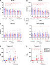
Conductance catheter derived analysis of right ventricular (RV) function and troponin-T plasma levels. A: RV SV, right ventricular stroke volume decreased through the experiment. B: RV SW, right ventricular stroke work decreased through the experiment. C: RV Ees, right ventricular end-systolic elastance showed no difference in time or between groups. D: RV Coupling, right ventricular-pulmonary artery coupling decrease at 6H suggesting uncoupling. E: Troponin-T increase in both control (CON) and clamping (CLA) groups with significant difference between groups at 6H; (F) Troponin-T increased in time in experiments resulting in right ventricular failure (RVF) but did not reach significance in experiments not resulting in right ventricular failure (NO RVF). BL, baseline; 1H, 1 Hour; 2H, 2 Hours; 3H, 3 Hours; 4H, 4 Hours; 5H, 5 Hours; 6H, 6 Hours; Clamp LL, left lung clamped after BL measurement; Rep LL, left lung reperfusion; Clamp RL, right lung clamped. Gray line indicates change of hemodynamic phase. *P < 0.05; **P < 0.01; ***P < 0.001; ****P < 0.0001. Data presented as individual values and means ±SD; two-way ANOVA was used. CON (n = 7), CLA (n = 9); RVF (n = 5), NO RVF (n = 4).
Figure 5.
Figure 5.
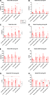
CLA group—comparing failed (RVF) vs. non-failed (NO-RVF) ventricles during the last experimental hour (6H). A: SW, stroke work decreased in RVF compared to No-RVF group after reaching peak. B: Ees, end-systolic elastance had an increasing tendency in RVF group to the point of failure resulting in Ees decrease. C: Ea, arterial elastance showing a different pattern between two groups. D: right ventricular-pulmonary artery coupling demonstrating a trend toward difference in two groups at end experiment. E: HR, heart rate remained comparable in both groups. F: mPAP, mean pulmonary arterial pressure remained comparable in both groups. G: CO, cardiac output decreased at end experiment in RVF group reflecting RV failure. H: SV, stroke volume demonstrating a different pattern between two groups from 10 min after right pulmonary artery (RPA) clamp. CLA, clamping. *P < 0.05; **P < 0.01; ***P < 0.001; ****P < 0.0001. Data presented as individual values and means ±SD; two-way ANOVA was used; RVF (n = 5), NO RVF (n = 4).
Figure 6.
Figure 6.
Correlating hemodynamic parameters, severity of pulmonary injury and cardiac ischemic injury in clamping (CLA) group [including experiments resulting in right ventricular failure (RVF) + experiments not resulting in RVF (NO RVF)]. A: correlation between mean pulmonary artery pressure (mPAP) and computed tomography (CT)-measured pulmonary density. B: correlation between left pulmonary artery (LPA) flow and CT-measured pulmonary density. C: correlation between heart rate (HR) and CT-measured pulmonary density. D: no correlation found between stroke volume (SV) and CT-measured pulmonary density. E: negative correlation between cardiac ischemic injury (Troponin-T) and CT-measured pulmonary density. F: strong negative correlation between left pulmonary artery (LPA) flow and cardiac ischemic injury (Troponin-T). Orange circles, experiment resulted in RVF; tan circles, experiment did not result in RVF. HU, Hounsfield Units; LPA Flow, flow in left pulmonary artery; RVF, right ventricular failure. *P < 0.05; **P < 0.01; ****P < 0.0001. Data presented as individual values; Pearson r was used for correlation assessment; RVF (n = 5), NO RVF (n = 4).

References

    1. Whitson BA, Prekker ME, Herrington CS, Whelan TP, Radosevich DM, Hertz MI, Dahlberg PS. Primary graft dysfunction and long-term pulmonary function after lung transplantation. J Heart Lung Transplant 26: 1004–1011, 2007. doi:10.1016/j.healun.2007.07.018. - DOI - PubMed
    1. Hamilton BC, Kukreja J, Ware LB, Matthay MA. Protein biomarkers associated with primary graft dysfunction following lung transplantation. Am J Physiol Lung Cell Mol Physiol 312: L531–L541, 2017. doi:10.1152/ajplung.00454.2016. - DOI - PMC - PubMed
    1. Den Hengst WA, Gielis JF, Lin JY, Van Schil PE, De Windt LJ, Moens AL. Lung ischemia-reperfusion injury: a molecular and clinical view on a complex pathophysiological process. Am J Physiol Heart Circ Physiol 299: H1283–H1299, 2010. doi:10.1152/ajpheart.00251.2010. - DOI - PubMed
    1. de Perrot M, Liu M, Waddell TK, Keshavjee S. Ischemia-reperfusion-induced lung injury. Am J Respir Crit Care Med 167: 490–511, 2003. doi:10.1164/rccm.200207-670SO. - DOI - PubMed
    1. Van Slambrouck J, Van Raemdonck D, Vos R, Vanluyten C, Vanstapel A, Prisciandaro E, Willems L, Orlitová M, Kaes J, Jin X, Jansen Y, Verleden GM, Neyrinck AP, Vanaudenaerde BM, Ceulemans LJ. A focused review on primary graft dysfunction after clinical lung transplantation: a multilevel syndrome. Cells 11: 745, 2022. doi:10.3390/cells11040745. - DOI - PMC - PubMed

Publication types

LinkOut - more resources