. 2023 Nov 4;33(22):11036-11046.
doi: 10.1093/cercor/bhad343.

Cross-modal exposure restores multisensory enhancement after hemianopia

Affiliations

Cross-modal exposure restores multisensory enhancement after hemianopia

Naomi L Bean et al. Cereb Cortex. .

Abstract

Hemianopia is a common consequence of unilateral damage to visual cortex that manifests as a profound blindness in contralesional space. A noninvasive cross-modal (visual-auditory) exposure paradigm has been developed in an animal model to ameliorate this disorder. Repeated stimulation of a visual-auditory stimulus restores overt responses to visual stimuli in the blinded hemifield. It is believed to accomplish this by enhancing the visual sensitivity of circuits remaining after a lesion of visual cortex; in particular, circuits involving the multisensory neurons of the superior colliculus. Neurons in this midbrain structure are known to integrate spatiotemporally congruent visual and auditory signals to amplify their responses, which, in turn, enhances behavioral performance. Here we evaluated the relationship between the rehabilitation of hemianopia and this process of multisensory integration. Induction of hemianopia also eliminated multisensory enhancement in the blinded hemifield. Both vision and multisensory enhancement rapidly recovered with the rehabilitative cross-modal exposures. However, although both reached pre-lesion levels at similar rates, they did so with different spatial patterns. The results suggest that the capability for multisensory integration and enhancement is not a pre-requisite for visual recovery in hemianopia, and that the underlying mechanisms for recovery may be more complex than currently appreciated.

Keywords: hemianopia; multisensory integration; rehabilitation; vision.


Figures

Fig. 1
Fig. 1
Behavioral apparatus, experimental series, and the cortical lesion. (A) Animals were trained and evaluated in a perimetry apparatus using target locations arranged along the azimuth spanning the central 180° of space in 15° intervals. Only the central 120° was used (the 0° target location was used for fixation only). Each location had LED (3) and speaker (2) complexes, but only the left-most components were used here (adapted from Gingras et al. 2009). (B) Two animals participated in multisensory testing prior to the lesion. Following that, all animals were trained and tested in the same manner in the same time span. Visual alone performance was assessed prior to and just after the lesion. Auditory performance was also assessed prior to post-lesion multisensory testing. During rehabilitation, a single multisensory session was conducted. Animals were assessed for multisensory integration a final time after rehabilitation (not to scale, see boxes for timeline). (C) The extent of the visual cortex lesion in an exemplar animal is indicated by shading shown in a dorsal view (left). Three coronal slices through the lesion (shading) are also shown to the right. A = anterior; P = posterior.
Fig. 2
Fig. 2
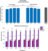
Pre-lesion performance. (A) All animals readily approached visual stimuli at each tested location (histogram bars) with no significant interhemispheric differences. They generally remained at the start location on catch trials (gray bar). (B) Shown are the percent of correct approach responses to the visual (V, blue, leftmost bar) and auditory (A, red, middle bar) stimuli, and their cross-modal combination (VA, purple, rightmost bar). Note that VA performance was significantly enhanced above statistical facilitation (SF, green dashed lines) at each location. The bars show the average group performance, and open circles show the performance of individual animals. **P < **P < 0.001; ns = non-significant.
Fig. 3
Fig. 3
Post-lesion visual and multisensory performance. Conventions are the same as in previous figures. (A) The visual performance rate was reduced to chance levels in the blinded hemifield but remained high in the intact hemifield (dashed line = spontaneous approach rate). (B) Multisensory enhancement was lost in the contralesional hemifield but retained in the ipsilesional hemifield. **P < 0.001; ns = non-significant.
Fig. 4
Fig. 4
Multisensory performance during rehabilitation. Conventions are the same as in previous figures. (A) Vision returned to the blinded hemifield in a central-to-peripheral pattern. Note that after five exposure sessions visual performance at −15° was higher than at any other location, and that performance was degraded at each 15° increment of eccentricity. Criterion performance was reached at all tested locations after 7–12 sessions. Shading indicates the timing of the multisensory probe tests. (B) Although visual recovery was evident at all locations, multisensory enhancement was not. It was elicited only when the cross-modal stimuli were presented at the exposure site (−45°). Its magnitude approached that found in the intact hemifield. *P < 0.05; **P < 0.001; ns = non-significant.
Fig. 5
Fig. 5
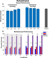
Visual and multisensory performance after rehabilitation. Conventions are the same as in previous figures. (A) Visual performance returned to pre-lesion levels for each animal and each tested location. (B) Multisensory enhancement was also restored in each animal at each location. **P < 0.001; ns = non-significant.

References

    1. Bean N, Stein B, Rowland B. Stimulus value gates multisensory integration. Eur J Neurosci. 2021:53(9):3142–3159. - PMC - PubMed
    1. Bean NL, Smyre SA, Stein BE, Rowland BA. Noise-rearing precludes the behavioral benefits of multisensory integration. Cereb Cortex. 2023:33(4):948–958. - PMC - PubMed
    1. Bell AH, Corneil BD, Meredith MA, Munoz DP. The influence of stimulus properties on multisensory processing in the awake primate superior colliculus. Can J Exp Psychol Rev Can Psychol Expérimentale. 2001:55(2):123–132. - PubMed
    1. Benedetti F. Orienting behaviour and superior colliculus sensory representations in mice with the vibrissae bent into the contralateral hemispace. Eur J Neurosci. 1995:7(7):1512–1519. - PubMed
    1. Burnett LR, Stein BE, Chaponis D, Wallace MT. Superior colliculus lesions preferentially disrupt multisensory orientation. Neuroscience. 2004:124(3):535–547. - PubMed

Publication types

LinkOut - more resources