. 2024 Jan;70(1):11-25.
doi: 10.1165/rcmb.2023-0127OC.

Amphipathic Helical Peptide L37pA Protects against Lung Vascular Endothelial Dysfunction Caused by Truncated Oxidized Phospholipids via Antagonism with CD36 Receptor

Affiliations

Amphipathic Helical Peptide L37pA Protects against Lung Vascular Endothelial Dysfunction Caused by Truncated Oxidized Phospholipids via Antagonism with CD36 Receptor

Pratap Karki et al. Am J Respir Cell Mol Biol. 2024 Jan.

Abstract

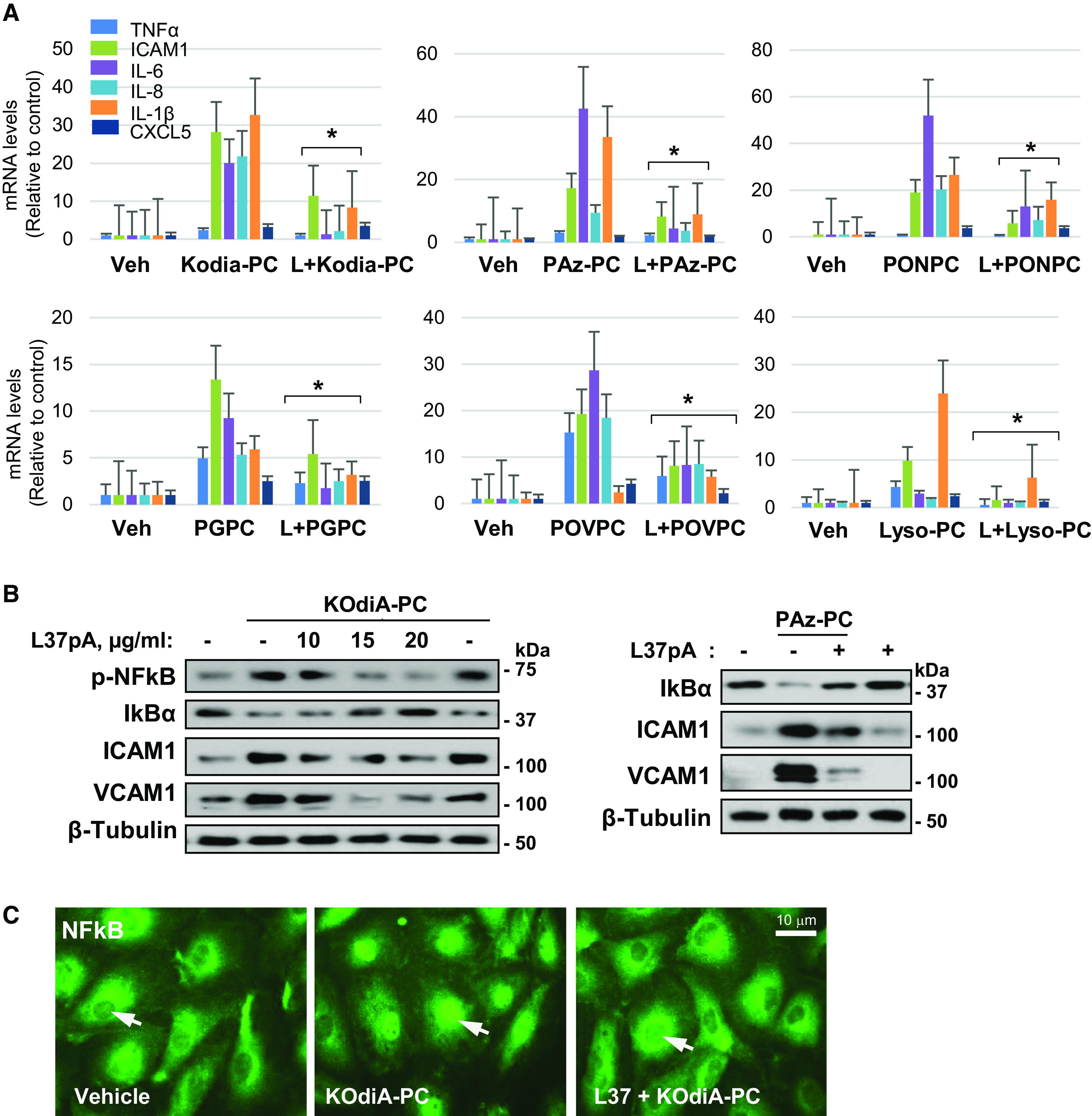
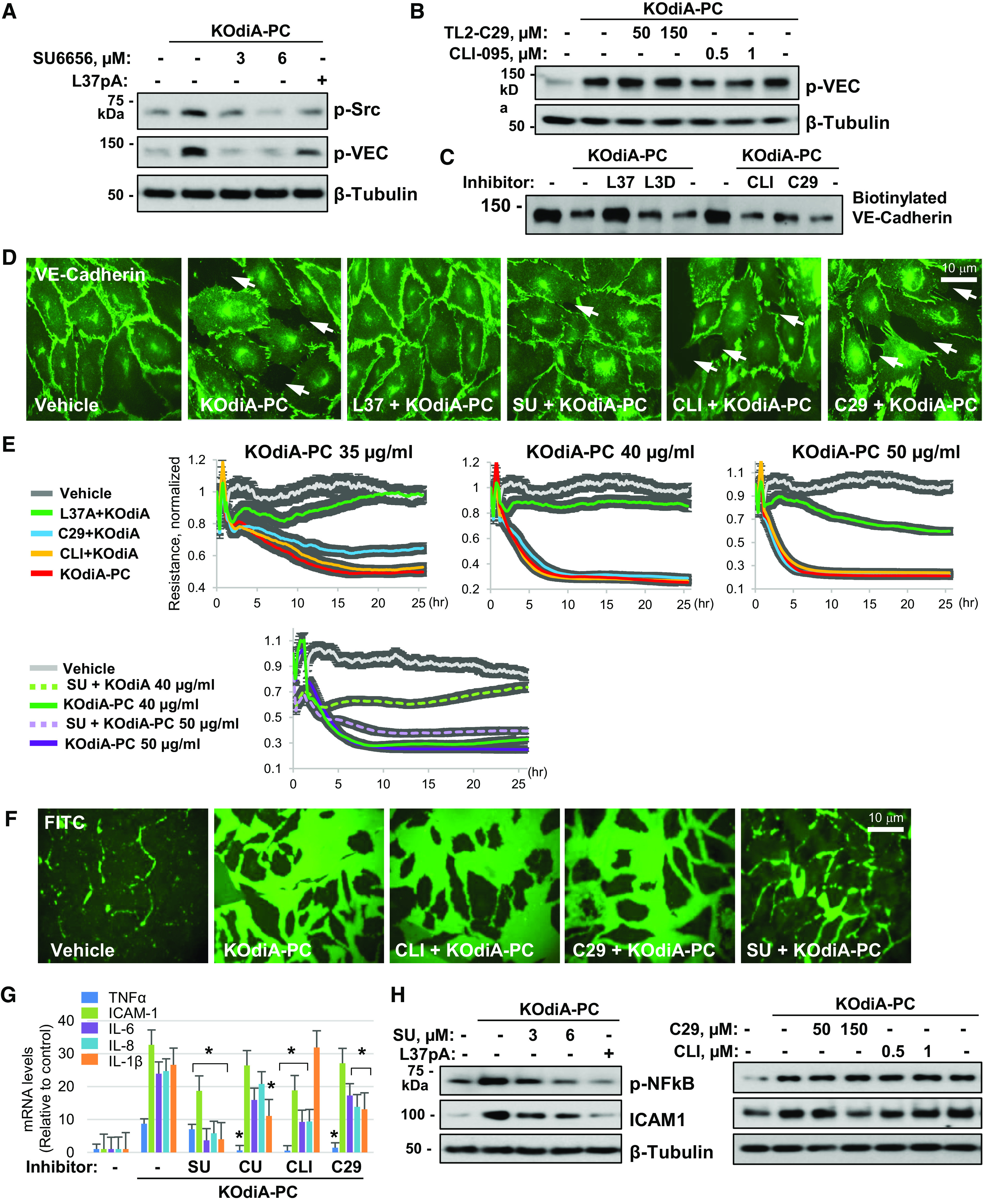
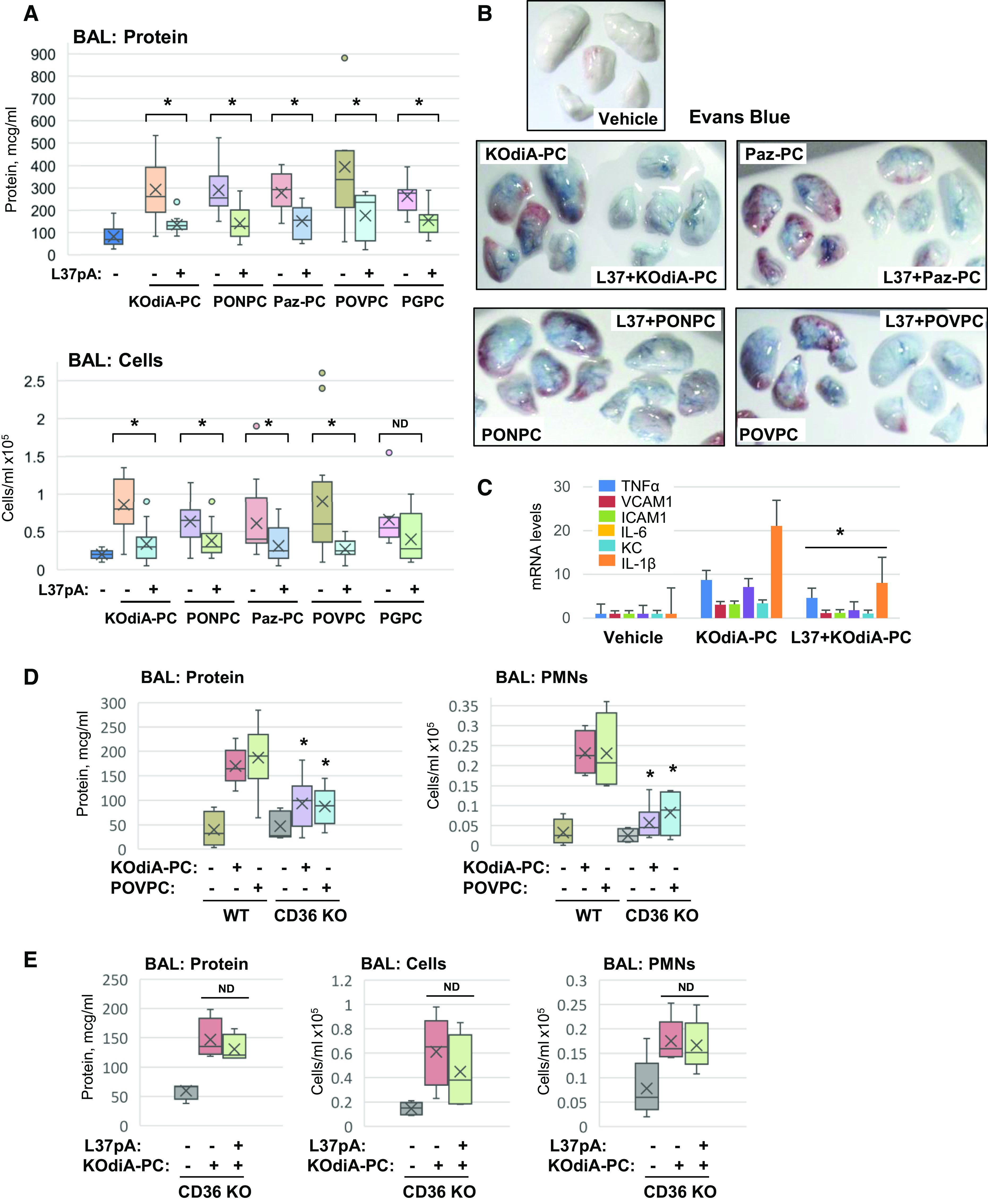
The generation of bioactive truncated oxidized phospholipids (Tr-OxPLs) from oxidation of cell-membrane or circulating lipoproteins is a common feature of various pathological states. Scavenger receptor CD36 is involved in lipid transport and acts as a receptor for Tr-OxPLs. Interestingly, Tr-OxPLs and CD36 are involved in endothelial dysfunction-derived acute lung injury, but the precise mechanistic connections remain unexplored. In the present study, we investigated the role of CD36 in mediating pulmonary endothelial cell (EC) dysfunction caused by Tr-OxPLs. Our results demonstrated that the Tr-OxPLs KOdia-PC, Paz-PC, PGPC, PON-PC, POV-PC, and lysophosphocholine caused an acute EC barrier disruption as revealed by measurements of transendothelial electrical resistance and VE-cadherin immunostaining. More importantly, a synthetic amphipathic helical peptide, L37pA, targeting human CD36 strongly attenuated Tr-OxPL-induced EC permeability. L37pA also suppressed Tr-OxPL-induced endothelial inflammatory activation monitored by mRNA expression of inflammatory cytokines/chemokines and adhesion molecules. In addition, L37pA blocked Tr-OxPL-induced NF-κB activation and tyrosine phosphorylation of Src kinase and VE-cadherin. The Src inhibitor SU6656 attenuated KOdia-PC-induced EC permeability and inflammation, but inhibition of the Toll-like receptors (TLRs) TLR1, TLR2, TLR4, and TLR6 had no such protective effects. CD36-knockout mice were more resistant to Tr-OxPL-induced lung injury. Treatment with L37pA was equally effective in ameliorating Tr-OxPL-induced vascular leak and lung inflammation as determined by an Evans blue extravasation assay and total cell and protein content in BAL fluid. Altogether, these results demonstrate an essential role of CD36 in mediating Tr-OxPL-induced EC dysfunction and suggest a strong therapeutic potential of CD36 inhibitory peptides in mitigating lung injury and inflammation.

Keywords: CD36; L37pA; acute lung injury; endothelial dysfunction; truncated oxidized phospholipids.


Figures

Figure 1.
Figure 1.
Truncated oxidized phospholipids (Tr-OxPLs) cause endothelial barrier disruption. (A) Chemical structures of the Tr-OxPLs used in this study. (B) Measurements of transendothelial electrical resistance (TER) in human pulmonary artery endothelial cells (HPAECs) stimulated with the indicated concentrations of Tr-OxPLs. Normalized resistance is plotted versus time to demonstrate dose-dependent endothelial permeability induced by Tr-OxPLs. (C) Western blot detection of phospho–VE-cadherin (Tyr731). HPAECs were exposed to 50 μg/ml 1-palmitoyl-2-glutaroyl-sn-glycero-phosphocholine (PGPC), 1-palmitoyl-2-azelaoyl-sn-glycero-3-phosphocholine (Paz-PC), 1-palmitoyl-2-(9-oxo-nonanoyl)-sn-glycero-3-phosphocholine (PONPC), O-1-O-Palmitoyl-2-O-(5,8-dioxo-8-hydroxy-6-octenoyl)-L-glycero-3-phosphocholine (KOdiA-PC), or 1-palmitoyl-2-(5-oxovaleroyl)-sn-glycero-phosphocholine (POVPC) or 20 μg/ml lysophosphocholine (Lyso-PC; 6 h; see Methods). Reprobing with pan–VE-cadherin antibody was used to verify equal protein loading. (D) VE-cadherin surface biotinylation assay of HPAECs treated with KOdiA-PC (50 μg/ml, 6 h). Equal amounts of cell lysates were run as input controls. (E) Immunofluorescence staining of VE-cadherin in HPAEC monolayers treated with 50 μg/ml KOdiA-PC for the indicated time periods or cells treated with PONPC, POVPC, PGPC, or Lyso-PC for 12 hours. Scale bars, 10 μm. WCL = whole cell lysates.
Figure 2.
Figure 2.
Tr-OxPLs induce endothelial inflammation. (A) Real-time quantitative PCR analysis of TNF-α, VCAM-1, ICAM-1, IL-6, IL-8, and CXCL5 transcripts in HPAECs stimulated with the indicated doses of Tr-OxPLs. Bar graphs present fold changes in mRNA expression against nonstimulated controls (n = 4; *P < 0.05). (B, C) Protein expression levels of IκBα, ICAM-1, and VCAM-1 in HPAECs treated with the indicated doses of KOdiA-PC for 6 hours (left) or 50 μg/ml KOdiA-PC for the indicated times (right). (D) Western analysis of ICAM-1 protein levels in HPAECs exposed to 30–50 μg/ml KOdiA-PC, Paz-PC, PGPC, POVPC, PONPC, or Lyso-PC (see Methods). Reprobing for β-tubulin was used as loading control.
Figure 3.
Figure 3.
CD36-inhibitory peptide L37pA rescues Tr-OxPL–induced endothelial hyperpermeability. (A) HPAECs were stimulated with the indicated doses of Tr-OxPLs (solid lines) with or without L37pA pretreatment (25 μg/ml, 30 min) (dotted lines). Endothelial permeability was determined by monitoring TER over time. (B) Cells were treated with L37pA (15 or 25 μg/ml) or L3D (25 μg/ml), followed by stimulation with KodiA-PC (50 μg/ml, 6 h). Express permeability testing was performed as described in Methods. FITC fluorescence images were taken to evaluate endothelial cell (EC) macromolecular permeability; DAPI staining was used to visualize cell nuclei. Scale bars, 20 μm. (C) VE-cadherin immunostaining of HPAECs exposed to the indicated Tr-OxPLs with or without L37A pretreatment. Scale bars, 10 μm. (D) Immunostaining of HPAEC monolayers treated with KodiA-PC (50 μg/ml, 15 min) with or without L37pA (25 μg/ml) with phospho–VE-cadherin antibody. Scale bars, 10 μm. (E) Western blot detection of phospho-Src and phospho–VE-cadherin levels in HPAECs pretreated with L37pA or L3D (25 μg/ml, 30 min) following KOdiA-PC (50 μg/ml, 6 h) exposure. Reprobing for β-actin or β-tubulin was used as loading controls.
Figure 4.
Figure 4.
L37pA attenuates Tr-OxPL–induced endothelial inflammation. (A) Effect of L37pA pretreatment (25 μg/ml, 30 min) on transcriptional activation of TNF-α, VCAM-1, ICAM-1, IL-6, IL-8, and CXCL5 by Tr-OxPL compounds (50 μg/ml, 6 h). Bar graphs depict fold change in mRNA expression compared with control nonstimulated cells. *P < 0.05 vs. respective OxPLs. (B) Effect of L37pA pretreatment (10, 15, or 20 μg/ml, 30 min) on protein levels of phospho–NF-κB, IκBα, ICAM-1, and VCAM-1 in HPAECs exposed to KOdiA-PC or Paz-PC (50 μg/ml, 6 h). Reprobing for β-tubulin was used to verify equal loading. (C) Immunofluorescence detection of NF-κB nuclear translocation in HPAECs treated with KOdiA-PC (50 μg/ml) in the absence or presence of L37pA (25 μg/ml). Scale bars, 10 μm. Veh = vehicle.
Figure 5.
Figure 5.
Posttreatment with L37pA rescues Tr-OxPL–induced endothelial dysfunction. (A) HPAECs were exposed to KOdiA-PC, Paz-PC, POVPC, PGPC, PONPC (50 μg/ml), or Lyso-PC (20 μg/ml), followed by the addition of L37pA (25 μg/ml; see Methods) 1, 2, or 6 hours after Tr-OxPL challenge. TER was monitored over time. The data are presented as normalized resistance. (B) Cells were treated with L37pA (25 μg/ml) 30 min before, concomitantly with, or 1, 2, or 4 hours after KOdiA-PC (50 μg/ml) challenge. Real-time PCR was performed to determine the mRNA levels of indicated inflammatory genes. *P < 0.05 vs. KOdia-PC. (C) Effects of L37pA before (pre), during (Co, co-treatment), and after treatment on KOdiA-PC–induced IκBα protein degradation detected by Western blot analysis.
Figure 6.
Figure 6.
Signaling mechanisms of Tr-OxPL–induced EC permeability and inflammation. (A) Western blot analysis of the effects of the Src inhibitor SU6656 (3 and 6 μM) or L37pA (25 μg/ml) on phospho-Src and phospho–VE-cadherin levels in HPAECs exposed to KOdiA-PC (50 μg/ml, 6 h). Reprobing for β-tubulin was used to verify equal loading. (B) Effects of TLR2 inhibitor TL2-C29 (50 and 150 μM) or TLR4 inhibitor CLI-095 (0.5 and 1 μM) on KOdiA-PC–induced VE-cadherin phosphorylation. (C) Surface biotinylation assay: effects of L37pA, L3D (25 μg/ml), CLI-095 (1 μM), and TL2-C29 (50 μM) on KOdiA-PC–induced VE-cadherin internalization. (D) Visualization and VE-cadherin–positive adherens junctions in KOdiA-PC–challenged HPAECs pretreated with L37pA, L3D, CLI-095, or TL2-C29 as described above. Representative immunostaining of HPAEC monolayers with VE-cadherin antibody is shown. Scale bars, 10 μm. (E) Measurements of TER in HPAEC monolayers stimulated with the indicated doses of KOdiA-PC in the presence of L37pA (25 μg/ml), TL2-C29 (50 μg/ml), CLI-095 (1 μM), or SU6656 (6 μM). (F) Express permeability testing for EC macromolecule permeability in KOdiA-PC–challenged HPAEC monolayers preincubated with CLI-095 (1 μM), TL2-C29 (50 μM), or SU6656 (3 μM) for 30 minutes. Scale bars, 10 μm. (G) Effects of Src, TLR2, and TLR4 inhibitors on KOdiA-PC–induced transcriptional activation of inflammatory marker genes (n = 4; *P < 0.05 vs. KOdiA-PC alone). (H) Western blot analysis of phospho–NF-κB and ICAM1 protein levels in KOdiA-PC–stimulated cells incubated in the presence of Src, TLR2, and TLR4 inhibitors.
Figure 7.
Figure 7.
L37pA rescues Tr-OxPL–induced acute lung injury in mice. C57/BL6J mice were injected with the indicated Tr-OxPLs (10 mg/kg; i.v., jugular vein) with or without L37pA (20 mg/kg, retroorbital sinus injection) cotreatment. (A) Analysis of BAL total cell counts and protein content 18 hours after treatments (n = 10; *P< 0.05). (B) Evans blue dye extravasation assay in mice challenged with Tr-OxPLs with and without L37pA cotreatment as described in Methods. Shown are representative lung images from four independent experiments. (C) Real-time-PCR analysis of inflammatory marker transcripts in lung tissue presented as fold change in mRNA expression over basal levels (n = 3; *P < 0.05). (D) Analysis of BAL protein and polymorphonuclear leukocyte content in wild-type and CD36-KO mice exposed to KOdiA-PC and PGPC (n = 6; *P < 0.05). (E) Analysis of BAL protein content and cell counts in CD36-knockout mice treated with KOdiA-PC with or without L37pA (n = 3). PMN = polymorphonuclear leukocyte; KO = knockout.

Comment in

References

    1. Bochkov VN, Oskolkova OV, Birukov KG, Levonen AL, Binder CJ, Stöckl J. Generation and biological activities of oxidized phospholipids. Antioxid Redox Signal . 2010;12:1009–1059. - PMC - PubMed
    1. Bochkov V, Gesslbauer B, Mauerhofer C, Philippova M, Erne P, Oskolkova OV. Pleiotropic effects of oxidized phospholipids. Free Radic Biol Med . 2017;111:6–24. - PubMed
    1. Watson AD, Leitinger N, Navab M, Faull KF, Hörkkö S, Witztum JL, et al. Structural identification by mass spectrometry of oxidized phospholipids in minimally oxidized low density lipoprotein that induce monocyte/endothelial interactions and evidence for their presence in vivo. J Biol Chem . 1997;272:13597–13607. - PubMed
    1. Witztum JL, Berliner JA. Oxidized phospholipids and isoprostanes in atherosclerosis. Curr Opin Lipidol . 1998;9:441–448. - PubMed
    1. Birukova AA, Starosta V, Tian X, Higginbotham K, Koroniak L, Berliner JA, et al. Fragmented oxidation products define barrier disruptive endothelial cell response to OxPAPC. Transl Res . 2013;161:495–504. - PMC - PubMed

LinkOut - more resources