

Androgen receptor and its correlation with estrogen and progesterone receptors, aimed for identification of cases for future anti-androgen therapy in endometrial cancers
- PMID: 37725629
- PMCID: PMC10508627
- DOI: 10.1371/journal.pone.0291361
Androgen receptor and its correlation with estrogen and progesterone receptors, aimed for identification of cases for future anti-androgen therapy in endometrial cancers
Abstract
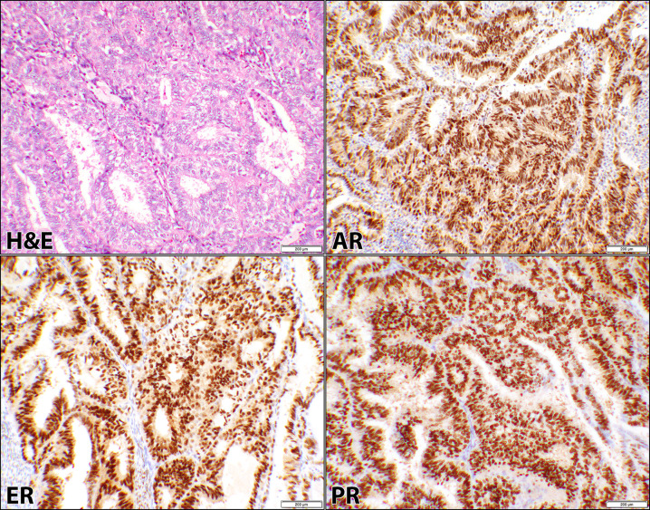
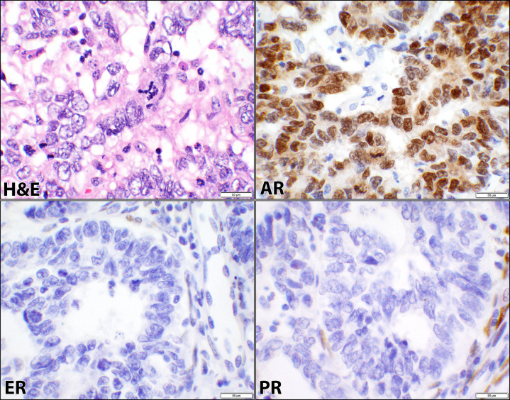
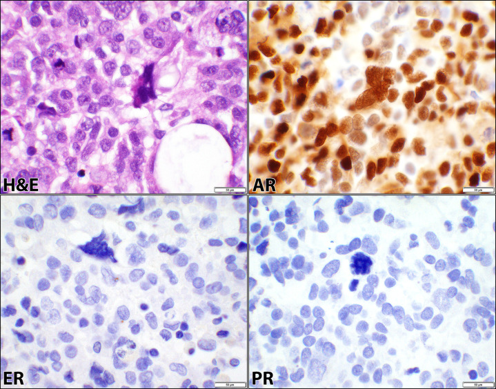
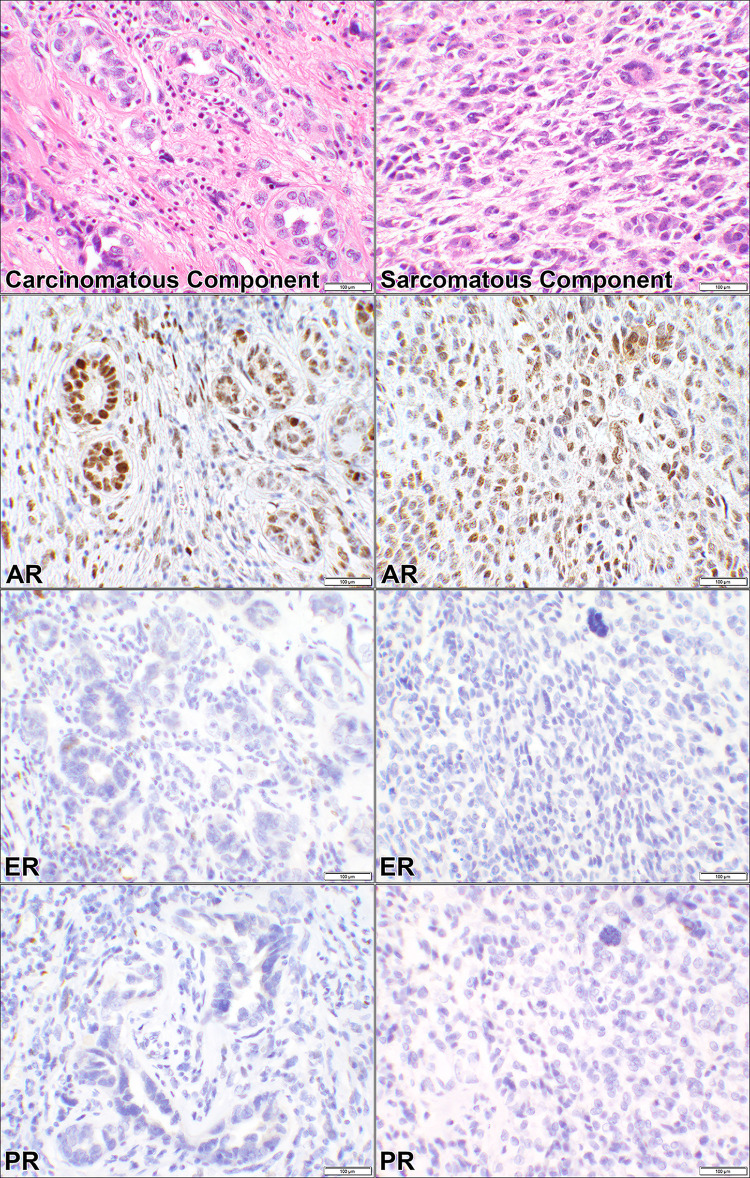
Introduction: The expression of androgen receptor (AR) is not commonly tested or studied in uterine cancers, unlike estrogen receptor (ER) and progesterone receptor (PR) which are positive in most endometrial carcinomas. In this series, we evaluated the expression of AR and its comparison to ER and PR in different types of endometrial cancers and have reviewed the literature.
Materials and methods: The status of AR, ER, and PR expression were evaluated in 71 cases which were categorized into endometrial endometrioid cancer (EEC), non-endometrioid endometrial cancers (NEEC), and metastatic carcinomas of endometrium. Expression of the receptors were compared to each other as well as to mismatch repair proteins (MMR), p53, and body mass index (BMI) using Fisher's Exact test in the StatPlus software.
Results: In EECs, the positivity was 97% for all the three receptors. In NEEC, positivity rates were 68%, 48%, and 35% for AR, ER, and PR respectively. In Metastatic carcinomas, AR and ER positivity was seen in 100% while PR was positive in 75% of the cases. In all cancers, the rates were 17% (11/66) for MMR loss, 57% (30/53) for p53 aberrant expression, and 76% (54/71) for the patients with BMI of ≥ 25 (kg/m2).
Conclusion: AR is expressed in a high percentage of endometrial cancers. Its significance is more evident in high-grade NEEC where ER and PR may not be expressed. These findings warrant further evaluation of AR expression and candidacy of this pathway as a potential therapeutic target in endometrial cancers.
Copyright: This is an open access article, free of all copyright, and may be freely reproduced, distributed, transmitted, modified, built upon, or otherwise used by anyone for any lawful purpose. The work is made available under the Creative Commons CC0 public domain dedication.
Conflict of interest statement
Authors have no competing interests.
Figures
Similar articles
-
Significance of androgen receptor and its potential for anti-androgen/androgen receptor-antagonist therapy in ovarian cancers.PLoS One. 2025 May 20;20(5):e0322744. doi: 10.1371/journal.pone.0322744. eCollection 2025. PLoS One. 2025. PMID: 40392873 Free PMC article.
-
Androgen Receptor Expression in Endometrial Carcinoma.Int J Gynecol Pathol. 2018 Mar;37(2):167-173. doi: 10.1097/PGP.0000000000000401. Int J Gynecol Pathol. 2018. PMID: 28582344
-
To study the expression of estrogen, progesterone receptor and p53 immunohistochemistry markers in subtyping endometrial carcinoma.Indian J Pathol Microbiol. 2024 Jan-Mar;67(1):62-67. doi: 10.4103/ijpm.ijpm_568_22. Indian J Pathol Microbiol. 2024. PMID: 38358190
-
Should progesterone and estrogen receptors be assessed for predicting the response to conservative treatment of endometrial hyperplasia and cancer? A systematic review and meta-analysis.Acta Obstet Gynecol Scand. 2019 Aug;98(8):976-987. doi: 10.1111/aogs.13586. Epub 2019 Mar 28. Acta Obstet Gynecol Scand. 2019. PMID: 30779338
-
Hormone receptors in breast cancer: more than estrogen receptors.Medicina (B Aires). 2019;79(Spec 6/1):540-545. Medicina (B Aires). 2019. PMID: 31864223 Review. English.
Cited by
-
Significance of androgen receptor and its potential for anti-androgen/androgen receptor-antagonist therapy in ovarian cancers.PLoS One. 2025 May 20;20(5):e0322744. doi: 10.1371/journal.pone.0322744. eCollection 2025. PLoS One. 2025. PMID: 40392873 Free PMC article.
-
Introducing the PLOS collection on rare cancer.PLoS One. 2024 Jul 31;19(7):e0308087. doi: 10.1371/journal.pone.0308087. eCollection 2024. PLoS One. 2024. PMID: 39083545 Free PMC article.
-
FBXW7 Directly Ubiquitinates and Degrades CTNNB1 Mediating the Suppression of ENKUR in Endometrial Cancer.Int J Biol Sci. 2025 Feb 10;21(4):1801-1818. doi: 10.7150/ijbs.104067. eCollection 2025. Int J Biol Sci. 2025. PMID: 39990660 Free PMC article.
References
-
- Matias-Guiu X, Longacre TA, McCluggage WG, Nucci MR, Oliva E. Tumours of the uterine corpus. In: Kim K-R, Lax SF, Lazar AJ, Longacre TA, Malpica A, Matias-Guiu X, Nucci MR, Oliva E, editors. Female Genital Tumors. 2020. p. 246–67.
Publication types
MeSH terms
Substances
LinkOut - more resources
Full Text Sources
Research Materials
Miscellaneous
